. 2013 Jul 12;288(28):20745-57.
doi: 10.1074/jbc.M112.410720. Epub 2013 May 29.

Accelerated development of pulmonary fibrosis via Cu,Zn-superoxide dismutase-induced alternative activation of macrophages

Affiliations

Accelerated development of pulmonary fibrosis via Cu,Zn-superoxide dismutase-induced alternative activation of macrophages

Chao He et al. J Biol Chem. .

Abstract

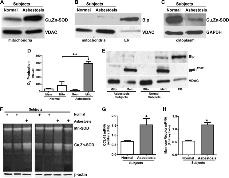
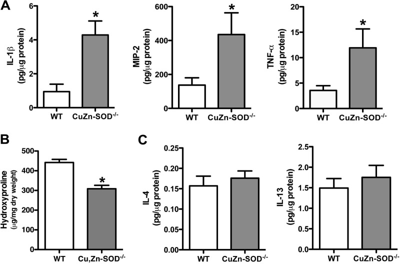
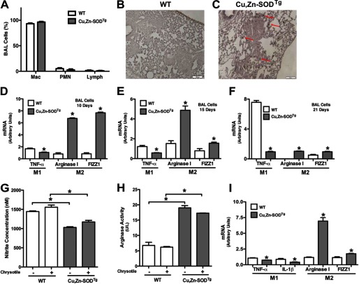
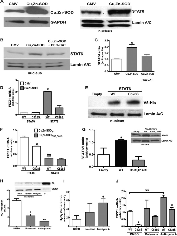
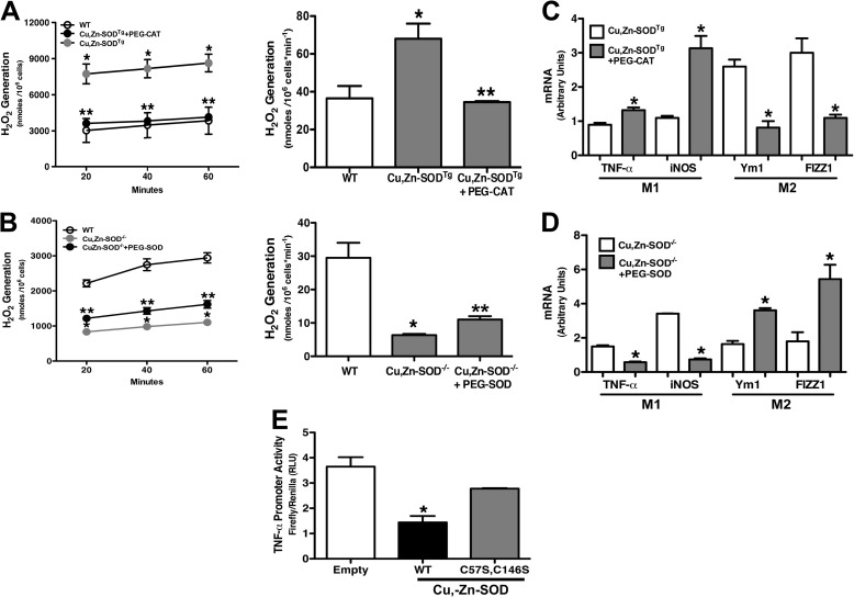
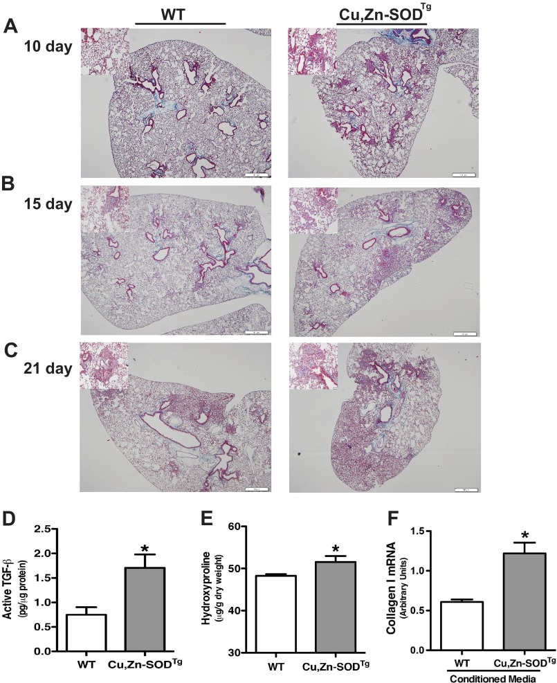
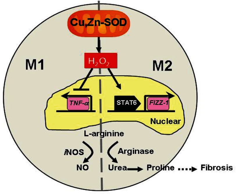
Macrophages not only initiate and accentuate inflammation after tissue injury, but they are also involved in resolution and repair. This difference in macrophage activity is the result of a differentiation process to either M1 or M2 phenotypes. M1 macrophages are pro-inflammatory and have microbicidal and tumoricidal activity, whereas the M2 macrophages are involved in tumor progression and tissue remodeling and can be profibrotic in certain conditions. Because mitochondrial Cu,Zn-superoxide dismutase (Cu,Zn-SOD)-mediated H2O2 is crucial for development of pulmonary fibrosis, we hypothesized that Cu,Zn-SOD modulated the macrophage phenotype. In this study, we demonstrate that Cu,Zn-SOD polarized macrophages to an M2 phenotype, and Cu,Zn-SOD-mediated H2O2 levels modulated M2 gene expression at the transcriptional level by redox regulation of a critical cysteine in STAT6. Furthermore, overexpression of Cu,Zn-SOD in mice resulted in a profibrotic environment and accelerated the development of pulmonary fibrosis, whereas polarization of macrophages to the M1 phenotype attenuated pulmonary fibrosis. Taken together, these observations provide a novel mechanism of Cu,Zn-SOD-mediated and Th2-independent M2 polarization and provide a potential therapeutic target for attenuating the accelerated development of pulmonary fibrosis.

Keywords: Hydrogen Peroxide; Macrophage Phenotype; Mitochondria; Oxidative Stress; Pulmonary Fibrosis; STAT6; Superoxide Dismutase (SOD).


Figures

FIGURE 1.
FIGURE 1.
Alveolar macrophages from asbestosis patients have an M2 phenotype. Mitochondria and cytoplasm were isolated from alveolar macrophages obtained from normal subjects (n = 3) and asbestosis patients (n = 3). A, a representative immunoblot analysis for Cu,Zn-SOD in mitochondria is shown. B, shown is a representative immunoblot analysis for Bip and VDAC in the mitochondrial fractionations. ER, endoplasmic reticulum. C, shown is a representative immunoblot analysis for Cu,Zn-SOD in cytoplasm in alveolar macrophages. D, membrane and mitochondria were isolated from alveolar macrophages from asbestosis patients and normal subjects. Membrane (Mem) and mitochondrial (Mito) protein lysates were used for measuring superoxide anion generation utilizing lucigenin assay. The superoxide generation rate (real light units s−1) was measured in both mitochondrial and membrane fractions. n = 3. *, p < 0.05 versus asbestosis membrane. **, p < 0.05 versus asbestosis mitochondria. E, mitochondria, membrane, and endoplasmic reticulum from asbestosis patients and normal volunteers were isolated, and immunoblot analysis for VDAC, gp91phox, and Bip was performed. F, Mn-SOD activity and Cu,Zn-SOD activity of alveolar macrophages from asbestosis patients (n = 3) and normal subjects (n = 4) were measured by native gel with nitroblue tetrazolium staining. Total RNA of alveolar macrophages from asbestosis patients and normal volunteers were isolated, and C-C motif ligand 18 (CCL-18; G) and mannose receptor gene expression (H) were measured. Results show arbitrary units normalized to hypoxanthine-guanine phosphoribosyltransferase mRNA. *, p < 0.05 versus normal subjects (n = 3).
FIGURE 2.
FIGURE 2.
Cu,Zn-SOD/ macrophages have increased M1 markers. A, WT and Cu,Zn-SOD−/− mice were exposed to 100 μg of chrysotile asbestos intratracheally. IL-1β, MIP-2, and TNF-α cytokine levels were measured in BAL fluid of WT and Cu,Zn-SOD−/− mice 21 days after asbestos exposure. *, p < 0.05 versus WT. (n = 6 per each group). B, extracellular collagen deposition in lung of WT and Cu,Zn-SOD−/− mice after initial asbestos exposure was determined using hydroxyproline assay. *, p < 0.05 versus WT. (n = 4 per each group). C, IL-4 and IL-13 cytokine levels were measured in BAL fluid of WT and Cu,Zn-SOD−/− 21 days after initial asbestos exposure (n = 11 per each group).
FIGURE 3.
FIGURE 3.
Cu,Zn-SOD induces macrophage M2 polarization. WT and Cu,Zn-SODTg mice were exposed to 100 μg of chrysotile asbestos intratracheally. A, BAL was performed 21 days after exposure, and cell differential was determined with Wright-Giemsa stain (n = 3). Mac, macrophage; PMN, polymorphonuclear cell. Lung sections of both WT (B) and Cu,Zn-SODTg (C) were processed for arginase I immunohistochemistry staining. Representative micrographs of one of three animals per genotype are shown. Magnification is 10×, and the bar indicates 100 μm. Macrophages isolated from WT and Cu,Zn-SODTg mice were cultured in the presence or absence of chrysotile asbestos overnight. 10 (D), 15 (E), or 21 (F) days after asbestos exposure the animals were euthanized, and alveolar macrophages were isolated by BAL and exposed in vitro to chrysotile for 4 h. Total RNA was isolated, and TNF-α, arginase I, and FIZZ1 gene expression were measured. Results show arbitrary units normalized to β-actin mRNA. *, p < 0.05 versus WT (n = 4 per each group). G, nitrite concentration was measured in cell culture medium. H, cell lysates were used to determine arginase activity by measuring urea synthesis and is expressed as units/liter of sample. U represents 1 unit of arginase that converts 1 μmol of l-arginine to urea per minute. *, p < 0.05 versus Cu,Zn-SODTg. (n = 4 per each group). I, macrophages isolated from WT and Cu,Zn-SODTg were cultured in BAL fluid from WT mice after asbestos exposure in the presence of chrysotile asbestos overnight. Total RNA was isolated, and TNF-α, IL-1β, arginase I, and FIZZ1 gene expression were measured. Results show arbitrary units normalized to β-actin mRNA. *, p < 0.05 versus WT. (n = 3 per each group).
FIGURE 4.
FIGURE 4.
Cu,Zn-SOD mediates macrophage M2 polarization via activation of STAT6. A, macrophages were infected with a replication-deficient adenovirus vector expressing either an empty vector (CMV) or Cu,Zn-SOD vector (Cu,Zn-SOD) for 48 h. Immunoblot analysis for Cu,Zn-SOD overexpression (left) is shown. Nuclear fractions were isolated, and immunoblot analysis for STAT6 was performed (right). B, macrophages were infected with a replication-deficient adenovirus vector expressing either an empty vector (CMV) or Cu,Zn-SOD vector (Cu,Zn-SOD) for 24 h. Cells were cultured for an additional day in the presence or absence of PEG-CAT. Nuclear fractions were isolated, and immunoblot analysis for STAT6 was performed. C, shown is the ratio of densitometry of nuclear STAT6 expression to lamin A/C. n = 4. *, p < 0.05 versus CMV and Cu,Zn-SOD+PEG-CAT. D, macrophages were infected with a replication-deficient adenovirus vector expressing either an empty vector (CMV) or Cu,Zn-SOD vector (Cu,Zn-SOD) for 24 h. After 24 h, cells were transfected with either STAT6WT or STAT6C528S vectors for 24 h. Total RNA from macrophages was isolated, and FIZZ1 gene expression was measured. Results show arbitrary units normalized to β-actin mRNA. n = 4. *, p < 0.05 Cu,Zn-SOD with STAT6WT versus all other groups. E, macrophages were transfected with either an empty, STAT6WT, or STAT6C528S vector, and nuclear fractions were isolated. Immunoblot analysis for V5-His was performed. F, macrophages were co-transfected with either Cu,Zn-SOD-V5-HisWT or Cu,Zn-SOD-V5-HisC57S,C147S vector with either STAT6WT or STAT6C528S vector for 24 h. Total RNA was isolated, and FIZZ1 gene expression was measured. Results show arbitrary units normalized to β-actin mRNA. n = 4. *, p < 0.05 Cu,Zn-SODWT+STAT6WT versus Cu,Zn-SODWT+STAT6C528S. **, p < 0.05 Cu,Zn-SODC57S,C147S+STAT6WT versus Cu,Zn-SODWT+STAT6WT. G, macrophages were transfected with STAT6WT vector with either an empty, Cu,Zn-SODWT, or Cu,Zn-SODC57S,C146S vector, and nuclear fractions were isolated. The ratio of densitometry of nuclear STAT6 expression to lamin A/C was performed. n = 3. *, p < 0.05 versus empty and Cu,Zn-SODC57S,C146S. Inset, shown is a representative immunoblot analysis for STAT6 and lamin A/C. H, macrophages were treated with either 10 μm rotenone or 10 μm antimycin A overnight. Cell lysates (10 μg) were used for measuring superoxide anion generation utilizing lucigenin assay. n = 3. *, p < 0.05, rotenone versus DMSO. n = 3. **, p < 0.05, antimycin A versus DMSO. Inset, a representative immunoblot analysis for Bip and VDAC in the mitochondrial fractionations is shown. I, macrophages were treated with DMSO vehicle, 10 μm rotenone, or 10 μm antimycin A overnight. H2O2 production was measured by pHPA assay. *, p < 0.05, antimycin A versus DMSO. J, macrophages were transfected with either STAT6WT or STAT6C528S vector. 12 h later cells were treated with either vehicle (DMSO), 10 μm rotenone, or antimycin A for another 12 h. Total RNA from macrophages was isolated, and FIZZ1 gene expression was measured. Results show arbitrary units normalized to β-actin mRNA. *, p < 0.05 versus STAT6WT in DMSO- and antimycin A-treated cells (n = 3). **, p < 0.05 versus STAT6WT+DMSO (n = 3).
FIGURE 5.
FIGURE 5.
Cu,Zn-SOD modulates M1/M2 polarization via H2O2 levels. A, macrophages isolated from WT and Cu,Zn-SODTg mice were cultured in the presence or absence of 100 units/ml PEG-CAT overnight and then treated with chrysotile asbestos. H2O2 generation was measured by pHPA assay (left). The rate of H2O2 generation is expressed in nmol/106 cells/min (right). n = 3. *, p < 0.05 Cu,Zn-SODTg versus WT; **, p < 0.05 Cu,Zn-SODTg+PEG-CAT versus Cu,Zn-SODTg. B, macrophages isolated from WT and Cu,Zn-SOD−/− mice were cultured in the presence or absence of 100 units/ml PEG-SOD for 3 h and then treated with chrysotile asbestos. H2O2 generation was measured by pHPA assay (left). Rate of H2O2 generation is expressed in nmol/106 cells/min (right). n = 3. *. p < 0.05 Cu,Zn-SOD−/− versus WT. **, p < 0.05 Cu,Zn-SOD−/−+PEG-SOD versus Cu,Zn-SOD−/−. C, total RNA from macrophages was isolated, and TNF-α, iNOS, FIZZ1, and Ym1 gene expression were measured. Results show arbitrary units normalized to β-actin mRNA. n = 4. *, p < 0.05 versus Cu,Zn-SODTg. D, total RNA from macrophages was isolated, and TNF-α, iNOS, FIZZ1, and Ym1 gene expression were measured. Results show arbitrary units normalized to β-actin mRNA. n = 4. *, p < 0.05 versus Cu,Zn-SOD−/−. E, macrophages were transfected with human TNF-α luciferase vector and either an empty vector, Cu,Zn-SOD-V5-HisWT, or Cu,Zn-SOD-V5-HisC57S,C147S vectors for 24 h and then exposed to chrysotile asbestos for 6 h. Firefly and Renilla luciferase activities were measured. Results are shown as firefly luciferase normalized to Renilla luciferase. n = 3. *, p < 0.05 WT versus empty and C57S,C146S mutant.
FIGURE 6.
FIGURE 6.
Cu,Zn-SOD-mediated M2 polarization accelerates pathogenesis of asbestos-induced pulmonary fibrosis in vivo. WT and Cu,Zn-SODTg mice were exposed to 100 μg of chrysotile asbestos intratracheally. 10 (A), 15 (B), and 20 (C) days later the animals were euthanized, and lungs were removed and processed for collagen deposition using Masson's trichome staining. Representative micrographs of one of five animals per genotype at each time point are shown. Magnification, 4×; the bar indicates 500 μm. Insets are representative micrographs at magnification 20×; the bar indicates 50 μm. D, active TGF-β in BAL fluid was measured. (n = 4 per each group. *, p < 0.05 versus WT. E, extracellular collagen deposition in lung of WT and Cu,Zn-SODTg mice 21 days after asbestos exposure were determined using hydroxyproline assay. *, p < 0.05 versus WT. n = 3 per each group. F, WT mouse lung fibroblasts were treated with conditioned media from either WT or Cu,Zn-SODTg macrophages overnight in the presence of asbestos. Total RNA from macrophages was isolated, and collagen I gene expression was measured. Results show arbitrary units normalized to β-actin mRNA. n = 3. *, p < 0.05 versus WT.
FIGURE 7.
FIGURE 7.
Schematic of Cu-Zn-SOD-dependent polarization of macrophages to the M2 phenotype. Cu,Zn-SOD-mediated H2O2 suppresses the M1 and activates the M2 macrophage phenotype by inhibiting the expression of proinflammatory cytokines (TNF-α) and promoting, via STAT6, the transcription of pro-fibrotic factors, respectively. In M2 macrophages, l-arginine is preferentially metabolized by arginase I to urea. The subsequent production of proline leads to increased collagen deposition and fibrosis.

References

    1. Selman M., King T. E., Pardo A. (2001) Idiopathic pulmonary fibrosis. Prevailing and evolving hypotheses about its pathogenesis and implications for therapy. Ann. Intern Med. 134, 136–151 - PubMed
    1. Martinez F. O., Helming L., Gordon S. (2009) Alternative activation of macrophages. An immunologic functional perspective. Annu. Rev. Immunol. 27, 451–483 - PubMed
    1. Gordon S., Martinez F. O. (2010) Alternative activation of macrophages. Mechanism and functions. Immunity 32, 593–604 - PubMed
    1. Fridovich I., Freeman B. (1986) Antioxidant defenses in the lung. Annu. Rev. Physiol. 48, 693–702 - PubMed
    1. Okado-Matsumoto A., Fridovich I. (2001) Subcellular distribution of superoxide dismutases (SOD) in rat liver. Cu,Zn-SOD in mitochondria. J. Biol. Chem. 276, 38388–38393 - PubMed

Publication types

MeSH terms