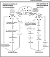
Dual effects of neuroprotection and neurotoxicity by general anesthetics: role of intracellular calcium homeostasis
- PMID: 23721657
- PMCID: PMC3791176
- DOI: 10.1016/j.pnpbp.2013.05.009
Dual effects of neuroprotection and neurotoxicity by general anesthetics: role of intracellular calcium homeostasis
Abstract
Although general anesthetics have long been considered neuroprotective, there are growing concerns about neurotoxicity. Preclinical studies clearly demonstrated that commonly used general anesthetics are both neuroprotective and neurotoxic, with unclear mechanisms. Recent studies suggest that differential activation of inositol 1,4,5-trisphosphate receptors, a calcium release channel located on the membrane of endoplasmic reticulum (ER), play important role on determining the fate of neuroprotection or neurotoxicity by general anesthetics. General anesthetics at low concentrations for short duration are sublethal stress factors which induce endogenous neuroprotective mechanisms and provide neuroprotection via adequate activation of InsP3R and moderate calcium release from ER. On the other hand, general anesthetics at high concentrations for prolonged duration are lethal stress factors which induce neuronal damage by over activation of InsP3R and excessive and abnormal Ca(2+) release from ER. This review emphasizes the dual effects of both neuroprotection and neurotoxicity via differential regulation of intracellular Ca(2+) homeostasis by commonly used general anesthetics and recommends strategy to maximize neuroprotective but minimize neurotoxic effects of general anesthetics.
Keywords: 3-(4,5-Dimethylthiazol-2-yl)-2,5-diphenyltetrazolium bromide; 5-HD; 5-Hydroxydecanoate; Akt; Anesthesia; Anesthetics; Calcium; ER; ERK; Endoplasmic reticulum; Extracellular signal-regulated kinases; GABA; GSK3β; Gamma-aminobutyric acid; Glycogen synthase kinase 3β; InsP(3) receptors; InsP(3)R; LDH; LTP; Lactate dehydrogenase; MAPK; MTT; Mitochondrial permeability transition pores; Mitogen-activated protein kinase; N-methyl-D-aspartate; NMDA; Neurodegeneration; Neuroprotection; Neurotoxicity; OGD; Oxygen-glucose deprivation; Protein kinase B; RYRs; Ryanodine receptors; VDCC; Voltage dependent Ca(2+) channels; Xc; Xestospongin C; long term potentiation; mPTP.
Copyright © 2013 Elsevier Inc. All rights reserved.
Figures

Similar articles
-
General Anesthetics Regulate Autophagy via Modulating the Inositol 1,4,5-Trisphosphate Receptor: Implications for Dual Effects of Cytoprotection and Cytotoxicity.Sci Rep. 2017 Sep 28;7(1):12378. doi: 10.1038/s41598-017-11607-0. Sci Rep. 2017. PMID: 28959036 Free PMC article.
-
Neurotoxicity versus Neuroprotection of Anesthetics: Young Children on the Ropes?Paediatr Drugs. 2017 Aug;19(4):271-275. doi: 10.1007/s40272-017-0230-8. Paediatr Drugs. 2017. PMID: 28466422 Review.
-
The acute neurotoxicity of mefloquine may be mediated through a disruption of calcium homeostasis and ER function in vitro.Malar J. 2003 Jun 12;2:14. doi: 10.1186/1475-2875-2-14. Epub 2003 Jun 12. Malar J. 2003. PMID: 12848898 Free PMC article.
-
Conflicting Actions of Inhalational Anesthetics, Neurotoxicity and Neuroprotection, Mediated by the Unfolded Protein Response.Int J Mol Sci. 2020 Jan 10;21(2):450. doi: 10.3390/ijms21020450. Int J Mol Sci. 2020. PMID: 31936788 Free PMC article.
-
Review of preclinical studies on pediatric general anesthesia-induced developmental neurotoxicity.Neurotoxicol Teratol. 2017 Mar-Apr;60:2-23. doi: 10.1016/j.ntt.2016.11.005. Epub 2016 Nov 18. Neurotoxicol Teratol. 2017. PMID: 27871903 Review.
Cited by
-
Perioperative changes of the linguistic functions in women after gynecological laparoscopic operations under propofol or sevoflurane-based anesthesia.Anaesthesiol Intensive Ther. 2024;56(5):305-315. doi: 10.5114/ait.2024.146710. Anaesthesiol Intensive Ther. 2024. PMID: 39917980 Free PMC article. Clinical Trial.
-
General anesthetic isoflurane modulates inositol 1,4,5-trisphosphate receptor calcium channel opening.Anesthesiology. 2014 Sep;121(3):528-37. doi: 10.1097/ALN.0000000000000316. Anesthesiology. 2014. PMID: 24878495 Free PMC article.
-
Notoginsenoside R1 Alleviates Oxygen-Glucose Deprivation/Reoxygenation Injury by Suppressing Endoplasmic Reticulum Calcium Release via PLC.Sci Rep. 2017 Nov 24;7(1):16226. doi: 10.1038/s41598-017-16373-7. Sci Rep. 2017. PMID: 29176553 Free PMC article.
-
Neurocognitive Adverse Effects of Anesthesia in Adults and Children: Gaps in Knowledge.Drug Saf. 2016 Jul;39(7):613-26. doi: 10.1007/s40264-016-0415-z. Drug Saf. 2016. PMID: 27098249 Review.
-
Postoperative Cognitive Dysfunction after Coronary Artery Bypass Grafting.Braz J Cardiovasc Surg. 2019 Jan-Feb;34(1):76-84. doi: 10.21470/1678-9741-2018-0165. Braz J Cardiovasc Surg. 2019. PMID: 30810678 Free PMC article. Review.
References
-
- Orrenius S, Zhivotovsky B, Nicotera P. Regulation of cell death: The calcium-apoptosis link. Nature Reviews Molecular Cell Biology. 2003;4:552–565. - PubMed
-
- Hajnoczky G, Davies E, Madesh M. Calcium signaling and apoptosis. [Review] [93 refs] Biochemical & Biophysical Research Communications. 2003;304(3):445–454. - PubMed
-
- Decuypere JP, Monaco G, Bultynck G, Missiaen L, De SH, Parys JB. The IP(3) receptor-mitochondria connection in apoptosis and autophagy. Biochimica et Biophysica Acta. 2011;1813:1003–1013. - PubMed
-
- Berridge MJ. Inositol trisphosphate and calcium signalling. Nature. 1993;361:315–325. - PubMed
Publication types
MeSH terms
Substances
Grants and funding
LinkOut - more resources
Full Text Sources
Other Literature Sources
Miscellaneous

