

Advances in NMR structures of integral membrane proteins
- PMID: 23721747
- PMCID: PMC3737378
- DOI: 10.1016/j.sbi.2013.05.002
Advances in NMR structures of integral membrane proteins
Abstract
Integral membrane proteins (IMPs) play a central role in cell communication with the environment. Their structures are essential for our understanding of the molecular mechanisms of signaling and for drug design, yet they remain badly underrepresented in the protein structure databank. Solution NMR is, aside from X-ray crystallography, the major tool in structural biology. Here we review recently reported solution NMR structures of polytopic IMPs and discuss the new approaches, which were developed in the course of these studies to overcome barriers in the field. Advances in cell-free protein expression, combinatorial isotope labeling, resonance assignment, and collection of structural data greatly accelerated IMP structure determination by solution NMR. In addition, novel membrane-mimicking media made possible determination of solution NMR structures of IMPs in a native-like lipid environment.
Copyright © 2013 Elsevier Ltd. All rights reserved.
Figures
References
-
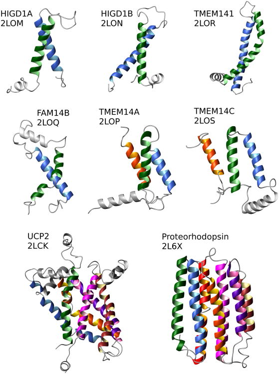
- Berardi MJ, Shih WM, Harrison SC, Chou JJ. Mitochondrial uncoupling protein 2 structure determined by NMR molecular fragment searching. Nature. 2011;476:109–113. The authors use molecular fragment replacement to define the local and secondary structures of UCP2 that best fit RDC data. The relative orientation of the secondary structural elements and their spatial arrangement in the 3D fold were determined from RDC and PRE constraints respectfully. - PMC - PubMed
-
- Reckel S, Gottstein D, Stehle J, Löhr F, Verhoefen MK, Takeda M, Silvers R, Kainosho M, Glaubitz C, Wachtveitl J, et al. Solution NMR Structure of Proteorhodopsin. Angewandte Chemie International Edition. 2011;50:11942–11946. Determination of the structure of CF expressed Proteorhodopsin in DHPC micelles. The assignment and collection of long-range NOEs assisted with CF selective isotope labeling. The structure with well-defined TM domain and extramembrane regions calculated using NOE, PRE, and RDC constraints. - PMC - PubMed
-
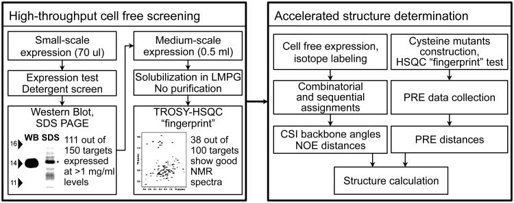
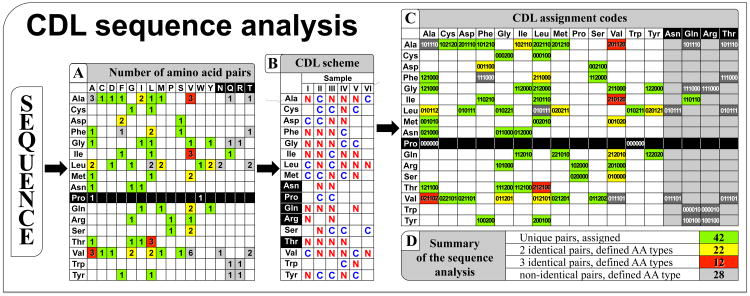
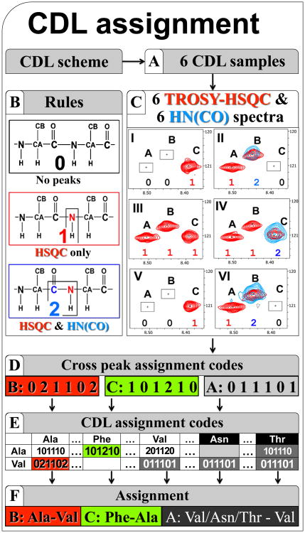
- Klammt C, Maslennikov I, Bayrhuber M, Eichmann C, Vajpai N, Chiu EJ, Blain KY, Esquivies L, Kwon JH, Balana B, et al. Facile backbone structure determination of human membrane proteins by NMR spectroscopy. Nat Methods. 2012;9:834–839. Combination of efficient CF expression and selective isotope labeling for fast assignment, data collection and analysis. Backbone structures of six human IMPs determined by fast analysis of PRE and CSI data. Efficient application of CF system for screening of NMR quality of >100 human IMPs. - PMC - PubMed
Publication types
MeSH terms
Substances
Grants and funding
LinkOut - more resources
Full Text Sources
Other Literature Sources
