. 2013 Aug;54(2):95-104.
doi: 10.1016/j.ceca.2013.05.002. Epub 2013 May 28.

Kainate-induced calcium overload of cortical neurons in vitro: Dependence on expression of AMPAR GluA2-subunit and down-regulation by subnanomolar ouabain

Affiliations

Kainate-induced calcium overload of cortical neurons in vitro: Dependence on expression of AMPAR GluA2-subunit and down-regulation by subnanomolar ouabain

Polina A Abushik et al. Cell Calcium. 2013 Aug.

Abstract

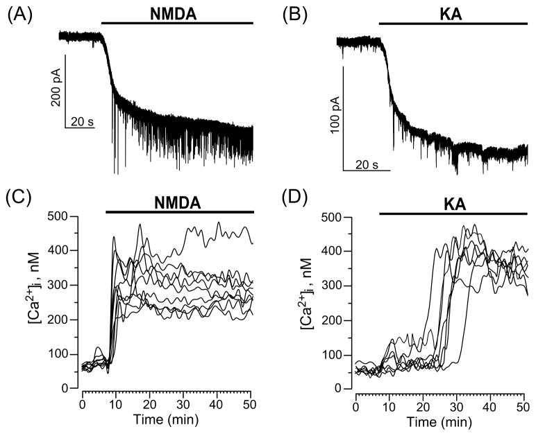
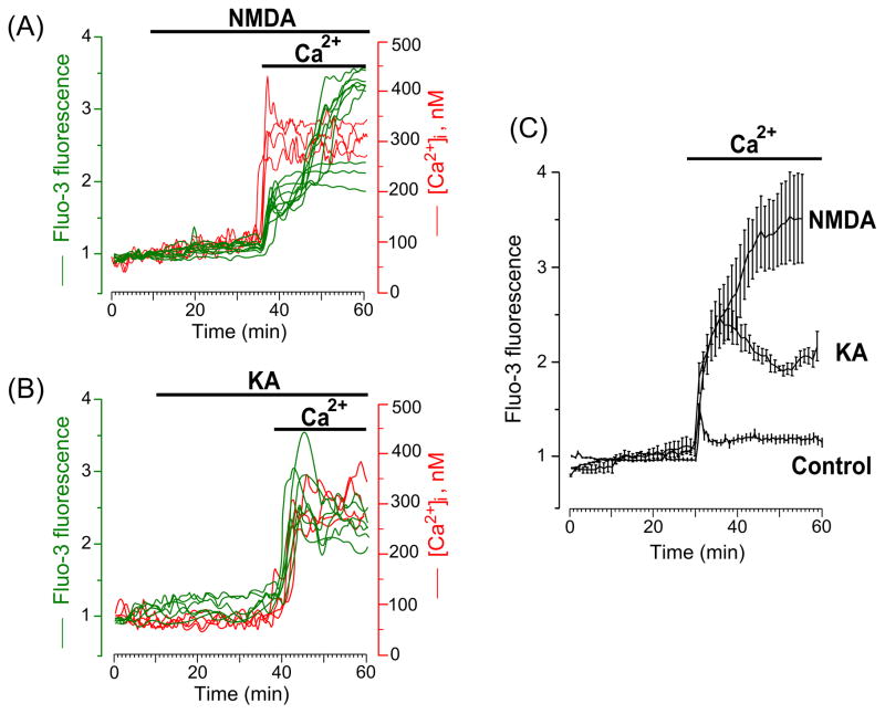
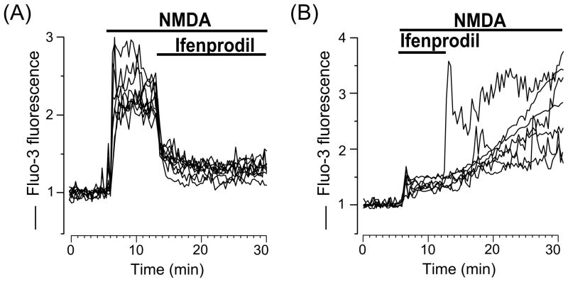
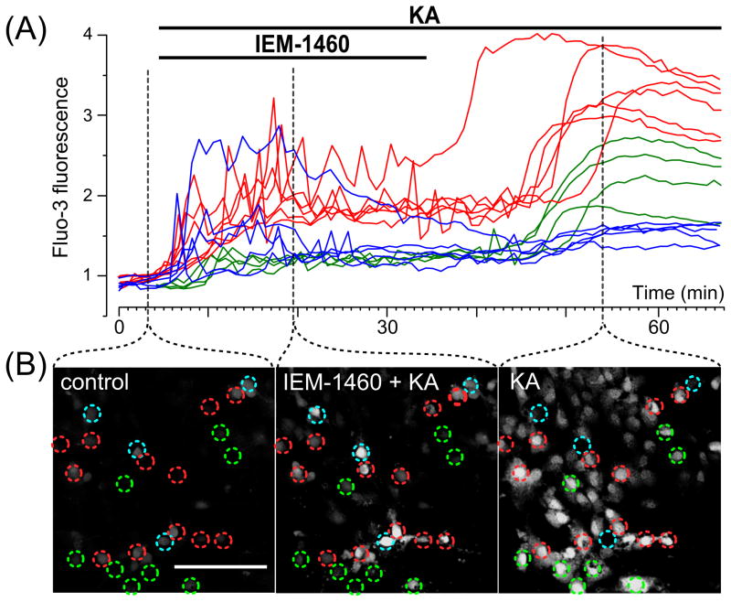
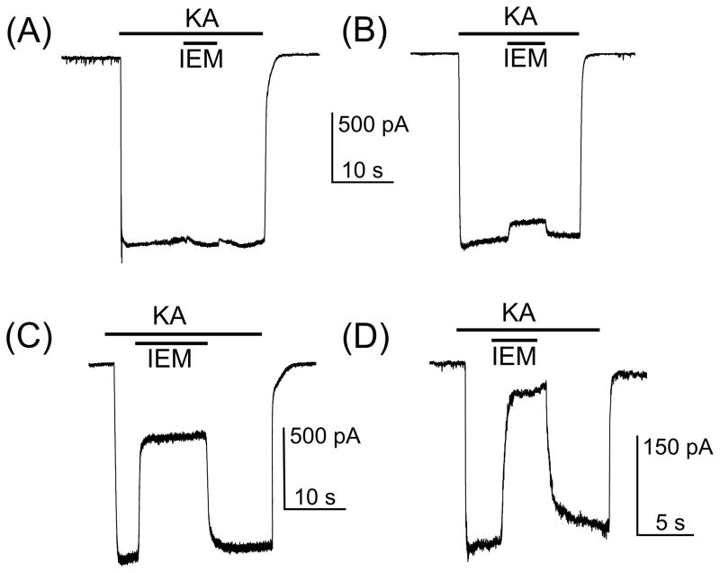
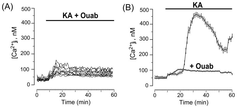
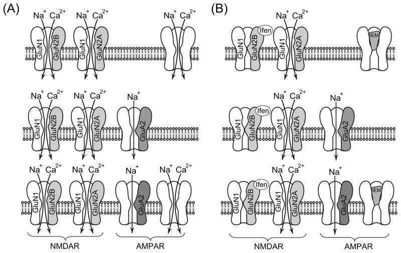
Whereas kainate (KA)-induced neurodegeneration has been intensively investigated, the contribution of α-amino-3-hydroxy-5-methyl-4-isoxazolepropionic acid receptors (AMPARs) in neuronal Ca2+ overload ([Ca2+]i) is still controversial. Using Ca2+ imaging and patch-clamp techniques, we found different types of Ca2+ entry in cultured rat cortical neurons. The presence of Ca2+ in the extracellular solution was required to generate the [Ca2+]i responses to 30 μM N-methyl-d-aspartate (NMDA) or KA. The dynamics of NMDA-induced [Ca2+]i responses were fast, while KA-induced responses developed slower reaching high [Ca2+]i. Ifenprodil, a specific inhibitor of the GluN2B subunit of NMDARs, reduced NMDA-induced [Ca2+]i responses suggesting expression of GluN1/GluN2B receptors. Using IEM-1460, a selective blocker of Ca(2+)-permeable GluA2-subunit lacking AMPARs, we found three neuronal responses to KA: (i) IEM-1460 resistant neurons which are similar to pyramidal neurons expressing Ca(2+)-impermeable GluA2-rich AMPARs; (ii) Neurons exhibiting nearly complete block of both KA-induced currents and [Ca2+]i signals by IEM-1460 may represent interneurons expressing GluA2-lacking AMPARs and (iii) neurons with moderate sensitivity to IEM-1460. Ouabain at 1 nM prevented the neuronal Ca2+ overload induced by KA. The data suggest, that cultured rat cortical neurons maintain functional phenotypes of the adult brain cortex, and demonstrate the key contribution of the Na/K-ATPase in neuroprotection against KA excitotoxicity.

Keywords: Calcium; Cortical neurons; Excitotoxicity; Glutamate receptors; Ouabain; Subunit selective antagonists; Whole-cell currents.


Figures

Fig. 1
Fig. 1
Current and Ca2+ responses of cultured cortical neurons to application of GluR agonists, NMDA and KA. (A) Representative whole-cell currents illustrating neuronal responses of cultured cortical neurons to application of 30 μM NMDA. (B) Representative whole-cell currents illustrating the response of cultured cortical neurons to application of 30 μM KA. The time of agonist application is indicated by the line above the traces. (C) Sustained Ca2+-responses of neurons loaded with Fura-2 upon application of 30 μM NMDA (time of application shown by the line above the traces). Each trace represents the response of one neuron. Data from one experiment are plotted. Number of experiments (N) = 4. (D) Ca2+-responses of neurons loaded with Fura-2 upon application of 30 μM KA (time of application shown by the line above the traces). Each trace represents the response of one neuron. Data from one experiment are plotted.(N = 4)
Fig. 2
Fig. 2
Intracellular Ca2+ signals induced by GluR agonists occur only when Ca2+ is present in the external solution. (A) Time course of the [Ca2+]i response after application of 30 μM NMDA in Ca2+-free extracellular solution, followed by the addition of 2 mM Ca2+ to the extracellular solution in the continued presence of 30 μM NMDA (the application episodes are marked with lines above the traces). Neurons were loaded with Fluo-3 (left ordinate, relative fluorescence intensity, green lines) and Fura-2 (right ordinate, [Ca2+]i, red lines). Each trace represents the response of one neuron. Data from two experiments (one with Fluo-3 and one with Fura-2) are plotted. Four (N=4) experiments for each Ca2+ indicator were performed. (B) Time course of the [Ca2+]i response after application of 30 μM KA in Ca2+-free extracellular solution, followed by the addition of 2 mM Ca2+ to the extracellular solution in the continued presence of 30 μM KA (the application episodes are marked with lines above the traces). Neurons were loaded with Fluo-3 (left ordinate, relative fluorescence intensity, green lines) and Fura-2 (right ordinate, [Ca2+]i, red lines). Each trace represents the response of one neuron. Data from two experiments (one with Fluo-3 and one with Fura-2) are plotted. Four (N=4) experiments for each Ca2+ indicator were performed. (C) Comparison of average time courses of the [Ca2+]i responses obtained in experiments with NMDA (N = 3, total number of analyzed neurons is 40), KA (N = 4, total number of analyzed neurons is 48) and in the absence of agonists (control, N = 4, total number of analyzed neurons is 83) upon an addition of 2 mM Ca2+ to the Ca2+-free extracellular solution. Mean values ± s.e. are plotted.
Fig. 3
Fig. 3
Ifenprodil, a GluN2B selective antagonist of NMDARs, inhibits intracellular Ca2+ responses induced by NMDA. (A) Ca2+ responses measured upon application of 30 μM NMDA following by the addition 10 μM ifenprodil (the application episodes are marked with lines above the traces). Neurons were loaded with Fluo-3 (ordinate, relative fluorescence intensity). Each trace represents the response of one neuron. (B) Ca2+ responses measured upon application of 30 μM NMDA with 10 μM ifenprodil following by ifenprodil washout (the application episodes are marked with lines above the traces). Neurons were loaded with Fluo-3 (ordinate, relative fluorescence intensity). Each trace represents the response of one neuron. Four experiments were performed.
Fig. 4
Fig. 4
Application of KA and IEM-1460, a subunit specific open channel blocker of AMPARs reveals three different types of intracellular Ca2+ responses in cultured cortical neurons. (A) Ca2+ responses upon application of 30 μM KA with 3 μM IEM-1460 followed by IEM-1460 washout (the application episodes are marked with lines above the traces). Neurons were loaded with Fluo-3 (ordinate, relative fluorescence intensity). Each trace represents the response of one neuron. Three (N = 3) experiments were performed. (B) Fluorescent images taken at different stages (indicated by dashed lines) of the experiment illustrated in (A). Ca2+ responses of neurons marked by circles are plotted in panel A. Red, blue and green traces (in A) were obtained from neurons marked by circles of the same color. Scale bar is 100 μm and valid for all images. Detailed description and interpretation of these data are presented in sections 3.4. and 4.4.
Fig. 5
Fig. 5
Inhibition by IEM-1460 of whole-cell currents induced in neurons by 30 μM KA. (A) KA-induced current recorded from a neuron which is insensitive to 10 μM IEM-1460. (B) KA-induced current recorded from a neuron which is weakly blocked (8 % of current is blocked) by 10 μM IEM-1460. (C) KA-induced current recorded from a neuron which is moderately blocked by IEM-1460. 60 % of current is blocked by 10 μM IEM-1460. (D) KA-induced current recorded from a neuron which is strongly blocked by IEM-1460. 85 % of current is blocked by 3 μM IEM-1460. The protocols of drug applications are shown above the current traces. The number of tested neurons is 24.
Fig. 6
Fig. 6
Ouabain at 1 nM prevents Ca2+ overload of neurons induced by KA. (A) Ca2+ responses measured upon simultaneous application of 30 μM KA and 1 nM ouabain. Each trace represents the response of one neuron. Data from one experiment are plotted. (B) Comparison of average time course of the [Ca2+]i responses obtained in experiments with 30 μM KA (N = 3, total number of analyzed neurons is 48) and 30 μM KA + 1 nM ouabain (N = 3, total number of analyzed neurons is 139). Mean values ± s.e. are plotted.
Fig. 7
Fig. 7
Schematic of the data interpretation. (A) In the presence of GluR agonists, extracellular Ca2+ can enter neurons through the channels of NMDARs and Ca2+-permeable channels of GluA2-lacking AMPARs. (B) Ifenprodil by inhibiting GluN1/GluN2B prevents Ca2+ entry through these particular NMDARs. Ca2+, therefore, may enter the cytoplasm through the channels of GluN1/GluN2A only. IEM-1460 by blocking the Ca2+ permeable channels of AMPARs abolishes Ca2+ signals in neurons expressing GluA2-lacking AMPARs. Detailed description of data interpretation is presented in the section 4.4.

References

    1. Traynelis SF, Wollmuth LP, McBain CJ, et al. Glutamate receptor ion channels: structure, regulation and function. Pharmacol Rev. 2010;62:405–496. - PMC - PubMed
    1. Choi DW. Excitotoxic cell death. J Neurobiol. 1992;23:1261–1276. - PubMed
    1. Olney JW. Excitatory transmitter neurotoxicity. Neurobiol Aging. 1994;15:259–260. - PubMed
    1. Sibarov DA, Bolshakov AE, Abushik PA, Krivoi II, Antonov SM. Na+, K+-ATPase functionally interacts with the plasma membrane Na+, Ca2+-exchanger to prevent Ca2+ overload and neuronal apoptosis in excitotoxic stress. J Pharmacol Exp Ther. 2012;343:596–607. - PubMed
    1. Choi DW. Glutamate neurotoxicity and diseases of the nervous system. Neuron. 1988;1:623–634. - PubMed

Publication types

MeSH terms