. 2013 Sep:38:51-60.
doi: 10.1016/j.neuro.2013.05.014. Epub 2013 May 27.

The Nrf2/SKN-1-dependent glutathione S-transferase π homologue GST-1 inhibits dopamine neuron degeneration in a Caenorhabditis elegans model of manganism

Affiliations

The Nrf2/SKN-1-dependent glutathione S-transferase π homologue GST-1 inhibits dopamine neuron degeneration in a Caenorhabditis elegans model of manganism

Raja Settivari et al. Neurotoxicology. 2013 Sep.

Abstract

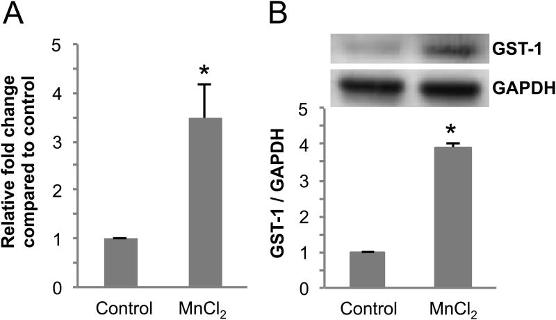
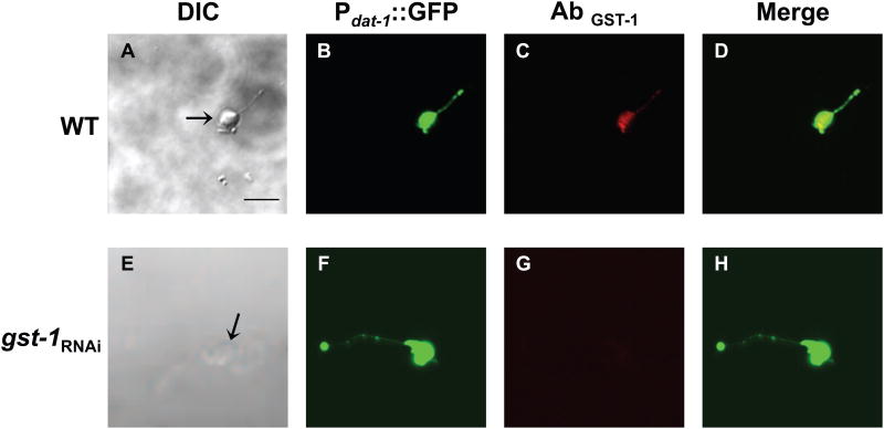
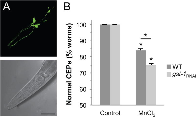
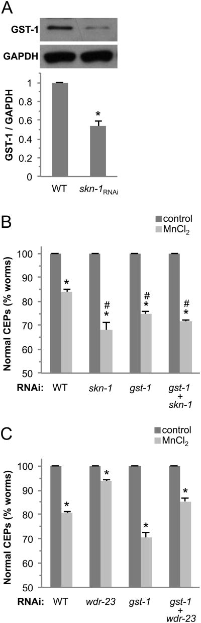
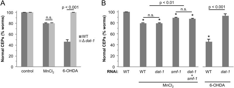
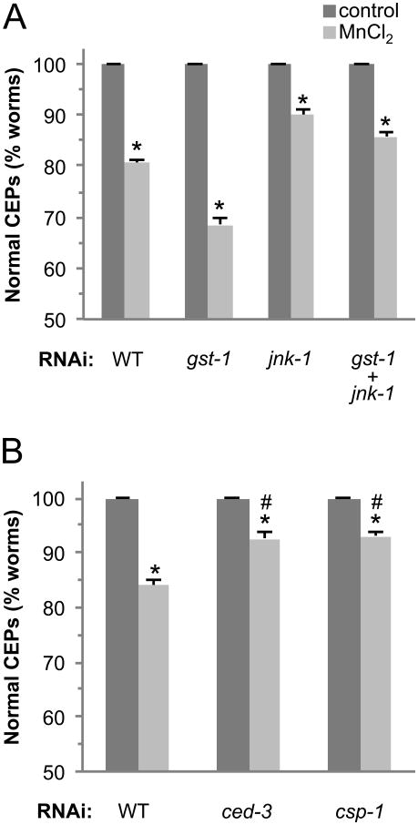
Exposure to high levels of manganese (Mn) results in a neurological condition termed manganism, which is characterized by oxidative stress, abnormal dopamine (DA) signaling, and cell death. Epidemiological evidence suggests correlations with occupational exposure to Mn and the development of the movement disorder Parkinson's disease (PD), yet the molecular determinants common between the diseases are ill-defined. Glutathione S-transferases (GSTs) of the class pi (GSTπ) are phase II detoxification enzymes that conjugate both endogenous and exogenous compounds to glutathione to reduce cellular oxidative stress, and their decreased expression has recently been implicated in PD progression. In this study we demonstrate that a Caenorhabditis elegans GSTπ homologue, GST-1, inhibits Mn-induced DA neuron degeneration. We show that GST-1 is expressed in DA neurons, Mn induces GST-1 gene and protein expression, and GST-1-mediated neuroprotection is dependent on the PD-associated transcription factor Nrf2/SKN-1, as a reduction in SKN-1 gene expression results in a decrease in GST-1 protein expression and an increase in DA neuronal death. Furthermore, decreases in gene expression of the SKN-1 inhibitor WDR-23 or the GSTπ-binding cell death activator JNK/JNK-1 result in an increase in resistance to the metal. Finally, we show that the Mn-induced DA neuron degeneration is independent of the dopamine transporter DAT, but is largely dependent on the caspases CED-3 and the novel caspase CSP-1. This study identifies a C. elegans Nrf2/SKN-1-dependent GSTπ homologue, cell death effectors of GSTπ-associated xenobiotic-induced pathology, and provides the first in vivo evidence that a phase II detoxification enzyme may modulate DA neuron vulnerability in manganism.

Keywords: Caspase; DA; ECL; GFP; Manganism; Neurodegeneration; Neurotoxicity; Nrf2; PAGE; PD; Parkinson's disease; ROS; SN; TH; WT; dopamine; enhanced chemiluminescence; green fluorescent protein; polyacrylamide gel electrophoresis; reactive oxygen species; substantia nigra; tyrosine hydroxylase; wild type.


Conflict of interest statement

Conflict of Interest Statement: The authors state that there are no conflicts of interest.

Figures

Fig 1
Fig 1. Exposure to Mn induces GST-1 gene and protein expression
(A) Synchronized L1 stage WT C. elegans were exposed to 50 mM MnCl2 for 30 mins, mRNA was extracted and reverse transcribed to cDNA. Relative gene expression changes of gst-1 were quantitated using real-time PCR. The fold change in gene expression relative to GAPDH was calculated following the ΔΔCt method. Shown are mean ± S.E. of three individual replicates. p value was calculated using t-test analysis. Asterisk indicates p ≤ 0.04 between control and toxicant-exposed group ΔCt values. (B) Synchronized L1 stage WT nematodes were exposed to 50 mM MnCl2 for 30 mins and allowed to recover for 24 h on NGM plates at 20°C. Following recovery/exposure, the worms were collected, homogenized and protein was quantified following standard a Bradford assay. For Western blot analysis, protein samples were separated by electrophoresis, transferred to a membrane, and probed with anti-GST-1 or GAPDH primary antibodies. Shown are mean ± SE of at least three individual replicates. p values were calculated using t-test analysis. Asterisks indicate p ≤ 0.03 between controls and Mn-exposed groups.
Fig 2
Fig 2. GST-1 is expressed in DA neurons
Primary C. elegans cultures expressing GFP in the dopamine neurons were generated with WT (A-D) or gst-1 knockdown animals (E-H). Primary cultures were incubated with GST-1 primary antibody followed by incubation with Texas Red conjugated goat-anti-rabbit secondary antibody. DIC images (A) and (E) of WT and gst-1RNAi cultures, respectively. DA neurons from WT and gst-1RNAi animals expressing GFP driven by the dat-1 promoter (B) and (F), respectively. GST-1 is expressed in DA neurons in WT animals (C), but not in gst-1RNAi (G). (D) Overlay of (B-C) and (H) overlay of (F-G). Images were observed under a Zeiss confocal microscope (Zeiss LSM 510). Scale bar represents 5 μm.
Fig 3
Fig 3. GST-1 inhibits dopamine neuron degeneration
Third generation L1 stage RJ928 animals were exposed to 50 mM MnCl2 for 30 mins and the dopamine neurons were visualized after a 72 h recovery on control or gst-1 RNAi plates at 20°C. DA neurons were considered degenerated when a break in or complete loss of GFP expression was observed in the CEPs (dendritic processes ending at the tip of the nose). An unexposed (normal) nematode has 4 complete CEP processes (A). Mn induces DA neuron degeneration (B) (arrows indicate the location of missing process), scale bar is 50 μm. Quantification of DA neuron degeneration is expressed as the percentage of worms with normal CEPs after exposure to MnCl2 in WT and gst-1RNAi animals (C). Shown are mean values ± SE of at least three individual replicates. Two-way ANOVA analysis was used and an asterisks indicates p ≤ 0.001. For both genotypes, Mn exposure significantly decreased the % normal CEPs, and the difference between WT and gst-1RNAi Mn exposed groups is significant.
Fig 4
Fig 4. GST-1 requires SKN-1 for inhibition of toxicant-associated DA neurodegeneration
(A) GST- 1 expression is partially dependent on SKN-1. Synchronized RJ928 animals were placed on RNAi plates spread with skn-1 RNAi bacteria. After 48 h, the worms were collected, homogenized and total protein was quantified following the standard Bradford assay. Protein samples were separated by electrophoresis, transferred to a membrane, and probed with anti-GST-1 or GAPDH primary antibodies. Band intensity was quantified relative to GAPDH expression and normalized to WT. p value was calculated with a t-test and the asterisk indicates p ≤ 0.0001 between WT and skn-1RNAi. skn-1RNAi results in increased DA neuron sensitivity to Mn (B), and wdr-23RNAi decreases sensitivity (C). L1 stage RJ928 worms were exposed to 50 mM MnCl2 for 30 mins with a 72 h recovery (B and C) on RNAi plates as indicated, and DA neurons were visualized for degeneration. Shown are mean values ± SE of at least three individual replicates. p values were calculated using two-way ANOVA analysis. Asterisks indicate p ≤ 0.01 between control and toxicant exposed groups within each genotype. Number sign indicates p ≤ 0.01 between toxicant-exposed WT and gene knockdown groups (B). In C, comparisons between all toxicant-exposed groups are statistically different with p ≤ 0.01.
Fig 5
Fig 5. Mn-induced cell death is not dependent on dat-1
WT (BY200) and dat-1 knockout (BY215) synchronized L1 worms (A) or third generation RJ928 nematodes grown on dat-1 or smf-1 RNAi or the combination (B) were exposed to 50 mM MnCl2 or 1 mM 6-OHDA for 30 mins and allowed to recover on NGM plates (A) or plates with RNAi bacteria (B) for 72 h. DA neurons expressing GFP were visualized under a fluorescent microscope following the exposure or recovery (> 50 worms/condition). Shown are mean ± SE of at least three individual replicates. Two-way ANOVA analysis indicates a significant difference between WT and dat-1 genetic knockout animals exposed to 6-OHDA (A). Oneway ANOVA was performed in (B). Asterisks indicate p ≤ 0.01 for all Mn-exposed groups compared to untreated. Within the Mn-treated groups, WT and dat-1RNAi are significantly different from smf-1RNAi and dat-1/smf-1RNAi. A t-test was used to compare 6-OHDA-treated WT animals vs dat-1RNAi.
Fig 6
Fig 6. Mn-induced cell death is dependent on apoptosis
Third generation RJ928 nematodes grown on gst-1 or jnk-1 RNAi or the combination (A) or ced-3 or csp-1 RNAi (B) were exposed to 50 mM MnCl2 for 30 mins and allowed to recover on plates with RNAi bacteria for 72 h. DA neurons expressing GFP were visualized under a fluorescent microscope following exposure and recovery (> 50 worms/condition). Shown are mean ± SE of at least three individual replicates. p values were calculated using two-way ANOVA. Asterisks indicate p ≤ 0.01 between control and toxicant-exposed groups. Comparisons between all Mn-treated groups are significant with p ≤ 0.001 (A) or p ≤ 0.01 (B).

Similar articles

Cited by

References

    1. An JH, Blackwell TK. SKN-1 links C. elegans mesendodermal specification to a conserved oxidative stress response. Genes Dev. 2003;17:1882–93. - PMC - PubMed
    1. Benedetto A, Au C, Avila DS, Milatovic D, Aschner M. Extracellular dopamine potentiates mn-induced oxidative stress, lifespan reduction, and dopaminergic neurodegeneration in a BLI-3-dependent manner in Caenorhabditis elegans. PLoS Genet. 2010;6 - PMC - PubMed
    1. Bianchi L, Driscoll M. Culture of embryonic C. elegans cells for electrophysiological and pharmacological analyses. WormBook. 2006:1–15. - PMC - PubMed
    1. Bove J, Prou D, Perier C, Przedborski S. Toxin-induced models of Parkinson's disease. NeuroRx. 2005;2:484–94. - PMC - PubMed
    1. Bradford M. A Rapid and Sensitive Method for the Quantitation of Microgram Quantities of Protein Utilizing the Principle of Protein-Dye Binding. Anal Biochem. 1976;72:248–254. - PubMed

Publication types

MeSH terms

LinkOut - more resources