

Differences between internal jugular vein and vertebral vein flow examined in real time with the use of multigate ultrasound color Doppler
- PMID: 23721896
- PMCID: PMC7965434
- DOI: 10.3174/ajnr.A3557
Differences between internal jugular vein and vertebral vein flow examined in real time with the use of multigate ultrasound color Doppler
Abstract
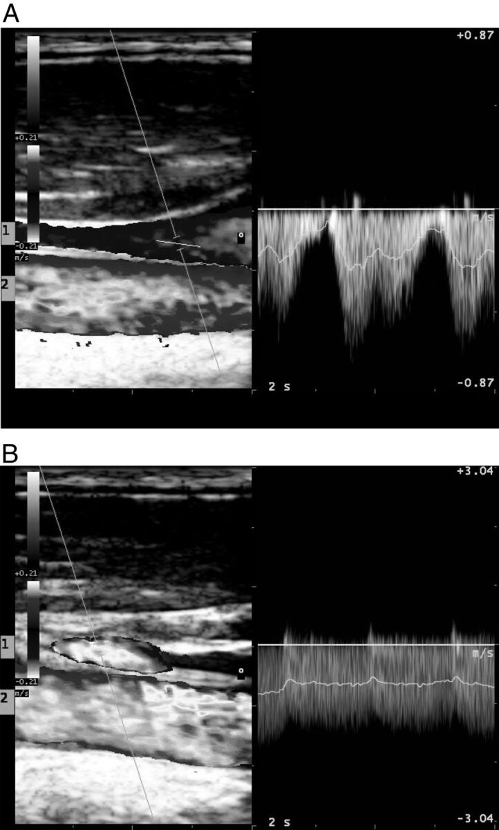
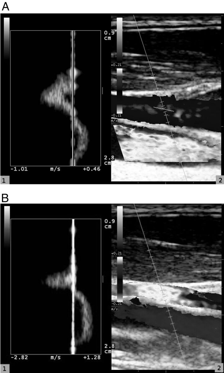
Background and purpose: The hypothesis that MS could be provoked by a derangement of the blood outflow from the brain has been largely discredited. In part, it was because data on the normal pattern of outflow are scarce and obtained with different methods. The aim of this study was to evaluate the normal pattern of outflow for the vertebral and internal jugular veins in healthy subjects with multigate color Doppler.
Materials and methods: Twenty-five volunteers were studied to assess vessel area, mean velocity, and flow for the vertebral and internal jugular veins in the supine and sitting positions.
Results: In the sitting position, flow decreases, both in vertebral veins and internal jugular veins, as the total vessel area decreases (from 0.46 ± 0.57 to 0.09 ± 0.08 cm(2)), even if the mean velocity increases (from 12.58 ± 10.19 to 24.14 ± 17.60 cm/s). Contrary to what happens to the blood inflow, outflow in the supine position, through vertebral and internal jugular veins, is more than twice the outflow in the sitting position (739.80 ± 326.32 versus 278.24 ± 207.94 mL/min). In the sitting position, on application of very low pressure to the skin with the sonography probe, internal jugular veins rarely appear to occlude. A pronounced difference of diameter between internal jugular veins was present in approximately one-third of subjects.
Conclusions: Our results support the view that other outflow pathways, like the vertebral plexus, play a major role in the normal physiology of brain circulation and must be assessed to obtain a complete picture of blood outflow.
Figures
Similar articles
-
Extracranial Doppler sonographic criteria of chronic cerebrospinal venous insufficiency in the patients with multiple sclerosis.Int Angiol. 2010 Apr;29(2):109-14. Int Angiol. 2010. PMID: 20351666
-
Collapsibility of the internal jugular veins in the lateral decubitus body position: A potential protective role of the cerebral venous outflow against neurodegeneration.Med Hypotheses. 2019 Dec;133:109397. doi: 10.1016/j.mehy.2019.109397. Epub 2019 Sep 11. Med Hypotheses. 2019. PMID: 31526984
-
Human cerebral venous outflow pathway depends on posture and central venous pressure.J Physiol. 2004 Oct 1;560(Pt 1):317-27. doi: 10.1113/jphysiol.2004.070409. Epub 2004 Jul 29. J Physiol. 2004. PMID: 15284348 Free PMC article.
-
Color Doppler sonography of the thoracic inlet veins.Radiographics. 1995 Nov;15(6):1357-71. doi: 10.1148/radiographics.15.6.8577962. Radiographics. 1995. PMID: 8577962 Review.
-
The Physical Examination to Assess for Anemia and Hypovolemia.Med Clin North Am. 2022 May;106(3):509-518. doi: 10.1016/j.mcna.2021.12.004. Epub 2022 Apr 4. Med Clin North Am. 2022. PMID: 35491070 Review.
Cited by
-
Real-Time Phase-Contrast MRI to Monitor Cervical Blood and Cerebrospinal Fluid Flow Beat-by-Beat Variability.Biosensors (Basel). 2022 Jun 15;12(6):417. doi: 10.3390/bios12060417. Biosensors (Basel). 2022. PMID: 35735564 Free PMC article.
-
Postural influence on intracranial fluid dynamics: an overview.J Physiol Anthropol. 2023 Apr 13;42(1):5. doi: 10.1186/s40101-023-00323-6. J Physiol Anthropol. 2023. PMID: 37055862 Free PMC article. Review.
-
Exploration of postural effects on the external jugular and diploic venous system using upright computed tomography scanning.Neuroradiology. 2024 Jun;66(6):963-971. doi: 10.1007/s00234-024-03357-4. Epub 2024 Apr 13. Neuroradiology. 2024. PMID: 38613702
-
Upright Catheter-Based Cerebral Angiography.J Vasc Interv Neurol. 2017 Dec;9(6):14-19. J Vasc Interv Neurol. 2017. PMID: 29445433 Free PMC article.
-
Internal Jugular Vein Cross-Sectional Area Enlargement Is Associated with Aging in Healthy Individuals.PLoS One. 2016 Feb 19;11(2):e0149532. doi: 10.1371/journal.pone.0149532. eCollection 2016. PLoS One. 2016. PMID: 26895434 Free PMC article.
References
-
- Zamboni P, Morovic S, Menegatti E, et al. . Screening for chronic cerebrospinal venous insufficiency (CCSVI) using ultrasound: recommendations for a protocol. Int Angiol 2011;306:571–97 - PubMed
-
- Doepp F, Paul F, Valdueza JM, et al. . No cerebrocervical venous congestion in patients with multiple sclerosis. Ann Neurol 2010;682:173–83 - PubMed
-
- Gius JA, Grier DH. Venous adaptation following bilateral radical neck dissection with excision of the jugular veins. Surgery 1950;282:305–21 - PubMed
-
- Fitz-Hugh GS, Robins RB, Craddock WD. Increased intracranial pressure complicating unilateral neck dissection. Laryngoscope 1966;765:893–906 - PubMed
MeSH terms
LinkOut - more resources
Full Text Sources
Other Literature Sources