Comparative Study
. 2013 Nov-Dec;34(11):2177-83.
doi: 10.3174/ajnr.A3589. Epub 2013 May 30.

In vitro and in vivo imaging characteristics assessment of polymeric coils compared with standard platinum coils for the treatment of intracranial aneurysms

Affiliations
Comparative Study

In vitro and in vivo imaging characteristics assessment of polymeric coils compared with standard platinum coils for the treatment of intracranial aneurysms

P Mordasini et al. AJNR Am J Neuroradiol. 2013 Nov-Dec.

Abstract

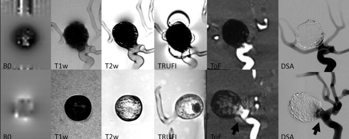
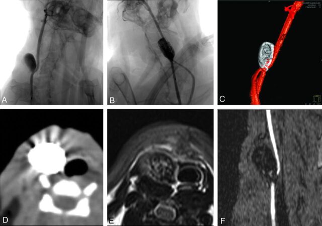
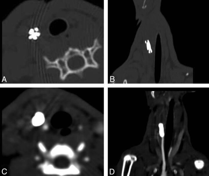
Background and purpose: Conventional platinum coils cause imaging artifacts that reduce imaging quality and therefore impair imaging interpretation on intraprocedural or noninvasive follow-up imaging. The purpose of this study was to evaluate imaging characteristics and artifact production of polymeric coils compared with standard platinum coils in vitro and in vivo.

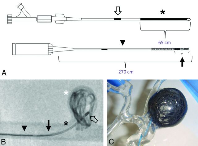
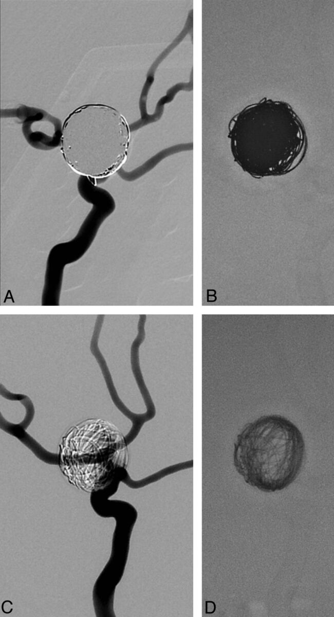
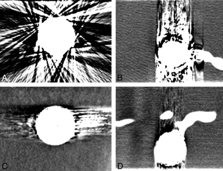
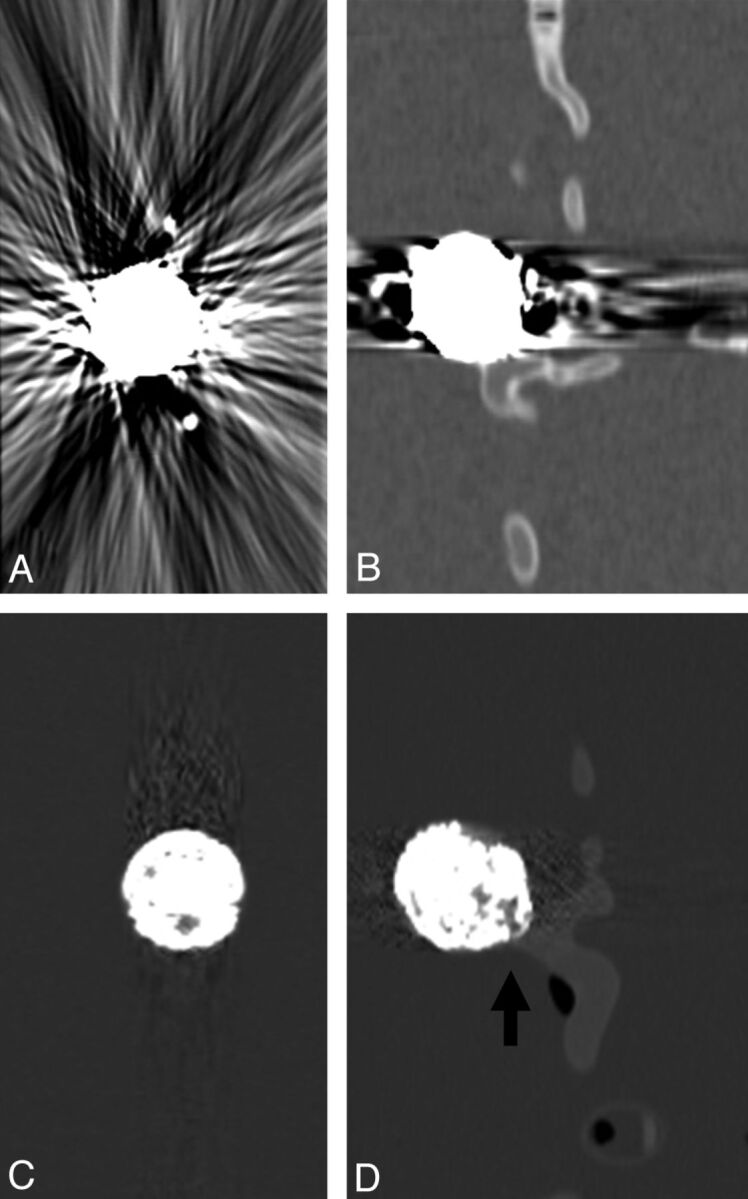
Materials and methods: Polymeric coils and standard platinum coils were evaluated in vitro with the use of 2 identical silicon aneurysm models coiled with a packing attenuation of 20% each. DSA, flat panel CT, CT, and MR imaging were performed. In vivo evaluation of imaging characteristics of polymeric coils was performed in experimentally created rabbit carotid bifurcation aneurysms. DSA, CT/CTA, and MR imaging were performed after endovascular treatment of the aneurysms. Images were evaluated regarding visibility of individual coils, coil mass, artifact production, and visibility of residual flow within the aneurysm.

Results: Overall, in vitro and in vivo imaging showed relevantly reduced artifact production of polymeric coils in all imaging modalities compared with standard platinum coils. Image quality of CT and MR imaging was improved with the use of polymeric coils, which permitted enhanced depiction of individual coil loops and residual aneurysm lumen as well as the peri-aneurysmal area. Remarkably, CT images demonstrated considerably improved image quality with only minor artifacts compared with standard coils. On DSA, polymeric coils showed transparency and allowed visualization of superimposed vessel structures.

Conclusions: This initial experimental study showed improved imaging quality with the use of polymeric coils compared with standard platinum coils in all imaging modalities. This might be advantageous for improved intraprocedural imaging for the detection of complications and posttreatment noninvasive follow-up imaging.


Figures

Fig 1.
Fig 1.
A, Schematic illustration of the polymeric coil system (above) and the delivery/detacher device (below). The coil system consists of a proximal shaft (open arrow, ink marker) attached to a hemostatic valve and a distal 65-cm-long radiopaque polymeric strand with an outer diameter of 0.018 inch (asterisk). The delivery/detacher device (total length, 270 cm) is introduced through the hemostatic valve into the inner lumen of the coil system. The delivery/detacher device consists of the microwire (arrowhead) and the heater coil attached to its distal tip (arrow). B, Fluoroscopy of an in vitro silicon aneurysm model showing the radiopaque polymeric strand (black asterisk) within the microcatheter placed in the aneurysm (microcatheter tip, open arrow) and its unfolded distal part within the aneurysm sac (white asterisk). Note the radiopaque section of the heater coil (arrow) at the distal tip of the delivery/detacher device (arrowhead) running within the coil strand. The polymeric strand can be detached at any point by advancing the radiopaque section of the heater coil at the level of the microcatheter tip and activating the thermal detachment. C, Photographic depiction of the silicon aneurysm model after introduction of the polymeric coil.
Fig 2.
Fig 2.
DSA of the in vitro silicon aneurysm model coiled with a calculated packing attenuation of 20%. Note the transparency of the polymeric coils (C) with persistent visualization of the superimposed vessel anatomy, which is completely obscured by standard platinum coils (A). Fluoroscopic images show the difference in radio-opacity of polymeric (D) and standard platinum coils (B).
Fig 3.
Fig 3.
Flat panel CT images of standard platinum coils (A and B) and polymeric coils (C and D). Note significant artifact reduction of polymeric coils compared with standard platinum coils; however, visualization of the peri-aneurysmal area is still significantly impaired.
Fig 4.
Fig 4.
CT scan of standard platinum coils (A and B) and polymeric coils (C and D). Note significant reduction of beam-hardening artifacts compared with standard platinum coils. In the aneurysm embolized with polymeric coils, individual coils as well as residual aneurysm lumen and neck (arrow) can be distinguished.
Fig 5.
Fig 5.
MR images and corresponding DSA of standard platinum coils (upper row) and polymeric coils (lower row) in vitro. Less magnetic field distortion and artifact production are seen with polymeric coils with the use of MRI. Individual coils can be distinguished in T2WI, TRUFI (true fast imaging with steady state precession), and TOF images as compared with standard coils. Note the visualization of residual flow within the aneurysm sac and the neck area in the aneurysm treated with polymeric coils in the TOF and DSA (arrows) compared with standard platinum coils at the same packing attenuation.
Fig 6.
Fig 6.
DSA demonstrates the anatomy of the venous patch carotid artery bifurcation model with the anastomosis of the left carotid artery proximal to the aneurysm (A and B). Posttreatment DSA (B) and 3D rotational angiography (C) show complete obliteration of the aneurysm (B). Note the persistent visibility of the right carotid artery despite superimposition by the coil mass. Note the minor effect of beam-hardening artifacts on the surrounding soft tissue on CT imaging (D). MR images demonstrate visibility of individual coil loops in the T2WI (E) and the lack of intra-aneurysmal flow in the TOF image (F).
Fig 7.
Fig 7.
CT images of a coiled carotid bifurcation aneurysm. Note the reduced artifact production and visibility of individual coils on the unenhanced CT scan (A and B). CTA shows complete occlusion of the aneurysm and minimal artifact production (C and D).

Similar articles

Cited by

References

    1. Molyneux AJ, Kerr RS, Yu LM, et al. . International Subarachnoid Aneurysm Trial (ISAT) of neurosurgical clipping versus endovascular coiling in 2143 patients with ruptured intracranial aneurysms: a randomised comparison of effects on survival, dependency, seizures, rebleeding, subgroups, and aneurysm occlusion. Lancet 2005;366:809–17 - PubMed
    1. Byrne JV, Sohn MJ, Molyneux AJ, et al. . Five-year experience in using coil embolization for ruptured intracranial aneurysms: outcomes and incidence of late rebleeding. J Neurosurg 1999;90:656–63 - PubMed
    1. Raymond J, Guilbert F, Weill A, et al. . Long-term angiographic recurrences after selective endovascular treatment of aneurysms with detachable coils. Stroke 2003;34:1398–403 - PubMed
    1. Kang HS, Han MH, Kwon BJ, et al. . Repeat endovascular treatment in post-embolization recurrent intracranial aneurysms. Neurosurgery 2006;58:60–70 - PubMed
    1. Campi A, Ramzi N, Molyneux AJ, et al. . Retreatment of ruptured cerebral aneurysms in patients randomized by coiling or clipping in the International Subarachnoid Aneurysm Trial (ISAT). Stroke 2007;38:1538–44 - PubMed

Publication types