. 2013 Sep;154(9):1528-1541.
doi: 10.1016/j.pain.2013.04.010. Epub 2013 Apr 8.

Diffusion tensor imaging detects microstructural reorganization in the brain associated with chronic irritable bowel syndrome

Affiliations

Diffusion tensor imaging detects microstructural reorganization in the brain associated with chronic irritable bowel syndrome

Benjamin M Ellingson et al. Pain. 2013 Sep.

Abstract

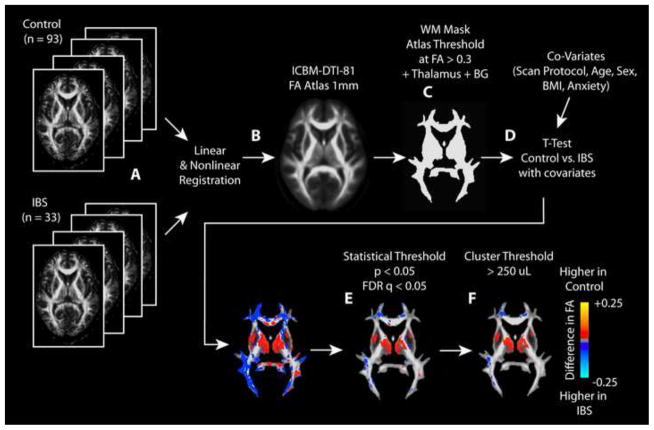
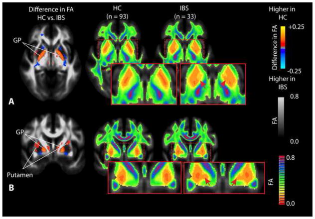
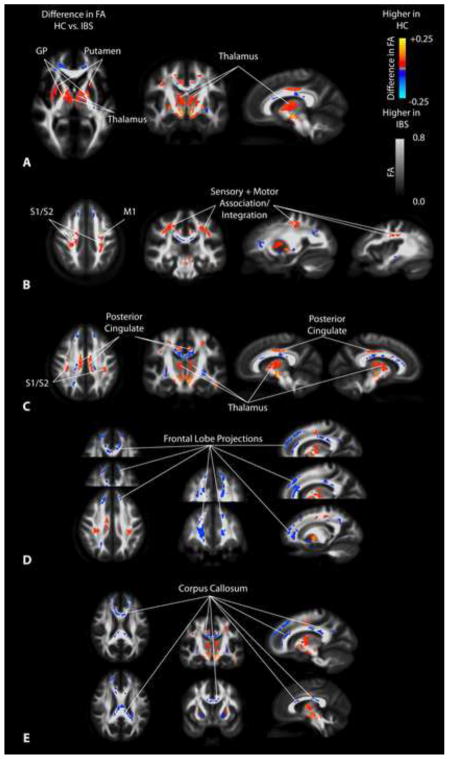
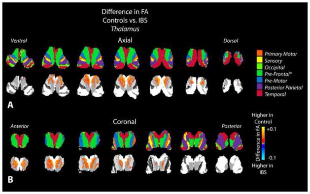
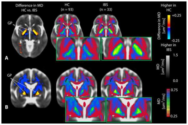
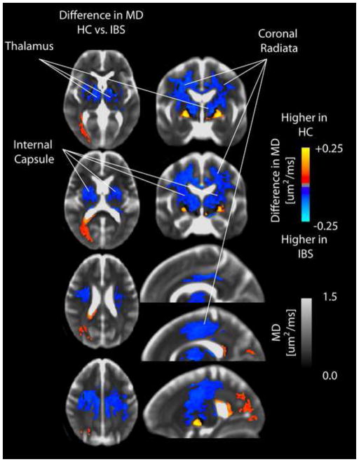
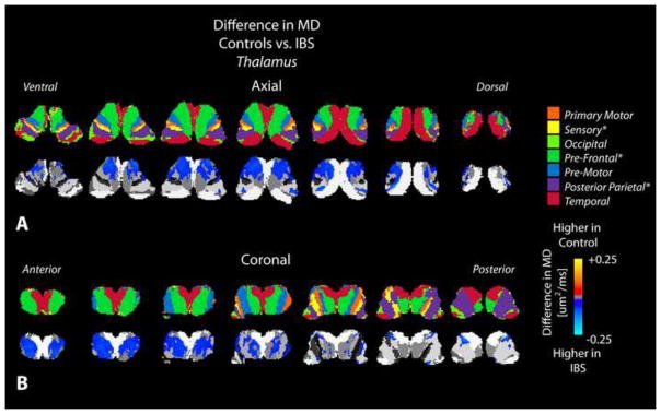
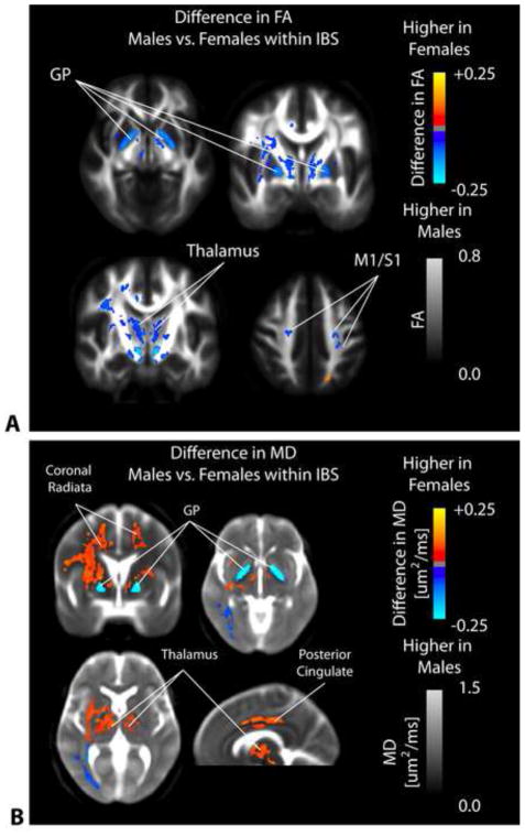
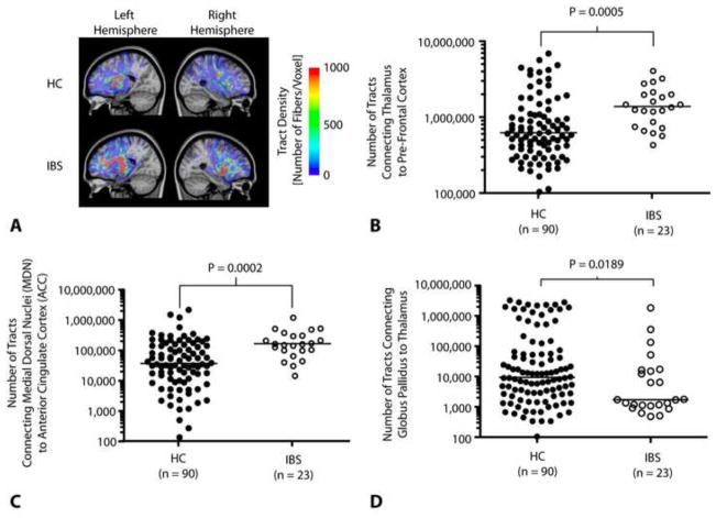
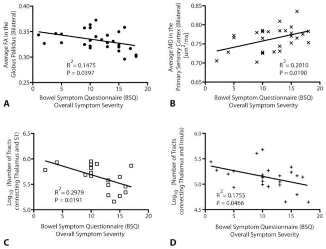
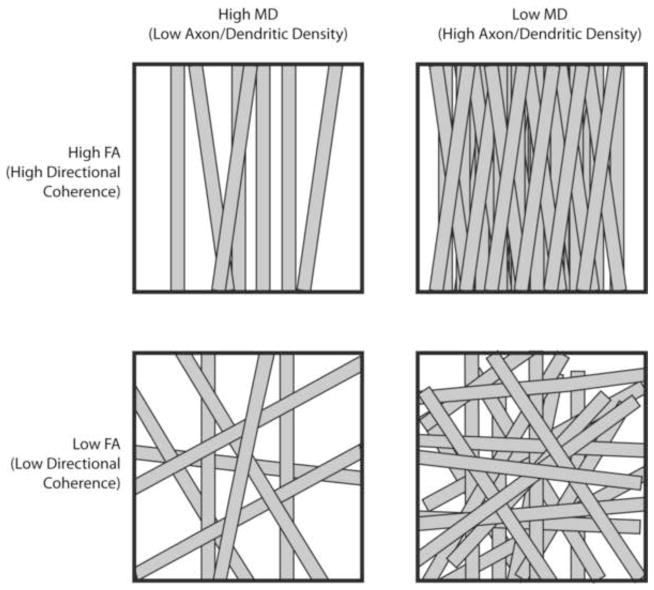
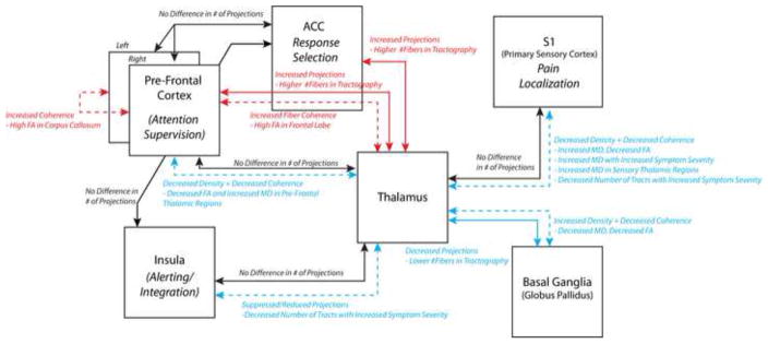
Irritable bowel syndrome (IBS) is a common gastrointestinal disorder characterized by recurring abdominal pain associated with alterations in bowel habits. We hypothesized that patients with chronic visceral pain associated with IBS may have microstructural differences in the brain compared with healthy control subjects (HCs), indicative of long-term neural reorganization of chronic pain pathways and regions associated with sensory integration. In the current study we performed population-based voxel-wise diffusion tensor imaging (DTI) comparisons and probabilistic tractography in a large sample of phenotyped patients with IBS (n=33) and in HCs (n=93). Patients had lower fractional anisotropy (FA) in thalamic regions, the basal ganglia (BG) and sensory/motor association/integration regions as well as higher FA in frontal lobe regions and the corpus callosum. In addition, patients had reduced mean diffusivity (MD) within the globus pallidus (GP) and higher MD in the thalamus, internal capsule, and coronal radiata projecting to sensory/motor regions, suggestive of differential changes in axon/dendritic density in these regions. Sex differences in FA and MD were also observed in the patients but not in HCs. Probabilistic tractography in patients confirmed a higher degree of connectivity between the thalamus and prefrontal cortex, as well as between the medial dorsal thalamic nuclei and anterior cingulate cortex, and a lower degree of connectivity between the GP and thalamus. Together, these results support the hypothesis that patients with chronically recurring visceral pain from IBS have long-term microstructural changes within the brain, particularly in regions associated with integration of sensory information and corticothalamic modulation.

Keywords: Chronic Pain; DTI; Diffusion Tensor Imaging; IBS; Irritable Bowel Syndrome; Reorganization.


Conflict of interest statement

Conflicts of Interest

No authors have any conflicts of interest regarding the subject matter in this manuscript.

Figures

Figure 1
Figure 1. Statistical parameter mapping (SPM) pipeline for DTI metrics
A) FA maps from each patient were calculated, then B) registered to the ICBM-DTI-81 atlas using both linear and nonlinear registration. All other DTI measurements were then aligned using these transformation matrices. C) Regions with FA greater than 0.3 were threshold on the FA atlas to isolate regions of white matter. Regions of the thalamus and basal ganglia (BG) were also included in the mask. D) A t-test with covariates were performed using AFNI (3dttest++) in order to compare HCs to IBS patients while considering the scan protocol, subject age, sex, body mass index (BMI), and anxiety score. E) A statistical threshold of P < 0.05 and a false discovery rate (FDR) q < 0.05 were used to identify regions of statistical significance. F) Additionally, a minimum cluster size of 250 uL was used to further isolate regions of statistical significance in the final SPMs.
Figure 2
Figure 2. Differences in fractional anisotropy (FA) within the basal ganglia
A) Axial SPMs showing a higher FA in the globus pallidus (GP) in HCs compared to patients with IBS, along with average color FA maps for both HCs and IBS patients showing dramatic changes within the basal ganglia. B) Coronal SPMs showing higher FA in the GP as well as the putamen in HCs when compared with IBS patients, which is also apparent when examining the average color FA maps for both HCs and IBS patients directly. Red arrows show regions of dramatic difference on color FA maps between HC and IBS patients.
Figure 3
Figure 3. SPMs showing regions of statistically different fractional anisotropy (FA) in IBS compared to HCs
A) SPMs showing higher FA in the globus pallidus (GP), putamen, and medial thalamus in HCs compared with IBS patients. B) SPMs demonstrating higher FA in sensory and motor association regions as well as primary cortical projections from the thalamus in HCs compared with IBS patients. C) SPMs illustrating higher FA in the thalamus and posterior cingulate white matter in HCs compared with IBS patients. D) SPMs demonstrating higher FA in the frontal lobe and the anterior cingulate white matter regions in IBS patients compared to HCs. E) SPMs illustrating higher FA in regions of the corpus callosum in IBS patients compared with HCs.
Figure 4
Figure 4. Statistical parameter maps (SPMs) for fractional anisotropy (FA) in the thalamus
A) Axial and B) coronal slices through the thalamus including the partitions proposed by Behrens et al. [12; 13] (top row) and SPMs (bottom row), suggesting a higher FA in thalamic areas connected to prefrontal regions in healthy controls compared to IBS patients.
Figure 5
Figure 5. Differences in mean diffusivity (MD) within the basal ganglia
A) Axial and B) coronal SPMs demonstrating elevated MD within the globus pallidus (GP) in HCs compared with IBS patients. This difference in MD between HCs and IBS patients can also be observed by visual examination of the average color MD for each group.
Figure 6
Figure 6. SPMs comparing mean diffusivity (MD) between healthy controls and IBS patients in the internal capsule, thalamus, and coronal radiata
SPMs demonstrate an elevated MD in patients with IBS compared to HCs in the internal capsule, thalamus, and coronal radiata.
Figure 7
Figure 7. Statistical parameter maps (SPMs) comparing mean diffusivity (MD) between healthy controls and IBS patients in the thalamus
A) Axial and B) coronal slices through the thalamus including the partitions proposed by Behrens et al. [12; 13] (top row) and SPMs (bottom row), suggesting a higher MD in thalamic areas connected to sensory, prefrontal, and posterior parietal regions in IBS patients compared to healthy controls.
Figure 8
Figure 8. SPMs comparing FA and MD between male and female IBS patients
A) SPMs clearly demonstrate a significantly higher FA in male compared with female IBS patients within the globus pallidus (GP), medial thalamic regions, and primary somatosensory and motor regions. B) SPMs examining the difference in MD between male and female IBS patients illustrate a significantly higher MD in female compared with male IBS patients in the coronal radiata, thalamus, and posterior cingulate white matter; however, male IBS patients had a significantly higher MD in the globus pallidus (GP) compared with female IBS patients.
Figure 9
Figure 9. Probabilistic tractography and structural connectivity in specific chronic pain pathways
A) Example DTI fiber tract density images connecting the thalamus to prefrontal cortex in a HC (top) and IBS patient (bottom). Results illustrate a lower tract density in these pathways in HCs compared with IBS patients. B) Number of tracts connecting the thalamus to prefrontal cortical regions were significantly higher (Mann-Whitney, P =0.0005) in IBS patients (median = 1,398,000 tracts) compared with HCs (median = 630,312 tracts). C) Number of tracts connecting the medial dorsal nuclei (MDN) to the anterior cingulate cortex (ACC) were significantly higher (Mann-Whitney, P = 0.0002) in IBS patients (median = 168,000 tracts) compared with HCs (median = 36,875 tracts). D) Number of tracts connecting the globus pallidus (GP) to the thalamus are significantly lower (Mann-Whitney, P = 0.0189) in IBS patients (median = 1,713 tracts) compared with HCs (median = 9,382 tracts).
Figure 10
Figure 10. Significant correlations between DTI metrics and symptom severity in patients with IBS
A) A significant negative correlation was detected between the average FA within the globus pallidus (GP), measured bilaterally, and the overall symptom severity on a BSQ (Pearson’s correlation coefficient, R2 = 0.1475, P = 0.0397). B) A significant positive correlation was also detected between average MD within the white matter regions adjacent to the primary sensory cortex, measured bilaterally, and the overall symptom severity on a BSQ (Pearson’s correlation coefficient, R2 = 0.2010, P = 0.0190). C) A significant negative correlation was found between the log-transformed number of fiber tracts connecting the thalamus and primary sensory cortex, as measured from probabilistic DTI tractography, and overall symptom severity on a BSQ (Pearson’s correlation coefficient, R2 = 0.2979, P = 0.0191). D) A significant negative correlation was also detected between the log-transformed number of fiber tracts connecting the thalamus and insula and overall symptom severity (Pearson’s correlation coefficient, R2 = 0.1755, P = 0.0466).
Figure 11
Figure 11. Microstructural interpretation of specific changes in fractional anisotropy (FA) and mean diffusivity (MD)
Top Left: In voxels with high MD and high FA, axons or dendritic arborizations are loosely packed (high MD) but maintain high directional coherence (high FA). Top Right: In voxels with low MD and high FA, axons or dendritic arborizations are tightly packed (low MD) and have a high degree of directional coherence (high FA). Bottom Left: In voxels with high MD and low FA, axons or dendritic arborizations are loosely packed (high MD) and lack directional coherence (low FA). Bottom Right: In voxels with low MD and low FA, axons or dendritic arborizations are tightly packed (low MD) but lack directional coherence (low FA).
Figure 12
Figure 12. DTI interpretation of connectivity in the major chronic pain pathways in IBS
Probabilistic tractography connectivity between regions, reflecting the relative number of fiber tracts, are shown in solid arrows; whereas interpretation of voxel-based analysis of DTI indices, e.g. FA and MD reflecting directional coherence and density of microstructure, are shown in dashed arrows. Red connections indicate increased connectivity, coherence, or density in IBS compared with HCs; blue connections indicate decreased connectivity, coherence, or density in IBS patients compared with HCs; and black connections indicate no difference in connectivity, coherence or density.

Comment in

Similar articles

Cited by

References

    1. Apkarian AV. The brain in chronic pain: clinical implications. Pain Manag. 2011;1(6):577–586. - PMC - PubMed
    1. Apkarian AV, Hashmi JA, Baliki MN. Pain and the brain: specificity and plasticity of the brain in clinical chronic pain. Pain. 2011;152(3 Suppl):S49–64. - PMC - PubMed
    1. Arendt-Nielsen L, Graven-Nielsen T, Petrini L. In: Experimental Human Models and Assessment of Pain in Non-pain Conditions. Giamberardino MA, Jenseneditors TS, editors. Book Title|, Vol. Volume|. City|: Publisher|, Year|. p.^pp. Pages|.
    1. As-Sanie S, Harris RE, Napadow V, Kim J, Neshewat G, Kairys A, Williams D, Clauw DJ, Schmidt-Wilcke T. Changes in regional gray matter volume in women with chronic pelvic pain: a voxel-based morphometry study. Pain. 2012;153(5):1006–1014. - PMC - PubMed
    1. Attree EA, Dancey CP, Keeling D, Wilson C. Cognitive function in people with chronic illness: inflammatory bowel disease and irritable bowel syndrome. Appl Neuropsychol. 2003;10(2):96–104. - PubMed

Publication types