Propofol-induced apoptosis of neurones and oligodendrocytes in fetal and neonatal rhesus macaque brain
- PMID: 23722059
- PMCID: PMC3667347
- DOI: 10.1093/bja/aet173
Propofol-induced apoptosis of neurones and oligodendrocytes in fetal and neonatal rhesus macaque brain
Abstract
Background: Exposure of the fetal or neonatal non-human primate (NHP) brain to isoflurane or ketamine for 5 h causes widespread apoptotic degeneration of neurones, and exposure to isoflurane also causes apoptotic degeneration of oligodendrocytes (OLs). The present study explored the apoptogenic potential of propofol in the fetal and neonatal NHP brain.
Method: Fetal rhesus macaques at gestational age 120 days were exposed in utero, or postnatal day 6 rhesus neonates were exposed directly for 5 h to propofol anaesthesia (n=4 fetuses; and n=4 neonates) or to no anaesthesia (n=4 fetuses; n=5 neonates), and the brains were systematically evaluated 3 h later for evidence of apoptotic degeneration of neurones or glia.
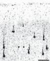
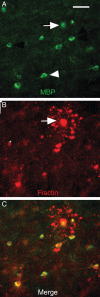
Results: Exposure of fetal or neonatal NHP brain to propofol caused a significant increase in apoptosis of neurones, and of OLs at a stage when OLs were just beginning to myelinate axons. Apoptotic degeneration affected similar brain regions but to a lesser extent than we previously described after isoflurane. The number of OLs affected by propofol was approximately equal to the number of neurones affected at both developmental ages. In the fetus, neuroapoptosis affected particularly subcortical and caudal regions, while in the neonate injury involved neocortical regions in a distinct laminar pattern and caudal brain regions were less affected.
Conclusions: Propofol anaesthesia for 5 h caused death of neurones and OLs in both the fetal and neonatal NHP brain. OLs become vulnerable to the apoptogenic action of propofol when they are beginning to achieve myelination competence.
Keywords: anaesthetics i.v., propofol; developing brain; neurones; non-human primates; oligodendroglia; toxicity.
Figures


Similar articles
-
Isoflurane-induced apoptosis of neurons and oligodendrocytes in the fetal rhesus macaque brain.Anesthesiology. 2014 Mar;120(3):626-38. doi: 10.1097/ALN.0000000000000037. Anesthesiology. 2014. PMID: 24158051 Free PMC article.
-
Alcohol-induced apoptosis of oligodendrocytes in the fetal macaque brain.Acta Neuropathol Commun. 2013 Jun 12;1:23. doi: 10.1186/2051-5960-1-23. Acta Neuropathol Commun. 2013. PMID: 24252271 Free PMC article.
-
Isoflurane-induced apoptosis of oligodendrocytes in the neonatal primate brain.Ann Neurol. 2012 Oct;72(4):525-35. doi: 10.1002/ana.23652. Ann Neurol. 2012. PMID: 23109147 Free PMC article.
-
Effect of Anesthesia on the Developing Brain: Infant and Fetus.Fetal Diagn Ther. 2018;43(1):1-11. doi: 10.1159/000475928. Epub 2017 Jun 7. Fetal Diagn Ther. 2018. PMID: 28586779 Review.
-
Developmental neurotoxicity of ketamine in pediatric clinical use.Toxicol Lett. 2013 Jun 20;220(1):53-60. doi: 10.1016/j.toxlet.2013.03.030. Epub 2013 Apr 6. Toxicol Lett. 2013. PMID: 23566897 Review.
Cited by
-
Behavioural impairments after exposure of neonatal mice to propofol are accompanied by reductions in neuronal activity in cortical circuitry.Br J Anaesth. 2021 Jun;126(6):1141-1156. doi: 10.1016/j.bja.2021.01.017. Epub 2021 Feb 26. Br J Anaesth. 2021. PMID: 33641936 Free PMC article.
-
Anaesthetic neurotoxicity and neuroplasticity: an expert group report and statement based on the BJA Salzburg Seminar.Br J Anaesth. 2013 Aug;111(2):143-51. doi: 10.1093/bja/aet177. Epub 2013 May 30. Br J Anaesth. 2013. PMID: 23722106 Free PMC article.
-
Construction and Characterization of a Population-Based Cohort to Study the Association of Anesthesia Exposure with Neurodevelopmental Outcomes.PLoS One. 2016 May 11;11(5):e0155288. doi: 10.1371/journal.pone.0155288. eCollection 2016. PLoS One. 2016. PMID: 27167371 Free PMC article.
-
Dexmedetomidine attenuates repeated propofol exposure-induced hippocampal apoptosis, PI3K/Akt/Gsk-3β signaling disruption, and juvenile cognitive deficits in neonatal rats.Mol Med Rep. 2016 Jul;14(1):769-75. doi: 10.3892/mmr.2016.5321. Epub 2016 May 23. Mol Med Rep. 2016. PMID: 27222147 Free PMC article.
-
Effect of cesarean section on the risk of autism spectrum disorders/attention deficit hyperactivity disorder in offspring: a meta-analysis.Arch Gynecol Obstet. 2024 Feb;309(2):439-455. doi: 10.1007/s00404-023-07059-9. Epub 2023 May 23. Arch Gynecol Obstet. 2024. PMID: 37219611
References
-
- Ikonomidou C, Bosch F, Miksa M, et al. Blockade of NMDA receptors and apoptotic neurodegeneration in the developing brain. Science. 1999;283:70–4. doi:10.1126/science.283.5398.70. - DOI - PubMed
-
- Rizzi S, Carter LB, Ori C, Jevtovic-Todorovic V. Clinical anesthesia causes permanent damage to the fetal guinea pig brain. Brain Pathol. 2008;18:198–210. doi:10.1111/j.1750-3639.2007.00116.x. - DOI - PMC - PubMed
-
- Satomoto M, Satoh Y, Terui K, et al. Neonatal exposure to sevoflurane induces abnormal social behaviors and deficits in fear conditioning in mice. Anesthesiology. 2009;110:628–37. doi:10.1097/ALN.0b013e3181974fa2. - DOI - PubMed
-
- Fredriksson A, Archer T, Alm H, Gordh T, Eriksson P. Neurofunctional deficits and potentiated apoptosis by neonatal NMDA antagonist administration. Behav Brain Res. 2004;153:367–76. doi:10.1016/j.bbr.2003.12.026. - DOI - PubMed
Publication types
MeSH terms
Substances
Grants and funding
LinkOut - more resources
Full Text Sources
Other Literature Sources

