

Review
doi: 10.1038/nmeth.2482.
Imaging human connectomes at the macroscale
Affiliations
- PMID: 23722212
- PMCID: PMC4096321
- DOI: 10.1038/nmeth.2482
Item in Clipboard
Review
Imaging human connectomes at the macroscale
Nat Methods.
2013 Jun.
Abstract
At macroscopic scales, the human connectome comprises anatomically distinct brain areas, the structural pathways connecting them and their functional interactions. Annotation of phenotypic associations with variation in the connectome and cataloging of neurophenotypes promise to transform our understanding of the human brain. In this Review, we provide a survey of magnetic resonance imaging–based measurements of functional and structural connectivity. We highlight emerging areas of development and inquiry and emphasize the importance of integrating structural and functional perspectives on brain architecture.
Figures
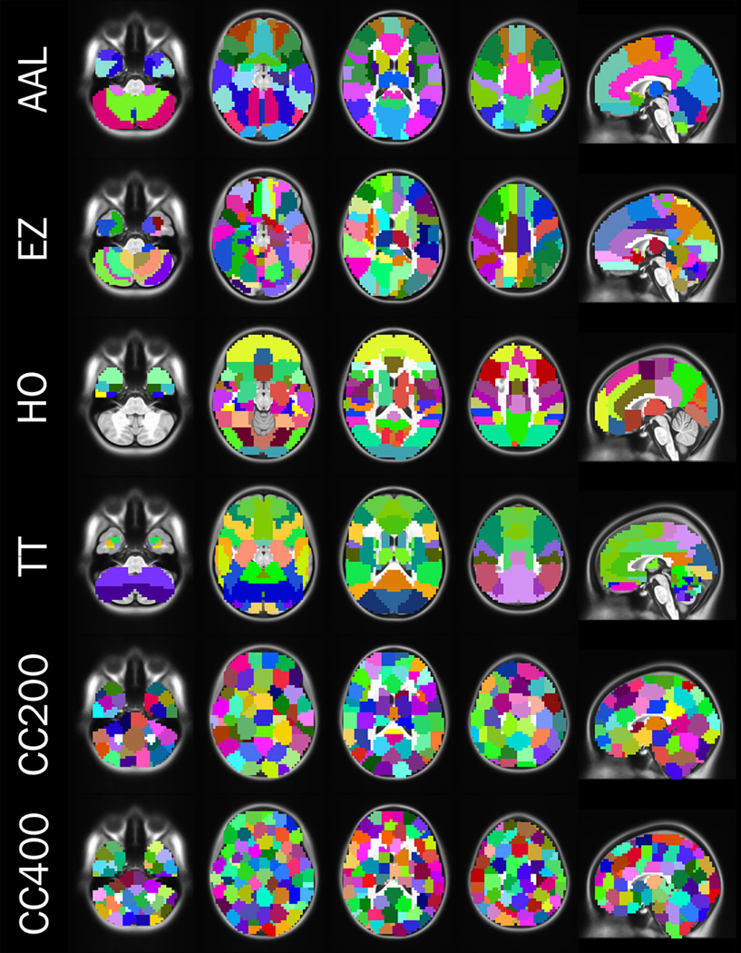
We depict several different atlases of brain areas generated using anatomical (rows 1 – 4) and functional (rows 5 – 6) parcellation schemes. AAL (automated anatomical labeling)and Harvard Oxford (HO)are derived from anatomical landmarks (sulci and gyral). The EZ (Eickhoff - Zilles)and TT (Talariach Daemon)atlases are derived from post-mortem cyto- and myelo-architectonic segmentations. The CC200 and CC400 atlases are derived from 200 and 400 unit functional parcellations.

(A) dMRI-based map of principal tensor orientations of the human brain viewed from below; blue: Superior-inferior, green: anterior-posterior and red: medial-lateral. (B) DTI fiber orientation estimates (red lines) from a region of corpus callosum superimposed on a fractional anisotropy image. Sample streamline depicted in yellow. (C) Pyramidal tract streamlines based on deterministic (left) and probabilistic (right) approaches are depicted (results superimposed on fractional anisotropy image). Image courtesy: Stam Sotiropoulos, http://etheses.nottingham.ac.uk/1164/ .
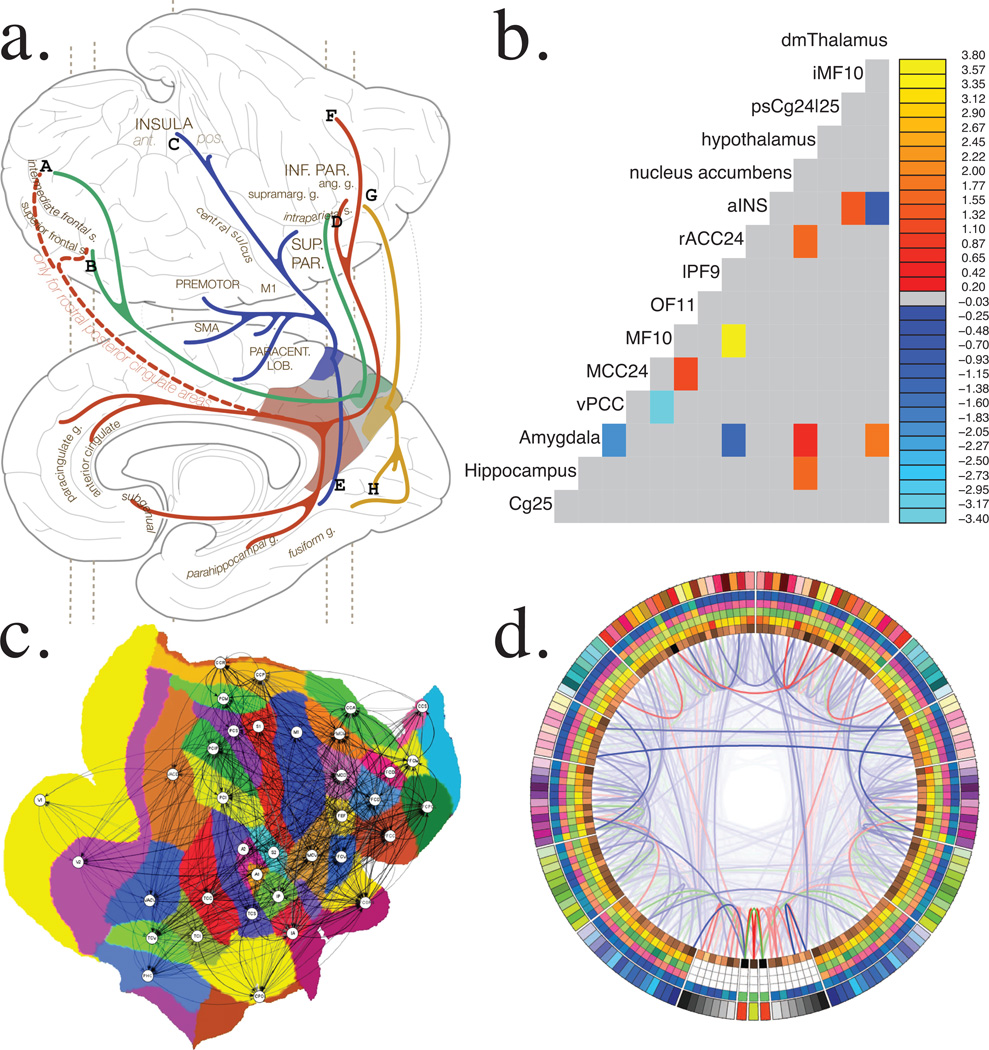
(A) Classical anatomical-tracing style depiction of iFC for posteromedial cortex subdivisions (reproduced with permission from Margulies et al., 2009). (B) Matrix representation of functional brain connections predictive of diagnostic status in depression (reproduced with permission from Craddock et al., 2009). (C) Flatmap-based representation of the CoCoMac atlas of the Macaque connectome (reproduced with permission from Knock et al., 2009). (D) Connectogram depicts brain areas (nodes) as columns in the circular band, differing connectivity metrics in separate layers and connections with lines, lobes are differentiated by color and left/right halves corresponds to hemispheres (reproduced with permission from van Horn et al., 2012).
References
-
- Varela F, Lachaux JP, Rodriguez E, Martinerie J. The brainweb: phase synchronization and large-scale integration. Nat. Rev. Neurosci. 2001;2:229–239. - PubMed
Publication types
MeSH terms
Grants and funding
LinkOut - more resources
Full Text Sources
Other Literature Sources
Medical
