. 2013 Jun;8(6):1249-59.
doi: 10.1038/nprot.2013.064. Epub 2013 May 30.

Preparation and use of MitoPY1 for imaging hydrogen peroxide in mitochondria of live cells

Affiliations

Preparation and use of MitoPY1 for imaging hydrogen peroxide in mitochondria of live cells

Bryan C Dickinson et al. Nat Protoc. 2013 Jun.

Abstract

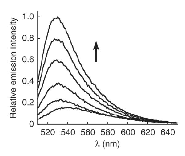
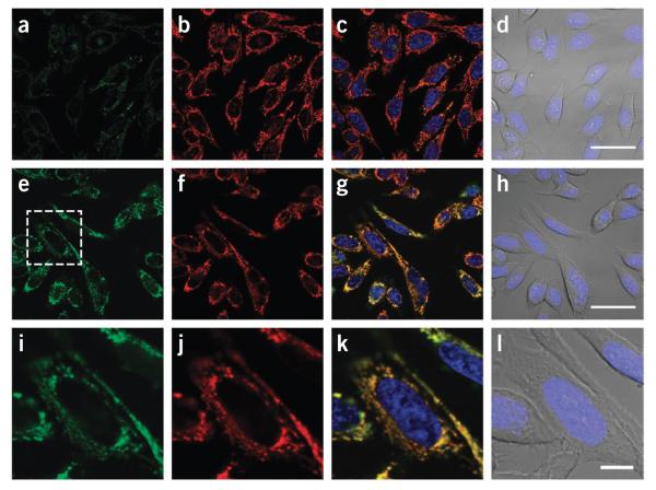
Mitochondria peroxy yellow 1 (MitoPY1) is a small-molecule fluorescent probe that selectively tracks to the mitochondria of live biological specimens and responds to local fluxes of hydrogen peroxide (H(2)O(2)) by a turn-on fluorescence enhancement. This bifunctional dye uses a triphenylphosphonium targeting group and a boronate-based molecular switch to selectively respond to H(2)O(2) over competing reactive oxygen species (ROS) within the mitochondria. MitoPY1 can be used to measure mitochondrial H(2)O(2) levels in both cell culture and tissue models. In this protocol, we describe the synthesis of MitoPY1 and how to use this chemical tool to visualize mitochondrial H(2)O(2) in live cells. The preparation of MitoPY1 is anticipated to take 7-10 d, and assays involving microscopy of cultured mammalian cells can be performed in 1-2 d.


Figures

Figure 1
Figure 1
Mitochondria Peroxy Yellow 1 (MitoPY1). (a) Scheme for the synthesis. (b) Scheme showing H2O2-mediated activation of MitoPY1.
Figure 2
Figure 2
Fluorescence turn-on response of 5 μM MitoPY1 to H2O2. Time points represent 0, 5, 15, 30, 45 and 60 min after the addition of 100 μM H2O2. Excitation at 503 nm.
Figure 3
Figure 3
Imaging mitochondrial H2O2 in live cells with MitoPY1. HeLa cells were loaded with 10 μM MitoPY1 in DPBS for 45 min at 37 °C. The medium was then exchanged for fresh DPBS containing 25 nM Mitotracker Deep Red and 1 μM Hoechst. After the addition of either H2O (control) or 100 μM H2O2, the cells were incubated for 60 min at 37 °C. (ad) Control cells were then imaged with MitoPY1 (a), Mitotracker Deep Red (b), overlay of MitoPY1 (green), Mitotracker Deep Red (red) and Hoechst (blue) (c), or overlay of bright-field and Hoechst (blue) (d). Scale bar, 40 μm. (eh) H2O2-treated cells were imaged with MitoPY1 (e), Mitotracker Deep Red (f), overlay of MitoPY1 (green), Mitotracker Deep Red (red) and Hoechst (blue) (g), or overlay of bright-field and Hoechst (blue) (h). Scale bar, 40 μm. (il) The region of H2O2-treated cells denoted in e is enlarged, showing MitoPY1 (i), Mitotracker Deep Red (j), overlay of MitoPY1 (green), Mitotracker Deep Red (red) and Hoechst (blue) (k), or overlay of bright field and Hoechst (blue) (l). Scale bar, 10 μm.

References

    1. Sundaresan M, Yu ZX, Ferrans VJ, Irani K, Finkel T. Requirement for generation of H2O2 for platelet-derived growth factor signal transduction. Science. 1995;270:296–299. - PubMed
    1. Niethammer P, Grabher C, Look AT, Mitchison TJ. A tissue-scale gradient of hydrogen peroxide mediates rapid wound detection in zebrafish. Nature. 2009;459:996–999. - PMC - PubMed
    1. Le Belle JE, et al. Proliferative neural stem cells have high endogenous ROS levels that regulate self-renewal and neurogenesis in a PI3K/Akt-dependant manner. Cell Stem Cell. 2011;8:59–71. - PMC - PubMed
    1. Dickinson BC, Peltier J, Stone D, Schaffer DV, Chang CJ. Nox2 redox signaling maintains essential cell populations in the brain. Nat. Chem. Biol. 2011;7:106–112. - PMC - PubMed
    1. O’Neill JS, Reddy AB. Circadian clocks in human red blood cells. Nature. 2011;469:498–503. - PMC - PubMed

Publication types