. 2013 Aug;20(8):1089-100.
doi: 10.1038/cdd.2013.44. Epub 2013 May 31.

A human iPSC model of Ligase IV deficiency reveals an important role for NHEJ-mediated-DSB repair in the survival and genomic stability of induced pluripotent stem cells and emerging haematopoietic progenitors

Affiliations

A human iPSC model of Ligase IV deficiency reveals an important role for NHEJ-mediated-DSB repair in the survival and genomic stability of induced pluripotent stem cells and emerging haematopoietic progenitors

K Tilgner et al. Cell Death Differ. 2013 Aug.

Abstract

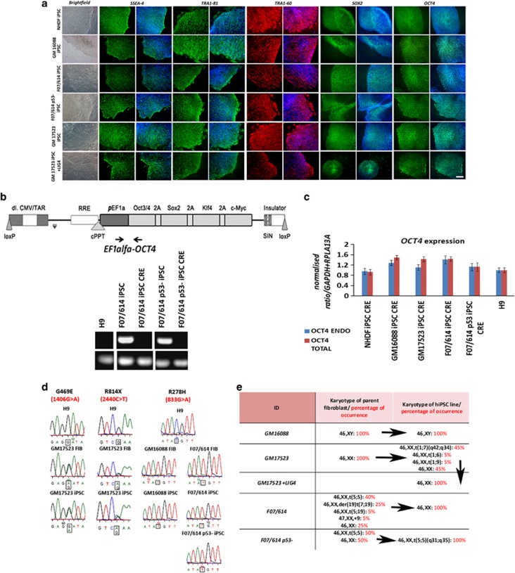
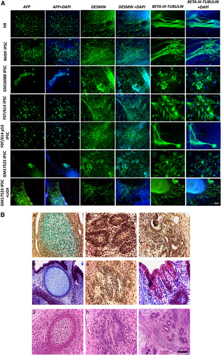
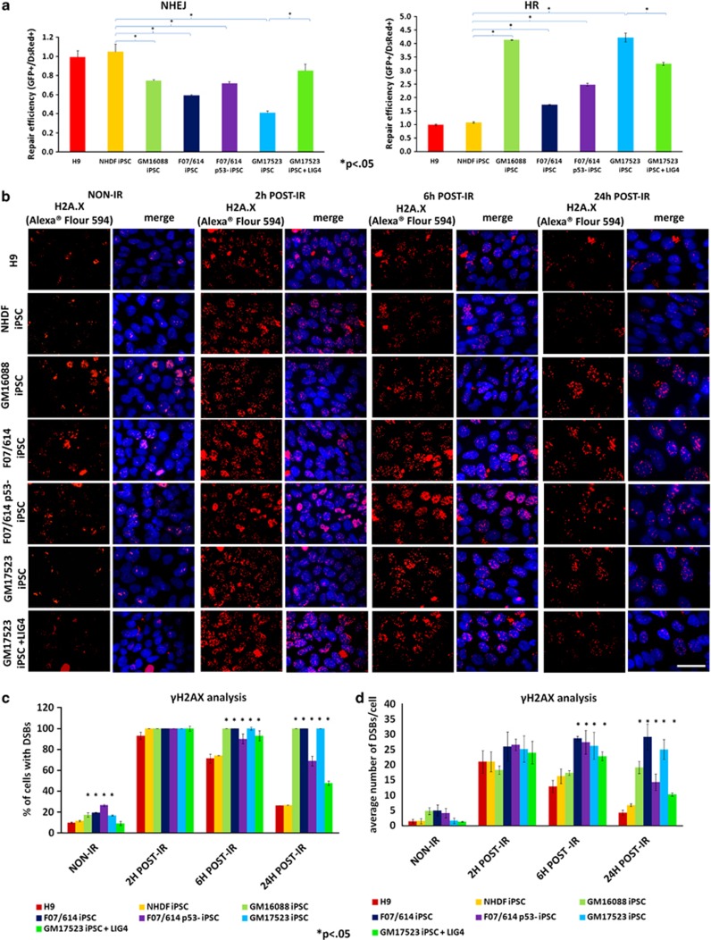
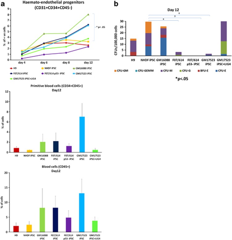
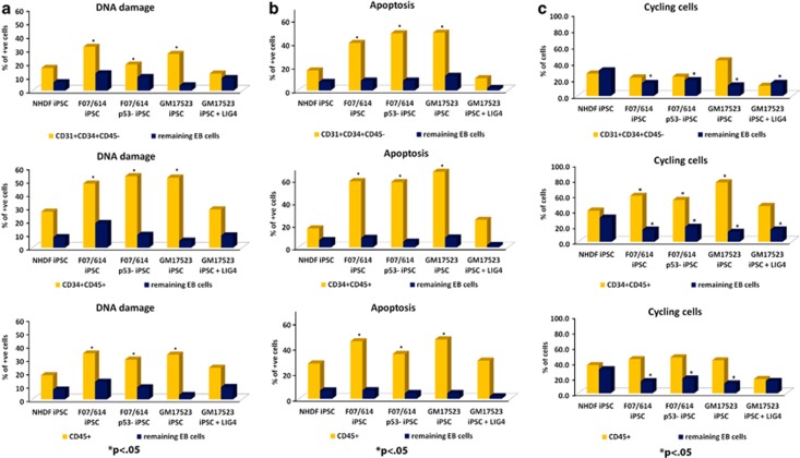
DNA double strand breaks (DSBs) are the most common form of DNA damage and are repaired by non-homologous-end-joining (NHEJ) or homologous recombination (HR). Several protein components function in NHEJ, and of these, DNA Ligase IV is essential for performing the final 'end-joining' step. Mutations in DNA Ligase IV result in LIG4 syndrome, which is characterised by growth defects, microcephaly, reduced number of blood cells, increased predisposition to leukaemia and variable degrees of immunodeficiency. In this manuscript, we report the creation of a human induced pluripotent stem cell (iPSC) model of LIG4 deficiency, which accurately replicates the DSB repair phenotype of LIG4 patients. Our findings demonstrate that impairment of NHEJ-mediated-DSB repair in human iPSC results in accumulation of DSBs and enhanced apoptosis, thus providing new insights into likely mechanisms used by pluripotent stem cells to maintain their genomic integrity. Defects in NHEJ-mediated-DSB repair also led to a significant decrease in reprogramming efficiency of human cells and accumulation of chromosomal abnormalities, suggesting a key role for NHEJ in somatic cell reprogramming and providing insights for future cell based therapies for applications of LIG4-iPSCs. Although haematopoietic specification of LIG4-iPSC is not affected per se, the emerging haematopoietic progenitors show a high accumulation of DSBs and enhanced apoptosis, resulting in reduced numbers of mature haematopoietic cells. Together our findings provide new insights into the role of NHEJ-mediated-DSB repair in the survival and differentiation of progenitor cells, which likely underlies the developmental abnormalities observed in many DNA damage disorders. In addition, our findings are important for understanding how genomic instability arises in pluripotent stem cells and for defining appropriate culture conditions that restrict DNA damage and result in ex vivo expansion of stem cells with intact genomes.


Figures

Figure 1
Figure 1
LIG4-iPSC display all the in vitro hallmarks of pluripotency and patient-specific genetic mutations. (a) Staining of control- and LIG4-iPSC lines with pluripotency cell surface markers (SSEA-4 (green), TRA1-81 (green) and TRA1-60 (red)) and transcription factors (SOX2 (green) and OCT4 (green)). DAPI staining of nuclei is shown in blue (Scale bar=100 μm); (b) Schematic presentation of polycystronic expression construct (OSKM) used for the induction of pluripotency and representative gel electrophoresis used to detect the transgene cassette prior and post Cre excision event; (c) Quantitative RT-PCR analysis for the expression of total and endogenous OCT4 showing removal of exogenous transgene post Cre expression. Data is presented as mean ± S.E.M., n=4. The value for H9 was set to 1 and all other values were calculated with respect to that. CRE=post Cre excision; (d) Direct sequencing of cloned PCR products spanning patient-specific mutations in H9 (human ESC control), patient-specific fibroblasts and iPSC lines shows that all LIG4-iPSC lines display the same mutations as fibroblasts isolated from LIG4 patients; (e) Summary of karyotype analysis performed in LIG4 fibroblasts and iPSC lines derived from them. Figures highlighted in red indicate the percentage of occurrence amongst 30 metaphases analysed for each sample. Karyotype analysis of fibroblasts was carried out at the same passage used for induction of pluripotency. Human iPSC lines were karyotyped at passage 5 and 15 postderivation. Identical results (shown in this figure) were obtained at both passages
Figure 2
Figure 2
In vitro and in vivo differentiation capacity of LIG4- and control-iPSC lines. (A) In vitro differentiation capacity to all three germ layers demonstrated by immunocytochemical staining with an anti-AFP antibody (marker of endoderm), anti-DESMIN antibody (marker of mesoderm) and anti-βIII-Tubulin antibody TUJ1 (marker of ectoderm). DAPI staining of nuclei is shown in blue (Scale bar=100 μm). (B) In vivo differentiation capacity of LIG4- and control-iPSC and human ESC lines. Histological analysis of tissue masses formed from grafted colonies of: (a–c) human ESC (H9, positive control); (d–f) NHDF-iPSC control; (g–i) LIG4 overexpressing iPSC line (GM17523+LIG4). Human ESC, control-iPSC control and DNA LIGASE IV overexpressing LIG4 patient-specific iPSC line (GM17523+LIG4) produced typical large (∼7–10 mm diameter) and diverse teratomas containing structures representative of each germ layer, notably cartilage (a, d, g); neuroepithelium (b, e, h); kidney (c, i) and profiles of gut wall (f). Histological staining: Weigert's (a–c, e); Masson's Trichrome (d, f), Hamatoxylin-Eosin (g, h, l). Scale bars: (a–c, e, f, g, h) 75 μm; (d, l) 150 μm
Figure 3
Figure 3
LIG4-iPSC lines display reduced NHEJ-mediated-DSB repair capacity and increased levels of DSBs under normal and IR conditions. (a) Schematic presentation of HR and NHEJ activity, indicating a significant downregulation of NHEJ-DSB repair capacity in LIG4-iPSC lines when compared with control NHDF-iPSC and human ESC lines at 16 h postIR. Data is presented as mean ± S.E.M., n=4. Student's t-test was carried out between each LIG4-iPSC and NHDF-iPSC control line to assess significant differences shown by *. (b) LIG4-iPSC lines show a greater level of DSB foci (marked by γH2A.X) under normal culture conditions (NON-IR), as well as 2, 6 and 24 h postIR. IR=ionising radiation. This is measured both in terms of cell percentage with DSB foci shown in (c) (only cells with more than five foci are counted) and average number of foci per cell shown in (d). Data shown in (c) and (d) is presented as mean ± S.E.M., n=4. Student's t-test was carried out between each LIG4-iPSC and NHDF-iPSC control line to assess significant differences shown by *. (ad), Overexpression of DNA LIGASE IV in GM17523-iPSC restores the NHEJ-DSB repair capacity and results in clearance of DSB caused by IR, albeit not the same extent as the control iPSC and human ESC line
Figure 4
Figure 4
Deficient NHEJ-mediated-DSB repair results in generation of haematopoietic progenitors with reduced clonogenic capacity and differentiation potential. (a) LIG4-iPSC undergo haematopoietic differentiation and give rise to haemato-endothelial progenitors (CD31+CD34+CD45−), primitive blood cells (CD34+CD45+ data shown from day 12 of differentiation) and mature blood cells (CD45+ data shown from day 12 of differentiation) with similar kinetics to human ESC (H9) and control NHDF-iPSC lines. Data is presented as mean ± S.E.M., n=4. Mann–Whitney U test was used to evaluate the differences in the haematopoietic differentiation efficiency between control and LIG4-iPSC lines; (b) LIG4-iPSC lines have lower capacity to give rise to haematopoietic colonies in cytokine supplemented semisolid methylcellulose assays. Data is presented at day 12 of differentiation as mean ± S.E.M., n=4. Student's t-test was carried out between each LIG4-iPSC and NHDF-iPSC control line to assess significant differences shown by *. Overexpression of DNA LIGASE IV restores the ability of GM17523-iPSC to give rise to haematopoietic colonies
Figure 5
Figure 5
Deficient NHEJ-DSB repair results in increased DNA damage and apoptosis in LIG4-iPSC derived haematopoietic progenitors. (ac) Haematopoietic progenitors derived from LIG4-iPSC lines show the highest level of DNA damage (a: flow cytometric analysis for phosphorylated H2A.X), highest level of apoptosis (b: flow cytometric analysis for measurement of apoptosis with Cleaved PARP) and in some subpopulations enhanced cell proliferation (c: flow cytometric analysis for detection of cell proliferation by BrdU) under normal culture conditions. Data is shown as mean of at least four independent experiments. Student's t-test was carried out between each LIG4-iPSC and NHDF-iPSC control for each select cell subpopulation to assess significant differences shown by *. All data presented in (ac) are from day 12 differentiation. (ac) Overexpression of DNA LIGASE IV in GM17523-iPSC reduces the accumulation of DSBs, lowers the proliferation and apoptotic haematopoietic progenitors back to levels comparable to progenitors derived from control NHDF-iPSC line

Comment in

References

    1. Hartlerode AJ, Scully R. Mechanisms of double-strand break repair in somatic mammalian cells. Biochem J. 2009;423:157–168. - PMC - PubMed
    1. Jeggo PA. DNA breakage and repair. Adv Genet. 1998;38:185–218. - PubMed
    1. Holthausen JT, Wyman C, Kanaar R. Regulation of DNA strand exchange in homologous recombination. DNA Repair (Amst) 2010;9:1264–1272. - PubMed
    1. Lieber MR, Ma Y, Pannicke U, Schwarz K. Mechanism and regulation of human non-homologous DNA end-joining. Nat Rev Mol Cell Biol. 2003;4:712–720. - PubMed
    1. Grawunder U, Grawunder U, Wilm M, Wu M, Kulesza P, Wilson TE, et al. Activity of DNA ligase IV stimulated by complex formation with XRCC4 protein in mammalian cells. Nature. 1997;388:492–495. - PubMed

Publication types

MeSH terms