

Antitumor activity of a humanized, bivalent immunotoxin targeting fn14-positive solid tumors
- PMID: 23722548
- PMCID: PMC3805367
- DOI: 10.1158/0008-5472.CAN-13-0187
Antitumor activity of a humanized, bivalent immunotoxin targeting fn14-positive solid tumors
Abstract
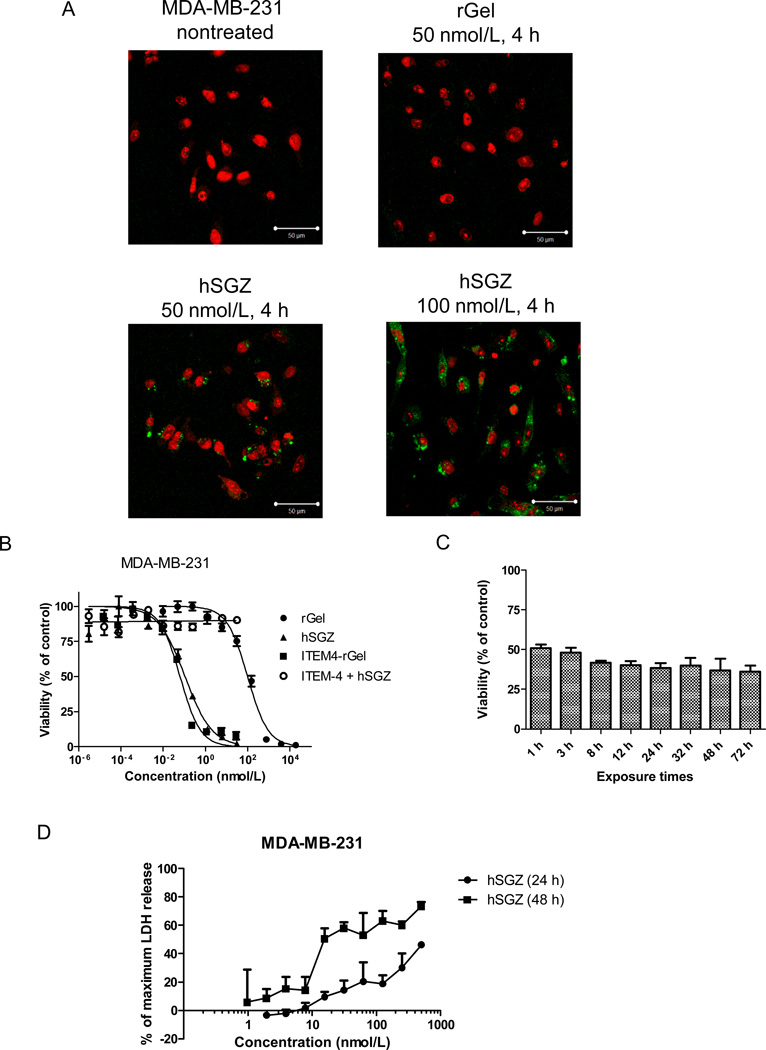
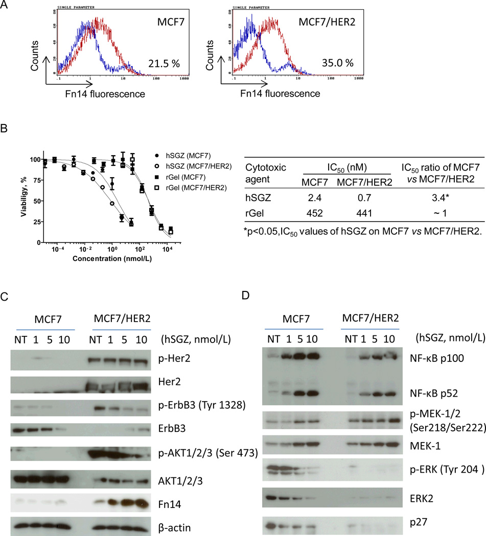
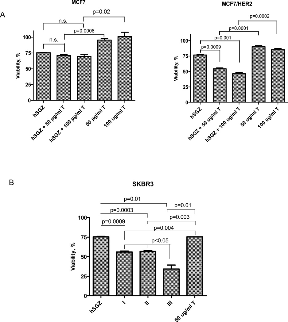
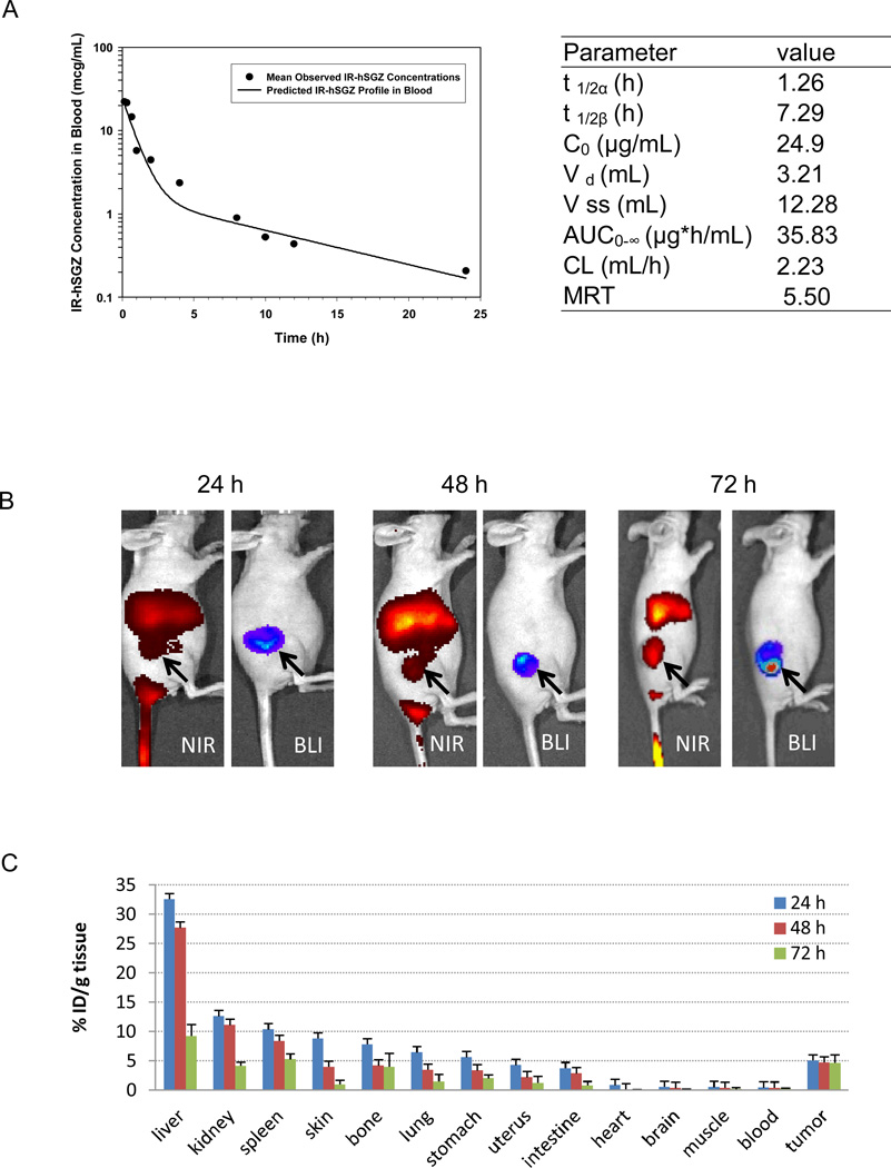
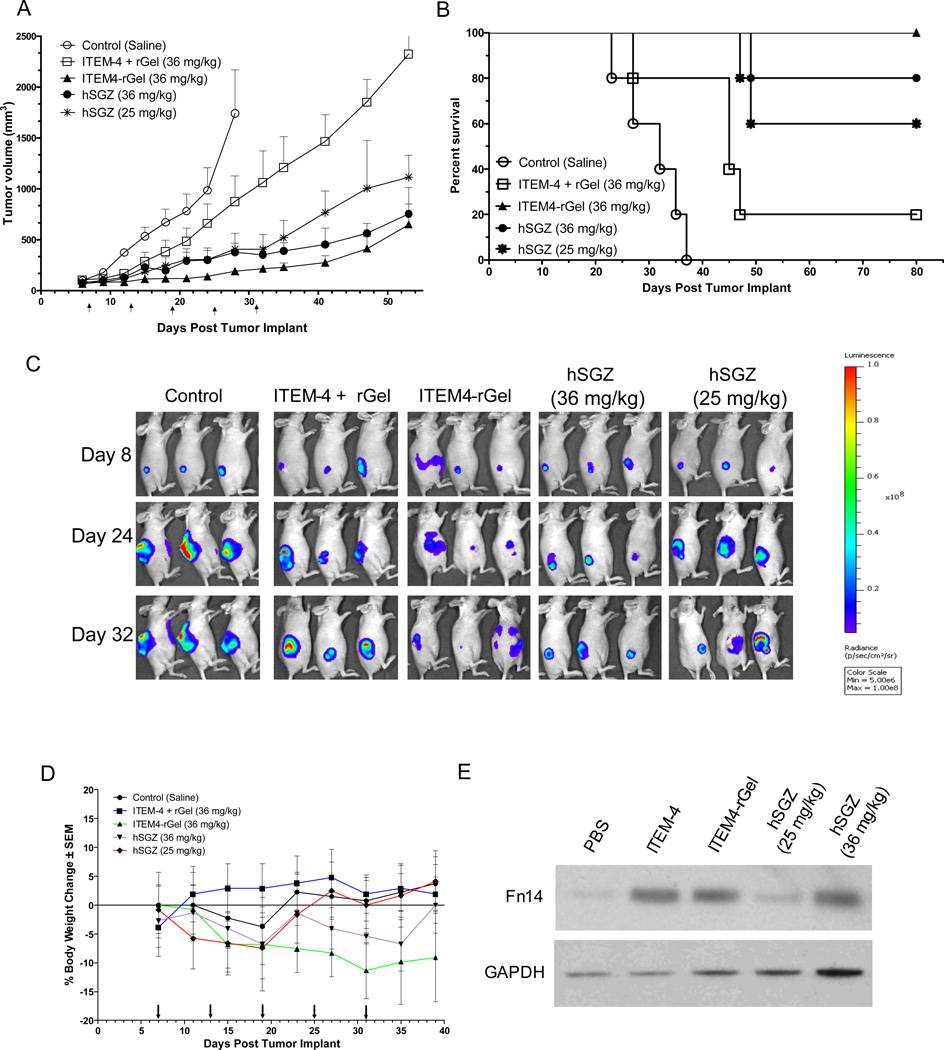
The TNF-like weak inducer of apoptosis (TWEAK; TNFSF12) receptor Fn14 (TNFRSF12A) is expressed at low levels in normal tissues but frequently highly expressed in a wide range of tumor types such as lung, melanoma, and breast, and therefore it is a potentially unique therapeutic target for these diverse tumor types. We have generated a recombinant protein containing a humanized, dimeric single-chain anti-fibroblast growth factor-inducible 14-kDa protein (Fn14) antibody fused to recombinant gelonin toxin as a potential therapeutic agent (designated hSGZ). The hSGZ immunotoxin is a highly potent and selective agent that kills Fn14-positive (Fn14(+)) tumor cells in vitro. Treatment of cells expressing the MDR protein MDR1 (ABCB1B) showed no cross-resistance to hSGZ. Induced overexpression of Fn14 levels in MCF7 cells through HER2 (ERBB2) signaling translated to an improved therapeutic index of hSGZ treatment. In combination with trastuzumab, hSGZ showed an additive or synergistic cytotoxic effect on HER2(+)/Fn14(+) breast cancer cell lines. Also, hSGZ treatment inhibited Erb3/Akt signaling in HER2-overexpressing breast cancer cells. Pharmacokinetic studies in mice revealed that hSGZ exhibited a biexponential clearance from plasma with a rapid initial clearance (t1/2α = 1.26 hours) followed by a seven-fold longer plasma half-life (t1/2β = 7.29 hours). At 24, 48, and 72 hours after injection, uptake of the hSGZ into tumors was 5.1, 4.8, and 4.7%ID/g, with a tumor-to-muscle ratio of 5.6, 6.2, and 9.0, respectively. Therapeutic efficacy studies showed significant tumor inhibition effects using an MDA-MB-231/Luc breast cancer xenograft model. Our findings show that hSGZ is an effective anticancer agent and a potential candidate for clinical studies.
©2013 AACR.
Conflict of interest statement
Figures
Similar articles
-
The TWEAK receptor Fn14 is a therapeutic target in melanoma: immunotoxins targeting Fn14 receptor for malignant melanoma treatment.J Invest Dermatol. 2013 Apr;133(4):1052-62. doi: 10.1038/jid.2012.402. Epub 2012 Nov 29. J Invest Dermatol. 2013. PMID: 23190886 Free PMC article.
-
Design optimization and characterization of Her2/neu-targeted immunotoxins: comparative in vitro and in vivo efficacy studies.Oncogene. 2014 Jan 23;33(4):429-39. doi: 10.1038/onc.2012.612. Epub 2013 Feb 4. Oncogene. 2014. PMID: 23376850 Free PMC article.
-
Development and characterization of a potent immunoconjugate targeting the Fn14 receptor on solid tumor cells.Mol Cancer Ther. 2011 Jul;10(7):1276-88. doi: 10.1158/1535-7163.MCT-11-0161. Epub 2011 May 17. Mol Cancer Ther. 2011. PMID: 21586630 Free PMC article.
-
TWEAK/Fn14 signaling in tumors.Tumour Biol. 2017 Jun;39(6):1010428317714624. doi: 10.1177/1010428317714624. Tumour Biol. 2017. PMID: 28639899 Review.
-
The TWEAK-Fn14 system as a potential drug target.Br J Pharmacol. 2013 Oct;170(4):748-64. doi: 10.1111/bph.12337. Br J Pharmacol. 2013. PMID: 23957828 Free PMC article. Review.
Cited by
-
Minimizing the non-specific binding of nanoparticles to the brain enables active targeting of Fn14-positive glioblastoma cells.Biomaterials. 2015 Feb;42:42-51. doi: 10.1016/j.biomaterials.2014.11.054. Epub 2014 Dec 13. Biomaterials. 2015. PMID: 25542792 Free PMC article.
-
Advances in anticancer immunotoxin therapy.Oncologist. 2015 Feb;20(2):176-85. doi: 10.1634/theoncologist.2014-0358. Epub 2015 Jan 5. Oncologist. 2015. PMID: 25561510 Free PMC article. Review.
-
Targeting fibroblast growth factor (FGF)-inducible 14 (Fn14) for tumor therapy.Front Pharmacol. 2022 Oct 21;13:935086. doi: 10.3389/fphar.2022.935086. eCollection 2022. Front Pharmacol. 2022. PMID: 36339601 Free PMC article. Review.
-
Antibody-based soluble and membrane-bound TWEAK mimicking agonists with FcγR-independent activity.Front Immunol. 2023 Jul 11;14:1194610. doi: 10.3389/fimmu.2023.1194610. eCollection 2023. Front Immunol. 2023. PMID: 37545514 Free PMC article.
-
AR-regulated TWEAK-FN14 pathway promotes prostate cancer bone metastasis.Cancer Res. 2014 Aug 15;74(16):4306-17. doi: 10.1158/0008-5472.CAN-13-3233. Epub 2014 Jun 26. Cancer Res. 2014. PMID: 24970477 Free PMC article.
References
-
- Jemal A, Bray F, Center MM, Ferlay J, Ward E, Forman D. Global cancer statistics. CA Cancer J Clin. 2011;61:69–90. - PubMed
-
- Wiley SR, Cassiano L, Lofton T, Davis-Smith T, Winkles JA, Lindner V, et al. A novel TNF receptor family member binds TWEAK and is implicated in angiogenesis. Immunity. 2001;15:837–846. - PubMed
-
- Burkly LC, Michaelson JS, Hahm K, Jakubowski A, Zheng TS. TWEAKing tissue remodeling by a multifunctional cytokine: role of TWEAK/Fn14 pathway in health and disease. Cytokine. 2007;40:1–16. - PubMed
Publication types
MeSH terms
Substances
Grants and funding
LinkOut - more resources
Full Text Sources
Other Literature Sources
Medical
Research Materials
Miscellaneous
