. 2013 Jul;123(7):3124-34.
doi: 10.1172/JCI67260. Epub 2013 Jun 3.

Dominant protein interactions that influence the pathogenesis of conformational diseases

Affiliations

Dominant protein interactions that influence the pathogenesis of conformational diseases

Jordan Wright et al. J Clin Invest. 2013 Jul.

Abstract

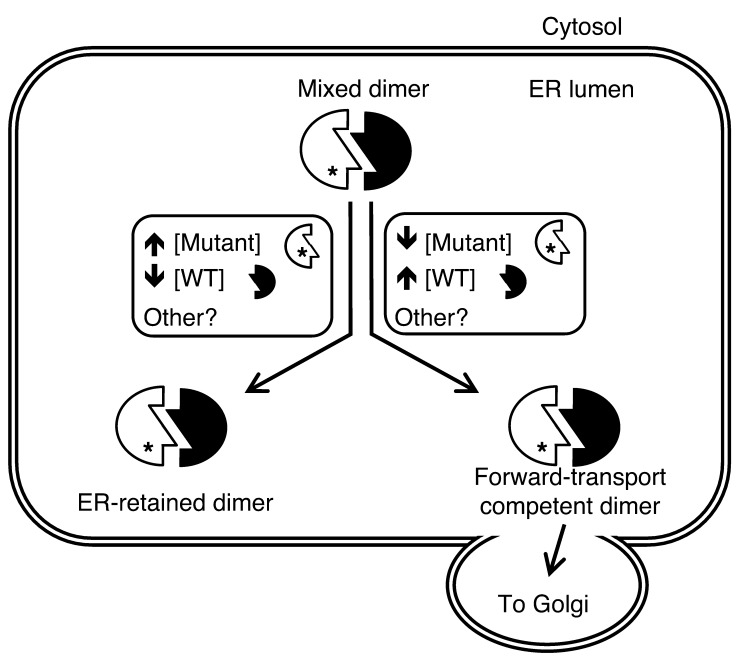
Misfolding of exportable proteins can trigger endocrinopathies. For example, misfolding of insulin can result in autosomal dominant mutant INS gene-induced diabetes of youth, and misfolding of thyroglobulin can result in autosomal recessive congenital hypothyroidism with deficient thyroglobulin. Both proinsulin and thyroglobulin normally form homodimers; the mutant versions of both proteins misfold in the ER, triggering ER stress, and, in both cases, heterozygosity creates potential for cross-dimerization between mutant and WT gene products. Here, we investigated these two ER-retained mutant secretory proteins and the selectivity of their interactions with their respective WT counterparts. In both cases and in animal models of these diseases, we found that conditions favoring an increased stoichiometry of mutant gene product dominantly inhibited export of the WT partner, while increased relative level of the WT gene product helped to rescue secretion of the mutant partner. Surprisingly, the bidirectional consequences of secretory blockade and rescue occur simultaneously in the same cells. Thus, in the context of heterozygosity, expression level and stability of WT subunits may be a critical factor influencing the effect of protein misfolding on clinical phenotype. These results offer new insight into dominant as well as recessive inheritance of conformational diseases and offer opportunities for the development of new therapies.


Figures

Figure 1
Figure 1. Proinsulin-KDEL interacts with and inhibits secretion of WT proinsulin.
293T cells transiently transfected with hPro-CpepMyc were cotransfected with plasmids as indicated. (A) Cell lysates (C) and media (M) were resolved by SDS-PAGE, electrotransfer, and immunoblotting (WB) with anti-Myc. The media/cell ratio of hPro-CpepMyc bands was decreased by 58.9% ± 12.8% (P = 0.003, n = 6) in cells coexpressing hPro-CpepSfGFP-KDEL compared with that in cells coexpressing WT hPro-CpepSfGFP. (B) Cells lysed in TX-CoIP buffer were immunoprecipitated with anti-GFP or anti-Myc and resolved by SDS-PAGE, electrotransfer, and immunoblotting with anti-GFP or anti-Myc as indicated. The top 2 rows demonstrate expression of the indicated proteins, and the bottom row demonstrates CoIP. Gels are representative of 3 independent experiments. (C) Cells transiently expressing WT hPro plus TgGFP were cotransfected with plasmids as indicated. The media collected overnight were analyzed by hPro-specific RIA. TgGFP in the same cell lysates and media was analyzed by SDS-PAGE, electrotransfer, and immunoblotting with anti-GFP. The media/cell ratio of TgGFP bands in cells coexpressing mPro-KDEL exhibited no significant change compared with that from cells coexpressing WT mPro (1.9 ± 0.2 vs. 2.1 ± 0.8; P = 0.3, n = 5). In B and C, noncontiguous lanes from the same gel are shown. (D) Cells transiently expressing hPro-CpepMyc were cotransfected with plasmids expressing mPro-KDEL or mPro. Media were collected overnight, and cell lysates were analyzed by hPro-specific RIA. The data in C and D represent mean ± SEM, each from ≥4 independent transfections. *P < 0.05.
Figure 2
Figure 2. Cross-dimerization of mutant/WT proinsulin and mutant/WT Tg.
293T cells were transiently cotransfected with plasmids expressing the indicated proinsulin or Tg variants. (A) At 48 hours after transfection, cell lysates and overnight media were collected; both were immunoprecipitated with anti-Myc (to prepurify the antigen) and then analyzed by SDS-PAGE, electrotransfer, and immunoblotting with anti-Myc. The media/cell ratio of hPro-CpepMyc bands from cells coexpressing mPro-G(B23)V decreased 66.1% ± 1.9% (P < 0.001, n = 4) compared with that of WT mPro. (B) At 48 hours after transfection, cell lysates and overnight media were collected, treated with or without EndoH, and were analyzed by immunoblotting with anti-Myc. The media/cell ratio of WT Tg3xMyc bands in cells coexpressing rdw-TgGFP decreased 74.1% ± 3.3% (P < 0.001, n = 4) compared with that of WT TgGFP. (C) At 48 hours after transfection, cells lysed in TX-CoIP buffer were immunoprecipitated with anti-GFP or anti-Myc and were analyzed by Western blotting with anti-GFP or anti-Myc, as indicated. (D) Cells transiently coexpressing WT secretory ChEL-HA plus secretory mutant rdw-ChEL-Myc or secretory WT ChEL-Myc (9) were cultured in the presence of brefeldin A (5 hours, 5 μg/ml) to allow intracellular coincubation of the coexpressed constructs. Cells were then lysed in NP40-CoIP buffer and analyzed by immunoblotting with or without immunoprecipitation, as indicated. For C and D, the top 2 rows demonstrate expression of protein partners, and the bottom row demonstrates CoIP. Gels in C and D are representative of n ≥ 3 experiments. In A and C, noncontiguous lanes from the same gel are shown.
Figure 3
Figure 3. Intracellular distribution of mutant proinsulins in regulated secretory cells coexpressing or not coexpressing WT proinsulin.
(A) Cultured INS1 pancreatic β cells (that express endogenous proinsulin) were transiently transfected to express WT or mutant hPro-CpepSfGFP, as indicated. Fixed cells (counterstained with DAPI) were examined by confocal microscopy for the distribution of SfGFP-containing peptides (scale bar: 20 μm). The cell lysates and overnight bathing media were collected, immunoprecipitated with anti-GFP, and analyzed by immunoblotting with anti-GFP to examine secretion efficiency. Noncontiguous lanes from the same gel are shown. The media/cell ratio for WT, G(B23)V, and C(A7)Y hPro-CpepSfGFP bands was 14.8 ± 3.8, 0.74 ± 0.04, and 0.16 ± 0.06, respectively (P < 0.05 for all groups, n = 4). (B) Cultured AtT20 pituitary corticotroph cells (that do not express endogenous proinsulin) were transiently cotransfected with one of three different plasmid combinations, as indicated. Fixed cells were examined by confocal fluorescence for the distribution of SfGFP-containing peptides (green) and immunofluorescence to localize ACTH-containing secretory granules (red) at the tips of cell (arrowheads; scale bar: 20 μm). Cell boundaries were defined from phase-contrast images (data not shown). Enrichment of average GFP intensity in the secretory granule region was compared with average GFP intensity in nongranule regions. Data represent mean ± SEM from 30 to 38 separately imaged cells for each of the 3 respective transfection conditions. *P < 0.05.
Figure 4
Figure 4. Cross-dimerization as a basis for secretory rescue of mutant proinsulin or Tg is specific to respective WT partners.
(A) 293T cells transiently expressing both mutant proinsulin and mutant Tg were cotransfected with either WT mPro or WT Tg. The media were collected overnight, and cells were lysed; proinsulin secretion was quantified by hPro-specific RIA. Data represent mean ± SEM relative to cells lacking mPro or WT Tg (P = 0.07, n = 3). From the same cell lysates and media, secretion of rdw-TgGFP was analyzed by SDS-PAGE, electrotransfer, and immunoblotting with anti-GFP. (B) At 48 hours after cotransfection, overnight secretion of mutant hProG(B23)V-CpepMyc (in duplicate) was measured by immunoprecipitation and immunoblotting with anti-Myc. The results shown in A and B are representative of 3 separate experiments. EV, empty vector. (C) At 48 hours after transfection, cells cotransfected as indicated were either untreated or treated with 5 μg/ml brefeldin A (BFA). After 5 hours, the media were collected and analyzed by hPro-specific RIA. The data shown are mean values ± range from 2 independent measurements.
Figure 5
Figure 5. Secretory rescue and stabilization of mutant proinsulin or Tg by their WT counterparts is linked to the WT/mutant expression ratio.
(AD) 293T cells were transiently cotransfected with the indicated plasmid combinations, with empty vector added to keep total DNA per well constant. (A) Total mutant proinsulin recovery at 20 hours after synthesis was measured by pulse chase (see Methods). Protein stability was quantified by band recovery at 20 hours chase relative to that at time 0 (mean ± SEM, n = 4, *P < 0.05). (B) Overnight media and cell lysates were analyzed by immunoblotting with anti-GFP (normalized to total cellular protein). Representative blots from 3 experiments are shown. (C) Overnight media were collected, and hPro secretion (normalized to total cellular protein) was measured by RIA. Data represent mean ± range from 2 independent experiments. (D) Overnight media and cell lysates (bottom 2 rows) or combined lysate and media (top row) were analyzed by immunoblotting with anti-GFP (normalized to total cellular protein). Representative blots from 3 experiments are shown. (E) INS1E cells were transfected with 2 or 0.5 μg plasmid expressing mutant proinsulin; the cellular levels of hProG(B23)V-CpepSfGFP are shown at left. Cell lysates and basal secretion (B) and glucose-stimulated secretion (S), normalized for hProG(B23)V-CpepSfGFP protein expression (1% of total for 2 μg transfection; 3% of total for 0.5 μg transfection), were analyzed by immunoblotting with anti-GFP. Percentage secretion was quantified as total GFP signal in media over total in cells. The data represent mean ± SEM, from 3 independent experiments. *P < 0.05.
Figure 6
Figure 6. Rescue of mutant Tg and blockade of WT proinsulin in primary tissue from animal models of disease.
(A) Lobules of thyroid glands were freshly prepared from mice of the indicated genotypes. Secretory proteins delivered for posttranslational iodination were labeled by incubation of thyroid lobules with 1.0 μCi/μl Na125I for 30 minutes, as described in Methods. The thyroid lobules were then lysed and immunoprecipitated with anti-Myc. The immunoprecipitates were either mock-digested or digested with EndoH, as in Figure 2B, and then analyzed by SDS-PAGE and autoradiography. *P < 0.05. (B) Pancreata from 6-week-old mice, with the genotypes indicated, were fixed in paraffin, sectioned, deparaffinized, and immunostained with antibodies specific to mPro (red) and calnexin to mark the ER (green). From confocal microscope images (scale bar: 10 μm), a blinded reader scored the localization of WT mPro in each β cell as either a predominant juxtanuclear crescent of increased intensity (Golgi, consistent with previous reports, refs. , ; e.g., see arrows) or mainly colocalized with calnexin (ER; e.g., see arrowheads). Quantitation of these data is shown as mean ± SEM from n = 5 mice with 5 islets per mouse. BG, blood glucose. *P < 0.05.
Figure 7
Figure 7. Bidirectional consequences of interactions between mutant and WT cross-dimerization partners.
(A) The overnight bathing media from cells cotransfected with mutant hPro-G(B23)V and WT mPro were selectively probed for simultaneous secretion of mPro (by ELISA) and hPro (by RIA); both assays (normalized to total cell protein) are entirely species specific. The data represent mean ± SEM of a minimum of 3 independent transfections; the arrows in the top and bottom panels demonstrate blockade and rescue, respectively. *P < 0.05. (B) The overnight bathing media from cells cotransfected with rdw-TgGFP and WT Tg3xMyc were resolved by SDS-PAGE and selectively probed for simultaneous secretion of mutant Tg (by specific immunoblotting with anti-GFP) and WT Tg (by specific immunoblotting with anti-Myc), normalized to total cellular protein. For the bottom panel, noncontiguous lanes from the same gel are shown. Note that, in cotransfection, rdw-Tg secretion becomes enhanced while WT Tg secretion becomes inhibited, whereas, in single transfections, rdw-Tg is not secreted and WT Tg is well secreted. The data shown are representative of 3 independent experiments.
Figure 8
Figure 8. Secretory rescue by WT proinsulin is restricted to a subset of MIDY mutants.
293T cells transiently cotransfected with plasmids expressing the indicated hPro mutants and either empty vector or WT mPro were incubated overnight in growth medium beginning at 24 hours after transfection. Media were collected, and hPro secretion was measured by RIA. The data shown (fold increase in mutant proinsulin secretion as a consequence of expressing WT mPro over empty vector) are mean values ± range from 2 independent experiments. X, undetectable.
Figure 9
Figure 9. Model of bidirectional intermolecular interactions of misfolded and native proteins.
Within the ER, many secretory proteins, including proinsulin and Tg, form homodimers. When mutant and WT alleles from the same gene cross-dimerize, there may be several outcomes, two of which are summarized in the figure. The WT gene product can assist the mutant partner to exit the ER, or the mutant protein can block anterograde transport of the WT protein. Among other protein-specific and general factors involved, the relative concentrations (i.e., stoichiometric ratio) of the 2 dimerization partners also contribute to the outcome, with lower WT/mutant ratios resulting in greater ER retention and higher WT/mutant ratios resulting in enhanced forward transport.

Similar articles

Cited by

References

    1. Aridor M, Hannan LA. Traffic jam: a compendium of human diseases that affect intracellular transport processes. Traffic. 2000;1(11):836–851. doi: 10.1034/j.1600-0854.2000.011104.x. - DOI - PubMed
    1. Kim PS, Arvan P. Endocrinopathies in the family of endoplasmic reticulum (ER) storage diseases: disorders of protein trafficking and the role of ER molecular chaperones. Endocr Rev. 1998;19(2):173–202. doi: 10.1210/er.19.2.173. - DOI - PubMed
    1. Dodson G, Steiner D. The role of assembly in insulin’s biosynthesis. Curr Opin Struct Biol. 1998;8(2):189–194. doi: 10.1016/S0959-440X(98)80037-7. - DOI - PubMed
    1. Liu M, et al. Proinsulin misfolding and diabetes: mutant INS gene-induced diabetes of youth. Trends Endocrinol Metab. 2010;21(11):652–659. - PMC - PubMed
    1. Liu M, et al. Mutant INS-gene induced diabetes of youth: proinsulin cysteine residues impose dominant-negative inhibition on wild-type proinsulin transport. PLoS One. 2010;5(10):e13333. doi: 10.1371/journal.pone.0013333. - DOI - PMC - PubMed

Publication types

MeSH terms