. 2013 Aug;43(2):405-15.
doi: 10.3892/ijo.2013.1968. Epub 2013 May 29.

Loss of p38δ mitogen-activated protein kinase expression promotes oesophageal squamous cell carcinoma proliferation, migration and anchorage-independent growth

Affiliations

Loss of p38δ mitogen-activated protein kinase expression promotes oesophageal squamous cell carcinoma proliferation, migration and anchorage-independent growth

Carol O'Callaghan et al. Int J Oncol. 2013 Aug.

Abstract

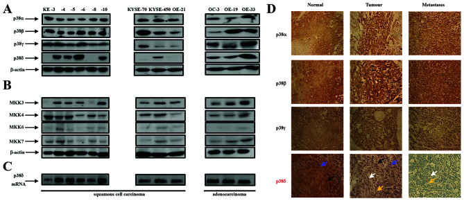
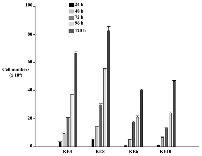
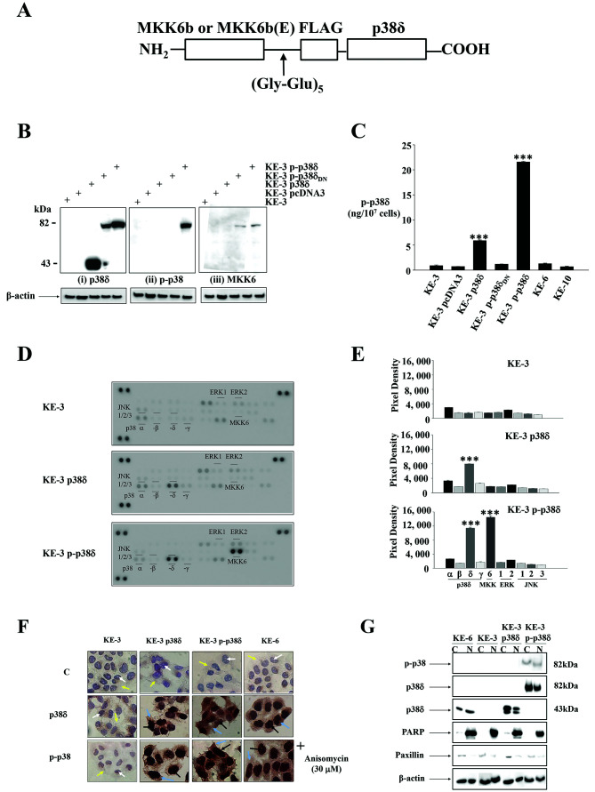
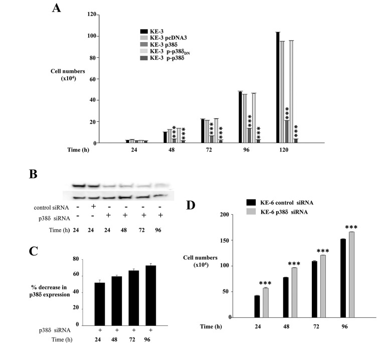
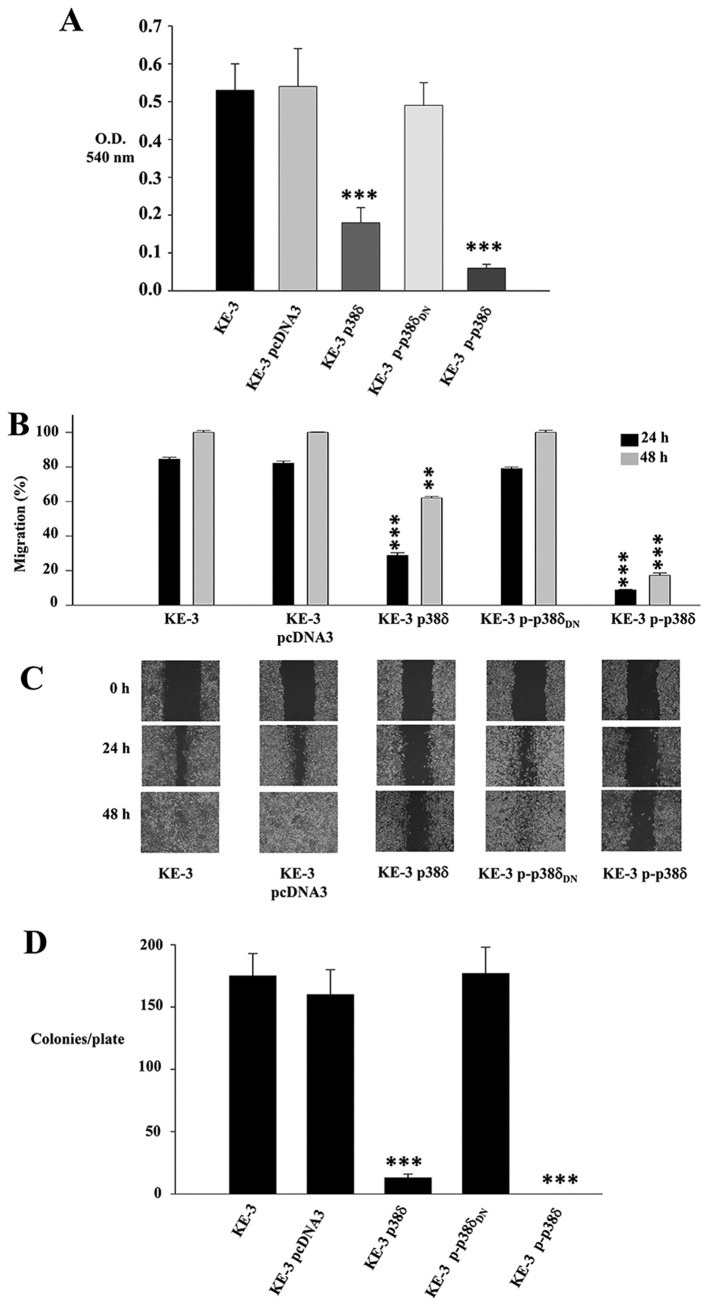
Oesophageal cancer is an aggressive tumour which responds poorly to both chemotherapy and radiation therapy and has a poor prognosis. Thus, a greater understanding of the biology of oesophageal cancer is needed in order to identify novel therapeutic targets. Among these targets p38 MAPK isoforms are becoming increasingly important for a variety of cellular functions. The physiological functions of p38α and -β are now well documented in contrast to -γ and -δ which are comparatively under-studied and ill-defined. A major obstacle to deciphering the role(s) of the latter two p38 isoforms is the lack of specific chemical activators and inhibitors. In this study, we analysed p38 MAPK isoform expression in oesophageal cancer cell lines as well as human normal and tumour tissue. We observed specifically differential p38δ expression. The role(s) of p38δ and active (phosphorylated) p38δ (p-p38δ) in oesophageal squamous cell carcinoma (OESCC) was delineated using wild-type p38δ as well as active p-p38δ, generated by fusing p38δ to its upstream activator MKK6b(E) via a decapeptide (Gly-Glu)5 linker. OESCC cell lines which are p38δ-negative (KE-3 and -8) grew more quickly than cell lines (KE-6 and -10) which express endogenous p38δ. Re-introduction of p38δ resulted in a time-dependent decrease in OESCC cell proliferation which was exacerbated with p-p38δ. In addition, we observed that p38δ and p-p38δ negatively regulated OESCC cell migration in vitro. Finally both p38δ and p-p38δ altered OESCC anchorage-independent growth. Our results suggest that p38δ and p-p38δ have a role in the suppression of OESCC. Our research may provide a new potential target for the treatment of oesophageal cancer.


Figures

Figure 1
Figure 1
Expression of p38 MAPK isoforms, MKK3, -4, -6 and -7 in oesophageal cancer. (A) Western blot analysis of p38 isoform expression in KE-3, -4, -5, -6, -8 and -10, KYSE-70, -450 and OE-21 (oesophageal squamous cell carcinoma cell lines) as well as OC-3, OE-19 and OE-33 (oesophageal adenocarcinoma cell lines). (B) Western blot analysis of MKK3, -4, -6 and -7 in the same twelve cell lines. Aliquots of 30 μg of protein lysate were loaded on a 10% SDS-PAGE gel and analyzed by immunoblotting using antibodies specific for p38α, -β2, -γ and -δ. β-actin analysis served as a loading control. The results shown are representative of four independent experiments. (C) Agarose gel electrophoresis analysis of DNA fragments produced by PCR amplification of p38δ mRNA from oesophageal squamous (KE3, -4, -5, -6, -8, 10, KYSE70, -450 and OE21) and adenocarcinoma (OC3, OE19 and -33) cell lines. (D) Immunohistochemical staining of p38α, -β2, -γ and -δ isoforms in normal, tumourigenic and metastatic (lymph node) oesophageal human tissue. Immunohistochemical staining was performed as outlined in Materials and methods. Blue arrow indicates cytoplasmic staining; black arrow indicates nuclear staining; white arrow indicates blue unstained nuclei and yellow arrow indicates blue unstained cytoplasm. Magnification, ×400. The results shown are representative of ten patients.
Figure 2
Figure 2
Oesophageal squamous cell carcinoma cell lines lacking endogenous expression of p38δ MAPK have a higher proliferation rate. KE-3 and -8 cell lines (lacking endogenous p38δ expression) and KE-6 and -10 cell lines (expressing endogenous p38δ expression) were seeded (3×10⁴) and counted for 24–120 h. The results shown are mean ± SE of three independent experiments.
Figure 3
Figure 3
Generation of active p38δ (p-p38δ) MAPK fusion proteins. (A) A schematic representation of MKK6b-p38δ or MKK6b(E)-p38δ MAPK fusion protein. The coding region of p38δ was fused in frame to the 3′-end of the stop codon-less MKK6b or MKK6b(E) through a peptide linker (Gly-Glu)5. (B) Western blot analysis of KE-3 cells stably transfected with empty vector (pcDNA3), wild-type p38δ, p-p38δ and p-p38δDN. Cells were analysed by immunoblot using antibodies specific for p38δ (i), p-p38 (ii) and MKK6 (iii). Aliquots of 30 μg protein lysate for each cell line were loaded on a 10% SDS-PAGE gel. The results shown are representative of four independent experiments. (C) Transfected and non-transfected KE-3, KE-6 and KE-10 cells were analysed to determine the amount of activated i.e., phosphorylated p38δ expression using the human phospho-p38δ (T180/Y182) ELISA commercial kit (R&D Systems). The ELISA assay was carried out according to the manufacturer’s protocol. The results shown are mean ± SE of three independent experiments. Significant (***p<0.001) changes from control non-transfected KE-3 cells. (D) The human phospho-MAPK array shows the effects of stably transfecting KE-3 cells with p38δ and p-p38δ. Arrays were incubated with 200 μg of cell lysate. (E) Corresponding pixel density for p38α, -β, -δ and -γ, MKK-6, ERK1/2 and JNK1/2/3 phosphorylation in non-transfected and transfected KE-3 cells. (F) Immunohistochemical subcellular localization of p38δ and p-p38 in KE-3 non-transfected cells and cells transfected with p38δ and p-p38δ. KE-6 cells were or were not treated with anisomycin (30 μM). (G) Nuclear and cytoplasmic localization of p38δ and p-p38 in KE-3 and KE-6 cells. The results shown are representative of four independent experiments (F and G).
Figure 4
Figure 4
Effect of p38δ and p-p38δ MAPK expression on cell proliferation. (A) KE-3, KE-3 pcDNA3, KE-3 p38δ, KE-3 p-p38δ and KE-3 p-p38δDN cells were seeded (3×10⁴) and counted for 24–120 h. The results shown are mean ± SE of three independent experiments. Significant (***p<0.001) changes from control non-transfected KE-3 cells. (B) Western blot analysis of KE-6 cells transiently transfected or not transfected with p38δ siRNA or control siRNA for 24–96 h. Cells were analysed by immunoblotting using a p38δ antibody. Aliquots of 30 μg protein lysate were loaded on a 10% SDS-PAGE gel. The results shown are representative of three independent experiments. (C) Densitometric analysis was performed to analyse % knockdown of KE-6 p38δ protein. (D) KE-6 cells (3×10⁴) transfected with p38δ siRNA or control siRNA were seeded and counted for 24–96 h. The results shown are mean ± SE of three independent experiments each done in triplicate. Significant (***p<0.001) changes from control siRNA transfected KE-6 cells.
Figure 5
Figure 5
Effect of p38δ and p-p38δ MAPK on KE-3 cell migration and anchorage-independent growth. KE-3, KE-3 pcDNA3, KE-3 p38δ, KE-3 p-p38δ and KE-3 p-p38δDN cells were analysed for cell migration (A–C) and anchorage-independent growth D). (A and B) p38δ and p-p38δ inhibit KE-3 migration at 24 h (A, Boyden Chamber) and 24 and 48 h (B, wound healing). (C) Representative wound healing images at 0, 24 and 48 h. Wound healing rates decrease in p38δ and p-p38δ transfected KE-3 cells. The results shown are representative of three independent experiments. (D) Anchorage-independent growth potential of KE-3 non-transfected and transfected cells were measured by their ability to form colonies on soft agar. Plates were stained with 3-(4,5-dimethylthiazol-2-yl)-2, 5-diphenyl tetrazolium bromide to visualize colonies. The number of colonies per plate is shown. The results shown are mean ± SE of four independent experiments (A, B and D). Significant (**p<0.01; ***p<0.001) changes from control non-transfected KE-3 cells.

References

    1. Kayani B, Zacharakis E, Ahmed K, Hanna GB. Lymph node metastases and prognosis in oesophageal carcinoma - a systematic review. Eur J Surg Oncol. 2011;37:747–753. - PubMed
    1. Enzinger PC, Mayer RJ. Esophageal cancer. N Engl J Med. 2003;349:2241–2252. - PubMed
    1. Almhanna K, Strosberg JR. Multimodality approach for locally advanced esophageal cancer. World J Gastroenterol. 2012;18:5679–5687. - PMC - PubMed
    1. Thallinger CM, Kiesewetter B, Raderer M, Hejna M. Preand postoperative treatment modalities for esophageal squamous cell carcinoma. Anticancer Res. 2012;32:4609–4627. - PubMed
    1. Klein CA, Stoecklein NH. Lessons from an aggressive cancer: evolutionary dynamics in esophageal cancer. Cancer Res. 2009;69:5285–5288. - PubMed

Publication types

MeSH terms

Substances