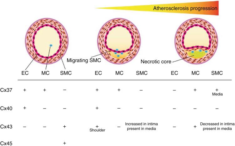
Review
. 2012 Jul;2(3):1981-2035.
doi: 10.1002/cphy.c110051.

Gap junctions

Affiliations
Review

Gap junctions

Morten Schak Nielsen et al. Compr Physiol. 2012 Jul.

Abstract

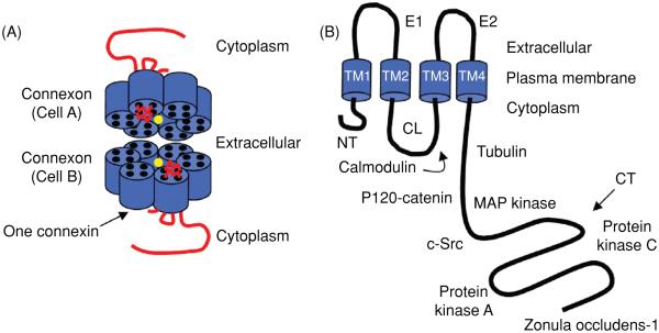
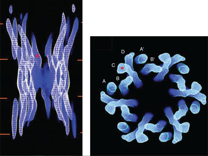
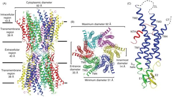
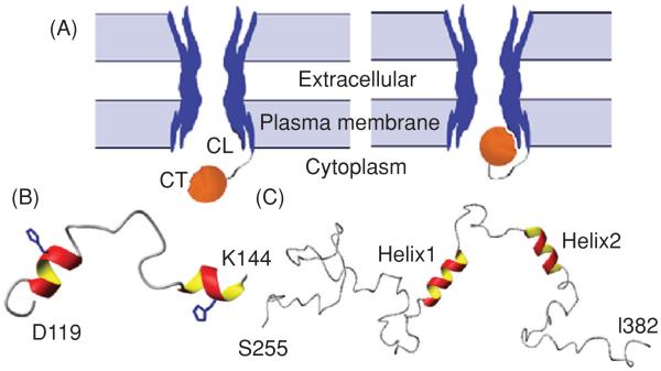
Gap junctions are essential to the function of multicellular animals, which require a high degree of coordination between cells. In vertebrates, gap junctions comprise connexins and currently 21 connexins are known in humans. The functions of gap junctions are highly diverse and include exchange of metabolites and electrical signals between cells, as well as functions, which are apparently unrelated to intercellular communication. Given the diversity of gap junction physiology, regulation of gap junction activity is complex. The structure of the various connexins is known to some extent; and structural rearrangements and intramolecular interactions are important for regulation of channel function. Intercellular coupling is further regulated by the number and activity of channels present in gap junctional plaques. The number of connexins in cell-cell channels is regulated by controlling transcription, translation, trafficking, and degradation; and all of these processes are under strict control. Once in the membrane, channel activity is determined by the conductive properties of the connexin involved, which can be regulated by voltage and chemical gating, as well as a large number of posttranslational modifications. The aim of the present article is to review our current knowledge on the structure, regulation, function, and pharmacology of gap junctions. This will be supported by examples of how different connexins and their regulation act in concert to achieve appropriate physiological control, and how disturbances of connexin function can lead to disease.


Figures

Figure 1
Figure 1
Model of a Cx43 gap junction channel and monomer. (A) The channel pore location has been indicated by the yellow circle. (B) The Cx43 monomer with protein partners. The abbreviations are as follows: NT, N-terminus; CL, cytoplasmic loop; CT, C-terminus; E1 and E2, extracellular loops 1 and 2; TM1-4, transmembrane segments 1-4.
Figure 2
Figure 2
Cx43 channel structure obtained by electron crystallography. The panel on the left shows a side view of the entire channel. The red lines represent the lipid bilayers. The red asterisk indicates the point at which the pore diameter is estimated to be the smallest. The panel on the right is a view from the cytoplasmic side. The channel is formed by six repeats of four identifiable densities (A-D), each density corresponding to one transmembrane domain. Modified, with permission, from Unger VM, Kumar NM, Gilula NB, Yeager M. Science 283: 1176-1180, 1999 (698). Reprinted, with permission, from AAAS.
Figure 3
Figure 3
Structure of the Cx26 gap junction channel and Cx26 protomer in ribbon representation. (A) Side view of the Cx26 gap junction channel. (B) Top view of the Cx26 gap junction channel showing the arrangement of the transmembrane helices TM1 to TM4. (C) Side view of the Cx26 protomer. Color code: red, NT; blue, TM1-TM4; green, E1; yellow, E2; gray, disulphide bonds; dashed lines, CL and CT, which were not visible in the map. E1 and E2 are the loops connecting TM1 and TM2, and TM3 and TM4, respectively. Modified, with permission, from Maeda et al. 2009 (409). Reprinted by permission from Macmillan Publishers Ltd: Nature (458: 597-602), copyright [2009].
Figure 4
Figure 4
“Ball-and-chain” model of Cx43 regulation. (A) Under normal conditions, the gate [cytoplasmic tail (CT)] is away from the pore. Under the appropriate stimulus, the gate swings toward the mouth of the channel, binds to a receptor [cytoplasmic loop (CL)] affiliated with the pore, and closes the channel. Lowest energy structure of the (B) Cx43CL and (C) Cx43CT domains; α-helices colored red and yellow. Figure is modified, with permission, from Delmar M, Coombs W, Sorgen P, Duffy HS, Taffet SM, Structural bases for the chemical regulation of Connexin43 channels, Cardiovasc.Res., 2003, 62(2): 268-275, (138) by permission of Oxford University Press, Duffy et al. 2002 (155), and Sorgen et al. 2004 (648), with permission.
Figure 5
Figure 5
Conformational changes in Cx26 hemichannels observed in low and high calcium buffers by atomic force microscopy (AFM). (A) AFM topograph showing the extracellular connexon surface imaged in a calcium-free buffer solution. Individual connexons exhibit defects in the number of subunits, as indicated by the circles. (B) Same connexon surface imaged in (A), but in the presence of 0.5 mmol/L calcium. The channel diameter has changed significantly as seen in the correlation averaged top view (inset) and the profile at the bottom of the inset. All images were displayed as relief tilted by 5°. (Modified, with permission, from Müller et al. 2002 (456). Reprinted by permission from Macmillan Publishers Ltd: EMBO J (21: 3598-3607), copyright [2002]).
Figure 6
Figure 6
Possible channels formed by multiple connexins. The figure shows gap junctional channels of different composition. Homomeric connexons are formed by a single connexin type whereas connexons containing more than one connexin type is heteromeric. When connexons of the same composition form a cell-cell channel it is homotypic and if the connexons differ in composition it is heterotypic. Reprinted from Cell, 84(3), Kumar NM, Gilula NB, The gap junction communication channel, 381-8, Copyright [1996], (338) with permission from Elsevier.
Figure 7
Figure 7
Voltage dependence of connexin channels. Upper left panel shows the current elicited by imposing a plus or minus 100 mV gradient across Cx43 channels. The initial current that decays over time until it reaches a lower steady-state current. Right panel shows a plot of the fractional conductance (steady-state conductance (Gss) divided by the initial conductance (Gi)] as a function of transjunctional voltage for Cx40, Cx43, and Cx45. Lower left panel demonstrates the concept of gating polarity of Cx43 (negative gating polarity). If the voltage gradient is sufficiently large the gating particle will close the connexion that is relatively negative on the cytoplasmatic side. Figure adapted from Moreno AP, Biophysical properties of homomeric and heteromultimeric channels formed by cardiac connexins, Cardiovasc.Res., 2002, 62(2):276-86, (448) by permission of Oxford University Press.
Figure 8
Figure 8
Effect of cytoplasmic tail (CT) truncation on fast Vj gating in Cx43. (A) Left: trace of channel activity recorded at a transjunctional voltage of −60 mV. In the expanded inset, shows clear transitions between the main open and residual state (fast gating). Right: all-events histogram showing the distribution of the observed conductance events. (B) Recording of activity by CT-truncated Cx43 channels at Vj = −60 mV. The trace and all-events histogram shows that only gating between the main open and closed state was observed. Figure adapted from Moreno AP, Chanson M, Elenes S, Anumonwo J, Scerri I, Gu H, Taffet SM, Delmar M, Role of the carboxyl terminal of connexin43 in transjunctional fast voltage gating, Circ.Res., 90(4):450-7, 2002 (449), with permission.
Figure 9
Figure 9
Role of ubiquitination in trafficking and degradation of Cx43. The left cell summarizes the events of polyubiquitination followed by ERAD. The right cells summarizes internalization and lysosomal degradation after Cx43 monoubiquitination. Reprinted from Cell Signal., Vol 22, Kjenseth A, Fykerud T, Rivedal E, Leithe E, Regulation of gap junction intercellular communication by the ubiquitin system, 1267-73, Copyright [2010] (317), with permission from Elsevier.
Figure 10
Figure 10
Expression of connexins in normal vessels and during progression of artherosclerosis. The expression of Cx37, Cx40, Cx43, and Cx45 is indicated for the different cell types involved [endothelial cells (ECs), smooth muscle cells (SMCs), and monocytes (MCs)]. Adapted with kind permission from Springer Science+Business Media: Semin.Immunopathol., Connexins participate in the initiation and progression of atherosclerosis, 31, 2009, 49-61, Morel S, Burnier L, Kwak BR, figure 3, (447).
Figure 11
Figure 11
RXP-E prevents particle-receptor interaction and channel closure. (A) Open channel. (B) Channel closed by particle-receptor interaction. (C) RXP-E binds the cytoplasmic tail (CT) and prevents closure by interrupting binding to the receptor. Modified from Delmar M, Coombs W, Sorgen P, Duffy HS, Taffet SM, Structural bases for the chemical regulation of Connexin43 channels, Cardiovasc.Res., 2004, 62(2): 268-75, (138), by permission of Oxford University Press.

References

    1. Ackert CL, Gittens JE, O’Brien MJ, Eppig JJ, Kidder GM. Intercellular communication via connexin43 gap junctions is required for ovarian folliculogenesis in the mouse. Dev Biol. 2001;233:258–270. - PubMed
    1. Ahmad S, Evans WH. Post-translational integration and oligomerization of connexin 26 in plasma membranes and evidence of formation of membrane pores: Implications for the assembly of gap junctions. Biochem J. 2002;365:693–699. - PMC - PubMed
    1. Ai X, Pogwizd SM. Connexin 43 Downregulation and dephosphorylation in nonischemic heart failure is associated with enhanced colocalized protein phosphatase Type 2A. Circ Res. 2005;96:54–63. - PubMed
    1. Ai Z, Fischer A, Spray DC, Brown AM, Fishman GI. Wnt-1 regulation of connexin43 in cardiac myocytes. J Clin Invest. 2000;105:161–171. - PMC - PubMed
    1. Al-Ubaidi MR, White TW, Ripps H, Poras I, Avner P, Gomes D, Bruzzone R. Functional properties, developmental regulation, and chromosomal localization of murine connexin36, a gap-junctional protein expressed preferentially in retina and brain. J Neurosci Res. 2000;59:813–826. - PubMed

Publication types

LinkOut - more resources