. 2013 Jul 19;288(29):20868-20882.
doi: 10.1074/jbc.M112.445734. Epub 2013 May 30.

Modulation of lipid kinase PI4KIIα activity and lipid raft association of presenilin 1 underlies γ-secretase inhibition by ginsenoside (20S)-Rg3

Affiliations

Modulation of lipid kinase PI4KIIα activity and lipid raft association of presenilin 1 underlies γ-secretase inhibition by ginsenoside (20S)-Rg3

Min Suk Kang et al. J Biol Chem. .

Abstract

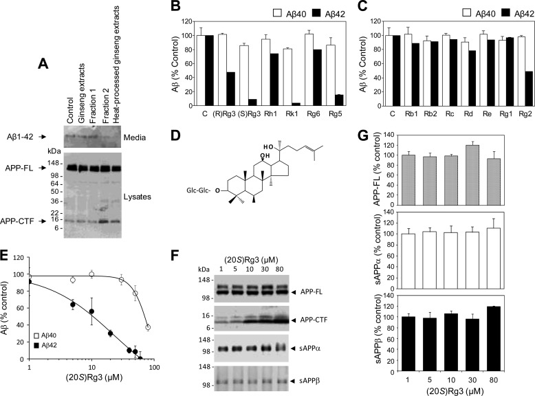
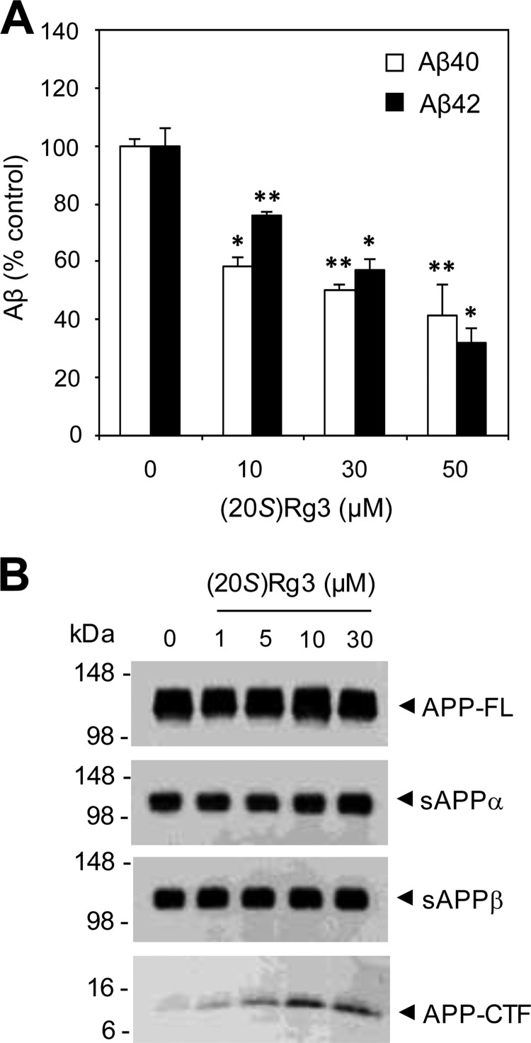
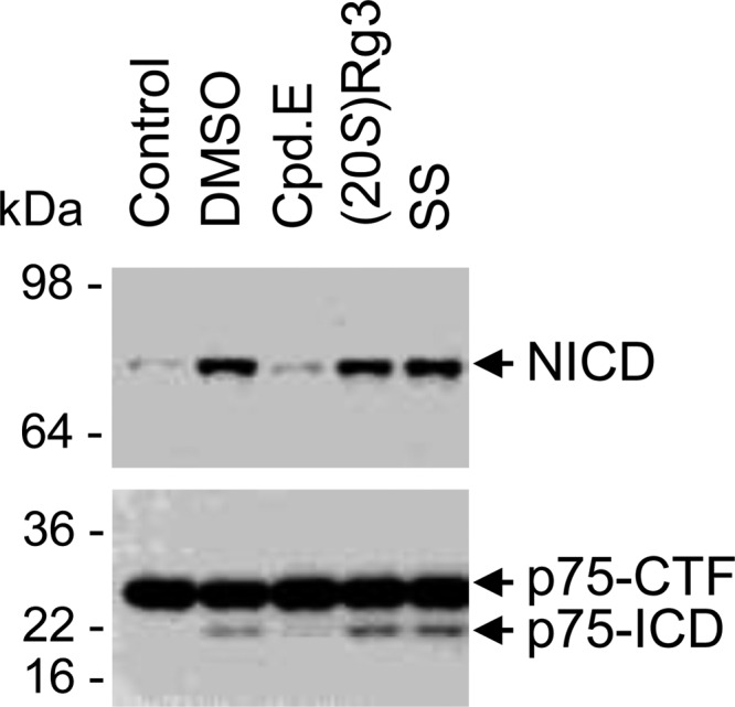
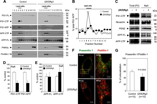
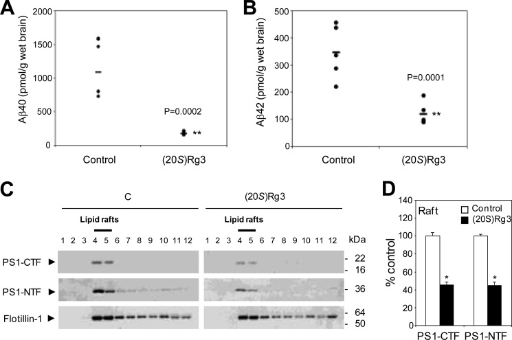
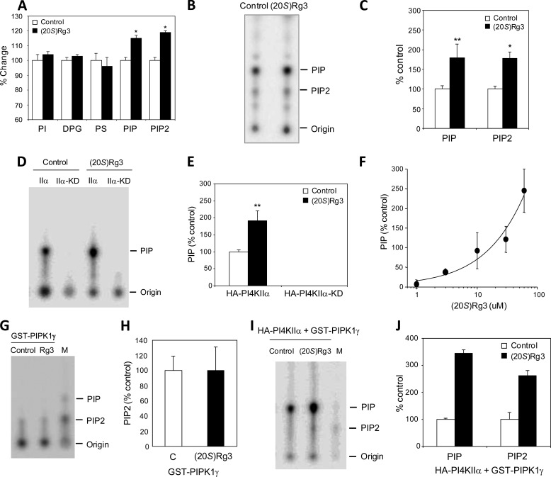
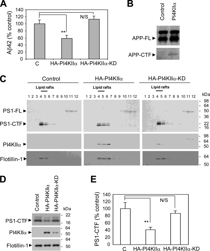
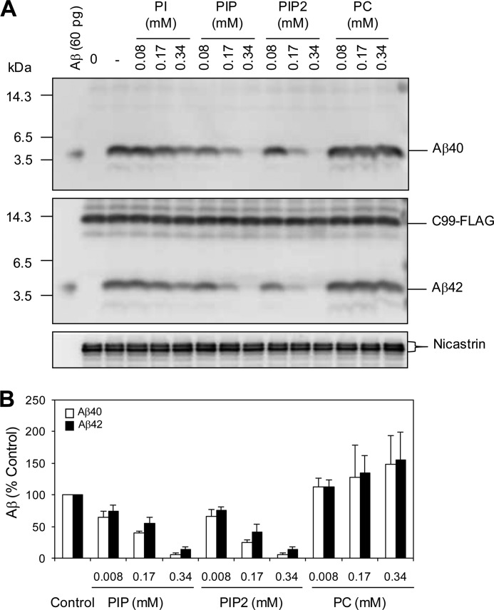
Amyloid β-peptide (Aβ) pathology is an invariant feature of Alzheimer disease, preceding any detectable clinical symptoms by more than a decade. To this end, we seek to identify agents that can reduce Aβ levels in the brain via novel mechanisms. We found that (20S)-Rg3, a triterpene natural compound known as ginsenoside, reduced Aβ levels in cultured primary neurons and in the brains of a mouse model of Alzheimer disease. The (20S)-Rg3 treatment induced a decrease in the association of presenilin 1 (PS1) fragments with lipid rafts where catalytic components of the γ-secretase complex are enriched. The Aβ-lowering activity of (20S)-Rg3 directly correlated with increased activity of phosphatidylinositol 4-kinase IIα (PI4KIIα), a lipid kinase that mediates the rate-limiting step in phosphatidylinositol 4,5-bisphosphate synthesis. PI4KIIα overexpression recapitulated the effects of (20S)-Rg3, whereas reduced expression of PI4KIIα abolished the Aβ-reducing activity of (20S)-Rg3 in neurons. Our results substantiate an important role for PI4KIIα and phosphoinositide modulation in γ-secretase activity and Aβ biogenesis.

Keywords: Alzheimer Disease; Lipid Raft; Lipids; Natural Products; Phosphatidylinositol; Presenilin.


Figures

FIGURE 1.
FIGURE 1.
Identification of active compounds that contribute to Aβ42-lowering activity of heat-processed ginseng. A, effects of ginseng extracts on Aβ42 production. CHO cells stably transfected with human APP (CHO-APP cells) were treated with a ginsenoside (saponin)-enriched fraction derived from either unprocessed ginseng (lane 2) or heat-processed ginseng (lane 5) along with fractions 1 and 2, which resulted from further chromatographic separation of heat-processed ginseng extracts. Fraction 2 is enriched for ginsenosides that are uniquely present in heat-processed ginseng but not in unprocessed ginseng (lane 4). Secreted Aβ42 was immunoprecipitated using Aβ42-specific antibody (FCA42) and probed with anti-Aβ antibody 6E10 (top panel, Media). Cell-associated holoAPP (APP-FL) and C-terminal fragments (APP-CTF) were analyzed by Western blot analysis using antibody specific to the APP C-terminal end (R1). B and C, effects of individual ginsenosides from white (B) or heat-processed (C) ginseng on the production of Aβ40 and Aβ42. CHO-APP cells were treated with the indicated compounds at 50 μg/ml for 8 h. Levels of secreted Aβ40 and Aβ42 were determined by ELISA and normalized to cell-associated full-length APP. In CHO-APP cells, average Aβ amounts in control samples were 320 pm for Aβ40 and 79 pm for Aβ42. The relative levels of Aβ40 and Aβ42 were normalized to values obtained from non-treated and vehicle-treated cells and are shown as percent of control. One of three representative experiments is shown. D, chemical structure of (20S)-Rg3. E, dose-dependent inhibition of Aβ secretion by (20S)-Rg3. CHO cells stably expressing human APP and PS1WT (CHO-APP/PS1WT cells) were treated with the indicated concentrations of (20S)-Rg3 for 8 h. Levels of secreted Aβ40 and Aβ42 were determined by ELISA and normalized to cell-associated full-length APP. The relative levels of Aβ40 and Aβ42 were normalized to values from vehicle-treated cells and are shown as percentage of control (data are expressed as mean ± S.D. (error bars); n = 4; *, p < 0.005). F, steady-state levels of full-length APP and APP C-terminal fragments (two upper panels) were examined by Western blot analysis using APP-CTmax antibody. Note that (20S)-Rg3 treatment resulted in a dose-dependent increase in APP C-terminal fragments. F, the effects of (20S)-Rg3 treatment on levels of secreted APP ectodomain. Soluble APP fragments that resulted from α-secretase cleavage of APP (sAPPα) were detected by immunoprecipitation (6E10) and Western blot analyses (LN27) in CHO-APP/PS1WT cells. sAPPβ was detected by immunoprecipitation (sAPPβ antibody) and Western blot analyses (LN27) (two lower panels). G, quantification of Western blot data in F. Note that (20S)-Rg3 treatment did not change the levels of either full-length APP (APP-FL) or secreted APP ectodomains (sAPPα and sAPPβ).
FIGURE 2.
FIGURE 2.
(20S)-Rg3 reduces Aβ42 secretion in cultured primary neurons. A, primary hippocampal neurons derived from Tg2575 APP transgenic mice were treated with (20S)-Rg3 for 6 h (at the indicated concentrations). The levels of secreted Aβ42 were measured by ELISA, and the values were normalized to the levels of full-length APP (APP-FL) (data are expressed as mean ± S.D. (error bars); n = 3; *, p < 0.05; **, p < 0.005). B, analysis of APP processing after (20S)-Rg3 treatment. Steady-state levels of full-length APP and APP C-terminal fragments were examined by Western blot analysis using APP-CTmax antibody. Soluble APP fragments that resulted from α-secretase cleavage of APP (sAPPα) were detected by immunoprecipitation (6E10) and Western blot analyses (LN27) in CHO-APP/PS1WT cells. sAPPβ was detected by immunoprecipitation (sAPPβ antibody) and Western blot analyses (LN27).
FIGURE 3.
FIGURE 3.
(20S)-Rg3 does not significantly affect the production of the ICDs of Notch1 (NICD) or p75NTR (p75-ICD). Membrane fractions isolated from 293 cells expressing either Notch-ΔE (upper) or p75-ΔE (lower) were incubated in the presence of the following compounds: Compound E (Cpd.E; general γ-secretase inhibitor), sulindac sulfide (SS; an Aβ42-lowering nonsteroidal anti-inflammatory drug), and (20S)-Rg3. p75-ICD was detected using antibodies raised against its cytoplasmic domains, and NICD was detected using a cleavage site-specific NICD antibody. Very low amounts of NICD and p75-ICD were detected in starting material (Control) or in samples treated with Compound E. Generation of NICD and p75-ICD was preserved in samples incubated with DMSO, (20S)-Rg3, and sulindac sulfide.
FIGURE 4.
FIGURE 4.
(20S)-Rg3 modulates the association of PS1 with lipid rafts. A, CHO-APP/PS1WT cells were treated with 50 μm (20S)-Rg3 at 37 °C for 6 h and compared with untreated cells. CHO-APP/PS1WT cells were solubilized with 1% Brij98-containing buffer at 37 °C for 10 min and subjected to discontinuous sucrose gradients. The distributions of PS1, APP, PI4KIIα, and flotillin-1 were determined by subjecting equal volumes of each fraction to SDS-PAGE followed by Western blot analysis using specific antibodies. Fractions 4 and 5 contain the detergent-insoluble proteins with light buoyant density and are enriched with a lipid raft marker, flotillin-1 (“Lipid rafts”). B, relative distribution of PS1-NTF in each sucrose density fraction is quantified and plotted as percent of total intensity. Note that the peak of PS1-NTF association in lipid raft is reduced by treatment with (20S)-Rg3 (50 μm) (closed symbol) as compared with control (open symbol). C, Western blot analysis of γ-secretase components and APP in total membrane preparation (P2) compared with pooled lipid raft fractions 4 and 5 from B in the presence or absence of (20S)-Rg3. D and E, quantification of PS1-CTF (D) and PS1-NTF (E) levels from Western blot is shown as percent of control (data are expressed as mean ± S.D. (error bars); n = 3; *, p < 0.005). F, confocal microscopic analysis of the subcellular distribution of PS1 and flotillin-1. CHO-APP/PS1WT cells were incubated in the presence or absence of (20S)-Rg3 and subjected to immunocytochemical analysis. Cells were double labeled with α-PS1-NT (green) and α-flotillin-1 (red) antibodies followed by Alexa Fluor 488-conjugated and Alexa Fluor 568-conjugated secondary antibodies, respectively. Magnification, 100×. Far right panels are 6× magnification of squares shown in the left panels. G, quantification of percent co-localization between PS1 and flotillin-1 using ImageJ. FL, full length.
FIGURE 5.
FIGURE 5.
Effect of (20S)-Rg3 on Aβ levels and the levels of PS1 fragments associated with lipid rafts in APP/PS1 mice. A and B, (20S)-Rg3 (n = 5) or control (n = 5) was administered daily via intraperitoneal injection into APP/PS1 mice (3 months old) for 3 weeks (10 mg/kg/day). Following treatment, the half-hemisphere of brain tissues from (20S)-Rg3- or vehicle-treated mice was extracted using formic acid, and the relative levels of Aβ40 and Aβ42 were determined by sandwich ELISA. Horizontal bars represent the average. C, the brain tissues were homogenized, solubilized by 1% Brij98 at 37 °C, and subjected to sucrose density gradient centrifugation. Lipid raft association of PS1-CTF, PS1-NTF, and flotillin-1 was analyzed by Western blotting. D, quantification of the Western blot data in C is shown as percent of control (data are expressed as mean ± S.D. (error bars); n = 3; *, p < 0.005).
FIGURE 6.
FIGURE 6.
(20S)-Rg3 increases PI(4,5)P2 by activating PI4KIIα. A, treatment of cortical neurons with (20S)-Rg3 (50 μm) leads to increased steady-state levels of PI(4)P and PI(4,5)P2 (n = 6 per treatment; *, p < 0.05). B, lipid kinase and TLC analysis of membranes prepared from stable PS1 Neuro2a APPsw cells treated with vehicle (Control) or 50 μm (20S)-Rg3. C, quantification of the TLC data on PI(4)P and PI(4,5)P2 in B. Treatment with (20S)-Rg3 promotes both PI(4)P and PI(4,5)P2 synthesis (data are expressed as mean ± S.D. (error bars); n = 3; *, p < 0.005; **, p < 0.01). D, (20S)-Rg3 enhances the PI4KIIα activity. A lipid kinase activity assay and TLC analysis were performed using either purified wild-type (IIα) or kinase-dead mutant (IIα-KD) forms of PI4KIIα. The reaction was performed in the absence or presence of 50 μm (20S)-Rg3. E, quantification of the TLC data in D (data are expressed as mean ± S.D. (error bars); n = 3; **, p < 0.01). F, dose-response curve of PI4KIIα activation by (20S)-Rg3. Increasing amounts of (20S)-Rg3 were included in the PI4KIIα lipid kinase assay to monitor the PI(4)P formation. G and H, (20S)-Rg3 has no effect on PIPK1γ. A lipid kinase assay and TLC analysis were performed using 2 μg of recombinant PIPK1γ (data are expressed as mean ± S.D. (error bars); n = 3). I, TLC analysis using either purified wild-type PI4KIIα or PIPk1γ. The reaction was performed in the absence or presence of 50 μm (20S)-Rg3. M, lipid markers run in parallel. J, quantification of the TLC data on PI(4)P and PI(4,5)P2 in I (data are expressed as mean ± S.D. (error bars); n = 3). PS, phosphatidylserine; DPG, diphosphatidylglycerol.
FIGURE 7.
FIGURE 7.
PI4KIIα modulates Aβ production and the association of PS1 with lipid rafts. A, overexpression of active PI4KIIα but not the kinase-dead mutant form of PI4KIIα (PI4KIIα-KD), leads to a reduction in Aβ42 secretion in stable PS1 Neuro2a APPWT cells. Aβ42 levels were normalized to APP full length (data are expressed as mean ± S.D. (error bars); n = 3; **, p < 0.01). B, effect of overexpression of PI4KIIα on the steady-state levels of APP full length (APP-FL) and C-terminal fragments (APP-CTF). C, the effects observed on the association of PS1-CTF with lipid rafts after the overexpression of PI4KIIα resemble the effects of (20S)-Rg3 treatment. CHO-APP/PS1WT cells were transiently transfected with HA-tagged PI4KIIα or PI4KIIα-KD. After a 24-h incubation, cells were solubilized with 1% Brij98 at 37 °C and subjected to sucrose density gradient centrifugation. Lipid raft association of PS1-CTF, PI4KIIα, and flotillin-1 was analyzed by Western blotting using anti-PS1-loop, anti-HA, and anti-flotillin antibodies. PI4KIIα and PI4KIIα-KD co-distribute with PS1-CTF and flotillin-1. D, Western analysis of pooled fractions 4 and 5 from C. Equal amounts of protein were analyzed per lane (20 μg). E, quantification of PS1-CTF levels from Western blot is shown as percent of control (data are expressed as mean ± S.D. (error bars); n = 3; **, p < 0.01). N/S, not significant.
FIGURE 8.
FIGURE 8.
Effects of PI(4)P and PI(4,5)P2 on γ-secretase activity. A, CHAPSO extracts of CHO cell membrane fractions were subjected to incubation with C99-FLAG for 4 h in the presence of the indicated phospholipids at the concentrations shown. Aβ40 and Aβ42 production was detected by Western blot analysis using BA27 and BC05, respectively. B, quantification of the data shown in A as percent of control. PC, phosphatidylcholine.
FIGURE 9.
FIGURE 9.
Reduced PI4KIIα leads to elevated Aβ42 production and abolishes the Aβ42-lowering activity of (20S)-Rg3 in neurons. A, directed differentiation of mouse ES cells into pyramidal neurons. Neuronal marker (TUJ1) reactivity and DAPI nuclear stain are shown in representative neurons derived from either wild-type (WT) or PI4KIIα heterozygous knock-out ES cells (+/−). B, Western blot analysis of lysates prepared from pyramidal neurons derived from WT or PI4KIIα+/− ES cells. Human APP was introduced by lentiviral infection of human APP (“None ” denotes no infection of APP virus). C and D, reduced PI 4-kinase activity in ES cell-derived neurons derived from WT or PI4KIIα+/− ES cells. A lipid kinase activity assay and TLC analysis were performed in neurons that were incubated with or without (20S)-Rg3. The quantification of [γ-32P]ATP incorporation into PI(4)P (representing PI4K activity) is shown in D (data are expressed as mean ± S.D. (error bars); n = 3; *, p < 0.005; **, p < 0.01). E, reduction of PI4KIIα results in elevated Aβ42 generation in neurons (white bars). (20S)-Rg3 reduces Aβ42 secretion in wild-type neurons but fails to reduce Aβ42 secretion in the neurons with reduced PI4KIIα (black bars) (data are expressed as mean ± S.D. (error bars); n = 3; *, p < 0.005; **, p < 0.01).

Similar articles

Cited by

References

    1. Hardy J., Selkoe D. J. (2002) The amyloid hypothesis of Alzheimer's disease: progress and problems on the road to therapeutics. Science 297, 353–356 - PubMed
    1. Tanzi R. E., Bertram L. (2005) Twenty years of the Alzheimer's disease amyloid hypothesis: a genetic perspective. Cell 120, 545–555 - PubMed
    1. O'Brien R. J., Wong P. C. (2011) Amyloid precursor protein processing and Alzheimer's disease. Annu. Rev. Neurosci. 34, 185–204 - PMC - PubMed
    1. Mangialasche F., Solomon A., Winblad B., Mecocci P., Kivipelto M. (2010) Alzheimer's disease, clinical trials and drug development. Lancet Neurol. 9, 702–716 - PubMed
    1. Bateman R. J., Xiong C., Benzinger T. L., Fagan A. M., Goate A., Fox N. C., Marcus D. S., Cairns N. J., Xie X., Blazey T. M., Holtzman D. M., Santacruz A., Buckles V., Oliver A., Moulder K., Aisen P. S., Ghetti B., Klunk W. E., McDade E., Martins R. N., Masters C. L., Mayeux R., Ringman J. M., Rossor M. N., Schofield P. R., Sperling R. A., Salloway S., Morris J. C.; Dominantly Inherited Alzheimer Network (2012) Clinical and biomarker changes in dominantly inherited Alzheimer's disease. N. Engl. J. Med. 367, 795–804 - PMC - PubMed

Publication types

MeSH terms

Substances

LinkOut - more resources