Multiple dermatofibromas: dermoscopic patterns
- PMID: 23723500
- PMCID: PMC3667312
- DOI: 10.4103/0019-5154.110862
Multiple dermatofibromas: dermoscopic patterns
Abstract
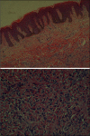
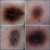
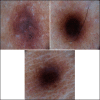
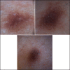
Dermatofibromas are benign skin lesions that consist of pigmented papules or nodules. They produce the dimple sign when laterally squeezed and are usually found on the legs. These clinical features lead to the diagnosis in most cases. However, the differential diagnosis with other lesions, such as atypical nevi and melanoma can be difficult, and the dermoscopy may help the diagnosis. There are several dermoscopic patterns associated with dermatofibromas, the most common being a central white scar like patch with delicate pigment network at the periphery. This article describes the case of a patient who had eleven clinically similar dermatofibromas, with four distinct patterns when submitted to dermoscopic examination.
Keywords: Benign fibrous; dermoscopy; histiocytoma; skin and connective tissue diseases; skin diseases.
Conflict of interest statement
Figures


References
-
- Zaballos P, Puig S, Llambrich A, Malvehy J. Dermoscopy of dermatofibromas: A prospective morphological study of 412 cases. Arch Dermatol. 2008;144:75–83. - PubMed
-
- Arpaia N, Cassano N, Vena GA. Dermoscopic patterns of dermatofibroma. Dermatol Surg. 2005;31:1336–9. - PubMed
-
- Canelas MM, Cardoso JC, Andrade PF, Reis JP, Tellechea O. Fibrous histiocytomas: Histopathologic review of 95 cases. An Bras Dermatol. 2010;85:211–5. - PubMed
-
- Yamamoto T. Dermatofibroma: A possible model of local fibrosis with epithelial/mesenchymal cell interaction. J Eur Acad Dermatol Venereol. 2009;23:371–5. - PubMed
LinkOut - more resources
Full Text Sources
Other Literature Sources
