. 2013 May 27;8(5):e64151.
doi: 10.1371/journal.pone.0064151. Print 2013.

Functional characterization of detergent-decellularized equine tendon extracellular matrix for tissue engineering applications

Affiliations

Functional characterization of detergent-decellularized equine tendon extracellular matrix for tissue engineering applications

Daniel W Youngstrom et al. PLoS One. .

Abstract

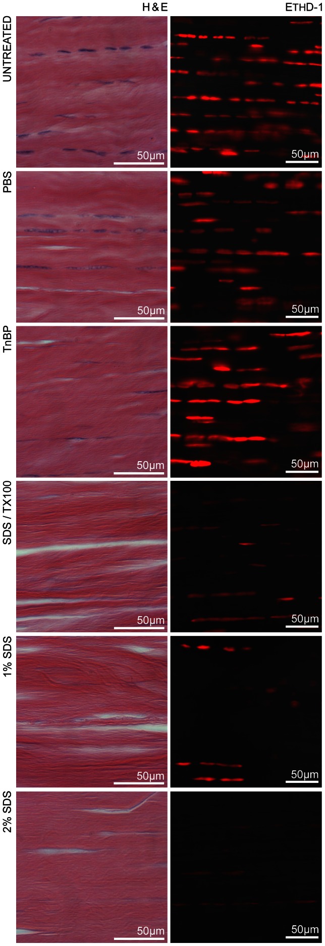
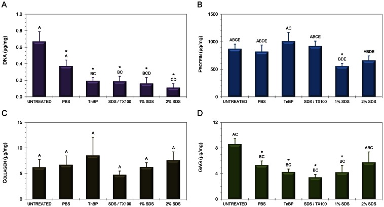
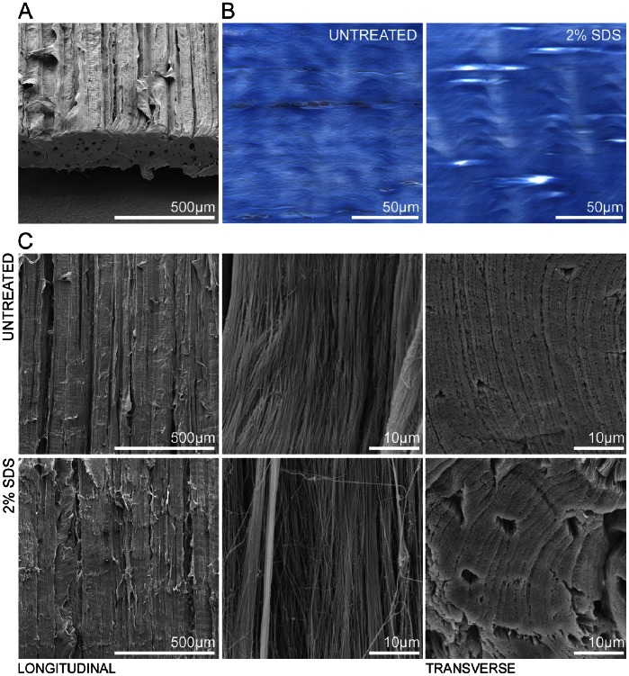
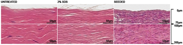
Natural extracellular matrix provides a number of distinct advantages for engineering replacement orthopedic tissue due to its intrinsic functional properties. The goal of this study was to optimize a biologically derived scaffold for tendon tissue engineering using equine flexor digitorum superficialis tendons. We investigated changes in scaffold composition and ultrastructure in response to several mechanical, detergent and enzymatic decellularization protocols using microscopic techniques and a panel of biochemical assays to evaluate total protein, collagen, glycosaminoglycan, and deoxyribonucleic acid content. Biocompatibility was also assessed with static mesenchymal stem cell (MSC) culture. Implementation of a combination of freeze/thaw cycles, incubation in 2% sodium dodecyl sulfate (SDS), trypsinization, treatment with DNase-I, and ethanol sterilization produced a non-cytotoxic biomaterial free of appreciable residual cellular debris with no significant modification of biomechanical properties. These decellularized tendon scaffolds (DTS) are suitable for complex tissue engineering applications, as they provide a clean slate for cell culture while maintaining native three-dimensional architecture.


Conflict of interest statement

Competing Interests: The authors have declared that no competing interests exist.

Figures

Figure 1
Figure 1. Loss of nuclear content following decellularization.
Brightfield images of H&E-stained histological cross-sections (5 µm-thick) and fluorescence micrographs of scaffolds imaged with EthD-1 (400 µm-thick). Marked loss of DNA content is evident in SDS-decellularized experimental groups, with 2% SDS resulting in the most dramatic decellularization. All images are representative and were acquired from the same horse for ease of comparison.
Figure 2
Figure 2. Treatment alters tendon matrix composition.
Biochemical analysis of tendon scaffolds, including DNA (A), total protein (B), soluble collagen (C), and GAG content (D). Statistical significance between untreated control and treatment is annotated by use of an asterisk. Statistical differences between treatments are indicated by different letters (repeated measures MANOVA).
Figure 3
Figure 3. Scaffold ultrastructure.
(A) Scanning electron micrograph of untreated tendon strip, angled to show both longitudinal and transverse section architecture. (B) Histological sections stained with Masson’s trichrome indicate maintenance of collagen content with a slight increase in porosity. (C) Comparison of SEM images obtained from untreated and 2% SDS decellularized tendon samples, shown at 100x and 5000x magnifications longitudinally, and 5000x transversely. Collagen ultrastructure is not adversely altered by detergent treatment.
Figure 4
Figure 4. Biocompatibility as assessed with MSC culture.
(A) Plating efficiency calculated as a ratio of MSCs adherent to scaffolds following a 48-hour incubation period. (B) Total cell count at 96 hours obtained via MTS assay, indicating no significant reduction in proliferation or metabolic activity on DTS. (C) Fluorescence micrographs portraying representative samples of untreated and decellularized tendon, with live cells stained green (calcein) and dead cells stained red (EthD-1).
Figure 5
Figure 5. Cellular integration.
H&E staining demonstrates infiltration of MSCs deep into DTS following 11 days of static tissue culture.

References

    1. Weber B, Emmert MY, Schoenauer R, Brokopp C, Baumgartner L, et al. (2011) Tissue engineering on matrix: future of autologous tissue replacement. Semin Immunopathol 33: 307–315. - PubMed
    1. Kolf CM, Cho E, Tuan RS (2007) Mesenchymal stromal cells. Biology of adult mesenchymal stem cells: regulation of niche, self-renewal and differentiation. Arthritis Res Ther 9: 204. - PMC - PubMed
    1. Guilak F, Cohen DM, Estes BT, Gimble JM, Liedtke W, et al. (2009) Control of stem cell fate by physical Interactions with the extracellular matrix. Cell Stem Cell 5: 17–26. - PMC - PubMed
    1. Zhang J, Li B, Wang JH (2011) The role of engineered tendon matrix in the stemness of tendon stem cells in vitro and the promotion of tendon-like tissue formation in vivo. Biomaterials 32: 6972–6981. - PMC - PubMed
    1. Ahmad Z, Wardale J, Brooks R, Henson F, Noorani A, et al. (2012) Exploring the application of stem cells in tendon repair and regeneration. Arthroscopy 28: 1018–1029. - PubMed

Publication types