

Sympathetic ophthalmia: to the twenty-first century and beyond
- PMID: 23724856
- PMCID: PMC3679835
- DOI: 10.1186/1869-5760-3-49
Sympathetic ophthalmia: to the twenty-first century and beyond
Abstract
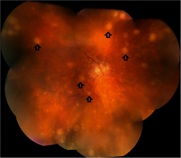
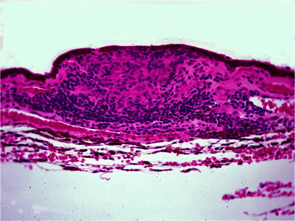
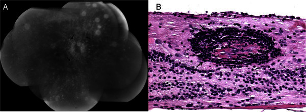
Sympathetic ophthalmia is a rare bilateral granulomatous inflammation that follows accidental or surgical insult to the uvea of one eye. Onset of sympathetic ophthalmia can be insidious or acute, with recurrent periods of exacerbation. Clinical presentation shows mutton-fat keratic precipitates, choroidal infiltrations, and Dalen-Fuchs nodules. Histopathology reveals diffuse or nodular granulomatous inflammation of the uvea. Prevention and treatment strategies for sympathetic ophthalmia are currently limited to two modalities, enucleation of the injured eye and immunosuppressive therapy, aimed at controlling inflammation. The etiology and pathophysiology of the disease is still unclear but is largely thought to be autoimmune in nature. Recent insight on the molecular pathology of the disease as well as developments in imaging technology have furthered both the understanding on the autoimmune process in sympathetic ophthalmia and the targeting of prevention and treatment strategies for the future.
Figures
References
-
- Duke-Elder S. In: Diseases of the uveal tract. Duke-Elder S, editor. St. Louis: Mosby; 1966. Sympathetic ophthalmitis; pp. 558–593.
-
- Chan CC. In: Ocular infection and immunity. Pepose JS GH, Wilhelmus KR, editor. St. Louis: Mosby; 1996. Sympathetic ophthalmia.
-
- Albert DM, Diaz-Rohena R. A historical review of sympathetic ophthalmia and its epidemiology. Surv Ophthalmol. 1989;3(1):1–14. - PubMed
LinkOut - more resources
Full Text Sources
Other Literature Sources
