. 2013 Oct;113(4):250-8.
doi: 10.1111/bcpt.12092. Epub 2013 Jun 27.

Alterations of N-3 polyunsaturated fatty acid-activated K2P channels in hypoxia-induced pulmonary hypertension

Affiliations

Alterations of N-3 polyunsaturated fatty acid-activated K2P channels in hypoxia-induced pulmonary hypertension

Gorm Nielsen et al. Basic Clin Pharmacol Toxicol. 2013 Oct.

Abstract

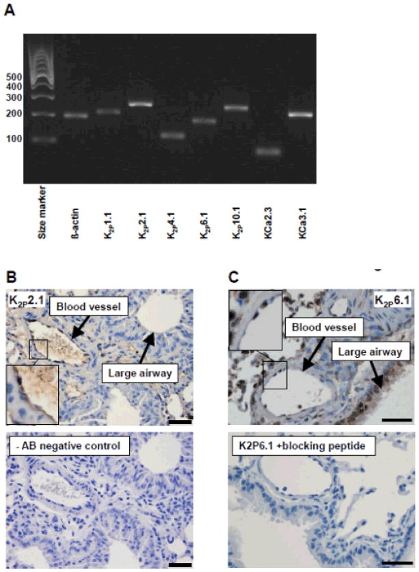
Polyunsaturated fatty acid (PUFA)-activated two-pore domain potassium channels (K2P ) have been proposed to be expressed in the pulmonary vasculature. However, their physiological or pathophysiological roles are poorly defined. Here, we tested the hypothesis that PUFA-activated K2P are involved in pulmonary vasorelaxation and that alterations of channel expression are pathophysiologically linked to pulmonary hypertension. Expression of PUFA-activated K2P in the murine lung was investigated by quantitative reverse-transcription polymerase chain reaction (qRT-PCR), immunohistochemistry (IHC), by patch clamp (PC) and myography. K2P -gene expression was examined in chronic hypoxic mice. qRT-PCR showed that the K2P 2.1 and K2P 6.1 were the predominantly expressed K2P in the murine lung. IHC revealed protein expression of K2P 2.1 and K2P 6.1 in the endothelium of pulmonary arteries and of K2P 6.1 in bronchial epithelium. PC showed pimozide-sensitive K2P -like K(+) -current activated by docosahexaenoic acid (DHA) in freshly isolated endothelial cells as well as DHA-induced membrane hyperpolarization. Myography on pulmonary arteries showed that DHA induced concentration-dependent instantaneous relaxations that were resistant to endothelial removal and inhibition of NO and prostacyclin synthesis and to a cocktail of blockers of calcium-activated K(+) channels but were abolished by high extracellular (30 mM) K(+) -concentration. Gene expression and protein of K2P 2.1 were not altered in chronic hypoxic mice, while K2P 6.1 was up-regulated by fourfold. In conclusion, the PUFA-activated K2P 2.1 and K2P 6.1 are expressed in murine lung and functional K2P -like channels contribute to endothelium hyperpolarization and pulmonary artery relaxation. The increased K2P 6.1-gene expression may represent a novel counter-regulatory mechanism in pulmonary hypertension and suggest that arterial K2P 2.1 and K2P 6.1 could be novel therapeutic targets.


Figures

Figure 1
Figure 1. Expression of K2P genes
A) RT-PCR showing mRNA-expression of K2P genes as well as KCa3.1 and KCa2.3 genes in murine lung tissue. The expected sizes of the RT-PCR products are noted in the materials and methods section. B) Immunohistology of the predominantly expressed K2P channel, K2P2.1 in murine lungs. Upper panel: K2P2.1 staining in larger airways and arteries of the lung. Lower panel: Immunohistology without the primary anti-K2P2.1 antibody. C) Immunohistology of the K2P channel, K2P6.1 in murine lungs. Upper panel: K2P6.1 staining in larger airways and arteries of the lung. Lower panel: Immunohistology where the primary anti-K2P6.1 antibody has been pre-incubated with a peptide blocking the antigen specific site of the antibody. D) Immunohistology in carotid artery of K2P2.1 (left) and K2P6.1 (right). The scale bar in each picture corresponds to 50 μm.
Figure 2
Figure 2. Electrophysiological characterization of K2P2.1-like channels in native endothelial cells
A) Whole-cell patch clamping in voltage-clamp mode showing the basal and DHA-induced current in murine carotid artery endothelial cells (mCAEC) B) Whole-cell patch clamping in voltage-clamp mode on mCAEC showing DHA-induced current blocked by pimozide (2 μM) C) Whole-cell patch clamping in voltage-clamp mode showing the basal and DHA-induced current in murine pulmonary artery endothelial cells (mPAEC). D) Whole-cell patch clamping of mPAEC in voltage-clamp mode. The graph shows the current over the cell membrane when clamped to a holding potential of −80 mV for 200 ms followed by 80 mV for 200 ms. E) Whole-cell patch clamping in voltage-clamp mode showing the basal and isoflurane-induced current in mPAEC. F) Whole-cell patch clamping of mPAEC in voltage-clamp mode, similar to Figure 2D but showing both basal and isoflurane induced current. G) Whole-cell patch clamping in current-clamp mode showing membrane potential changes to DHA (10 μM). H) Whole-cell patch clamping in current-clamp mode showing membrane potential changes in response to DHA (10 μM) and to DHA in combination with pimozide (2 μM).
Figure 3
Figure 3. Vasorelaxing effect of DHA
All measurements were done in the presence of L-NAME (100 μM) and indomethacin (10 μM). A) Isometric tension recordings in murine pulmonary artery, showing the relaxing effect of increasing concentrations of DHA both without KCa blockers (circles) as well as in the presence of 100 nM Iberiotoxin, 1 μM TRAM-34 and 1 μM UCL1684 (squares) and, finally, after removal of the endothelium (triangles). B) Isometric tension recordings in murine pulmonary artery, showing the relaxing effect of 50 μM of DHA in the presence of control (5.9 mM) and high (30 mM) potassium. ***, p < 0.001.
Figure 4
Figure 4. Expression profile of K2P-channels in murine lung under hypoxia induced pulmonary hypertension
A) QRT-PCR showing K2P2.1 mRNA to be predominantly expressed ahead of K2P10.1 and K2P4.1 mRNA during both hypoxia induced pulmonary hypertension as well as under normal conditions (normoxia). B) QRT-PCR showing the levels of K2P1.1 and K2P6.1 mRNA under the same conditions as in figure 4A. The K2P6.1 mRNA is significantly higher during hypoxia induced pulmonary hypertension compared to normoxic conditions. C) Immunohistology showing similar pattern of expression of K2P2.1 under both hypoxia and normoxia. D) Immunohistology of K2P6.1 under both hypoxia and normoxia. A more intense staining of the bronchiolar epithelium is seen in lungs from mice with hypoxia induced pulmonary hypertension compared to the normoxic control group. The scale bar in each picture corresponds to 50 μm. ***, p < 0.001.

References

    1. Gardener MJ, Johnson IT, Burnham MP, Edwards G, Heagerty AM, Weston AH. Functional evidence of a role for two-pore domain potassium channels in rat mesenteric and pulmonary arteries. Br J Pharmacol. 2004;142:192–202. - PMC - PubMed
    1. Lembrechts R, Pintelon I, Schnorbusch K, Timmermans JP, Adriaensen D, Brouns I. Expression of mechanogated two-pore domain potassium channels in mouse lungs: special reference to mechanosensory airway receptors. Histochem Cell Biol. 2011;136:371–385. - PubMed
    1. Garry A, Fromy B, Blondeau N, Henrion D, Brau F, Gounon P, Guy N, Heurteaux C, Lazdunski M, Saumet JL. Altered acetylcholine, bradykinin and cutaneous pressure-induced vasodilation in mice lacking the TREK1 potassium channel: the endothelial link. EMBO Rep. 2007;8:354–359. - PMC - PubMed
    1. Blondeau N, Petrault O, Manta S, Giordanengo V, Gounon P, Bordet R, Lazdunski M, Heurteaux C. Polyunsaturated fatty acids are cerebral vasodilators via the TREK-1 potassium channel. Circ Res. 2007;101:176–184. - PubMed
    1. Namiranian K, Lloyd EE, Crossland RF, Marrelli SP, Taffet GE, Reddy AK, Hartley CJ, Bryan RM., Jr Cerebrovascular responses in mice deficient in the potassium channel, TREK-1. Am J Physiol Regul Integr Comp Physiol. 2010;299:R461–R469. - PMC - PubMed

Publication types

MeSH terms

LinkOut - more resources