. 2013 Jun 2:12:51.
doi: 10.1186/1476-4598-12-51.

MutS homologue hMSH4: interaction with eIF3f and a role in NHEJ-mediated DSB repair

Affiliations

MutS homologue hMSH4: interaction with eIF3f and a role in NHEJ-mediated DSB repair

Yen-Lin Chu et al. Mol Cancer. .

Abstract

Background: DNA mismatch repair proteins participate in diverse cellular functions including DNA damage response and repair. As a member of this protein family, the molecular mechanisms of hMSH4 in mitotic cells are poorly defined. It is known that hMSH4 is promiscuous, and among various interactions the hMSH4-hMSH5 interaction is involved in recognizing DNA intermediate structures arising from homologous recombination (HR).

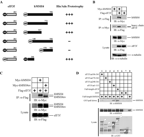
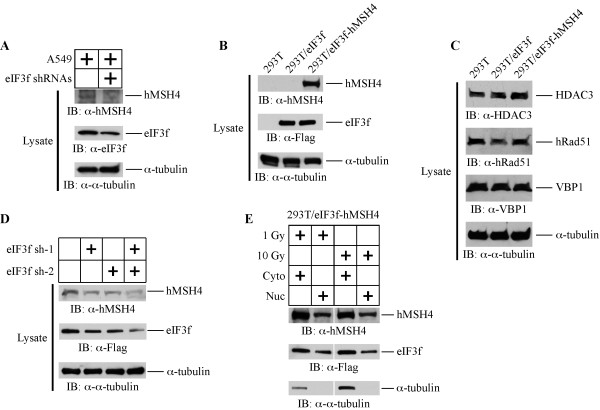
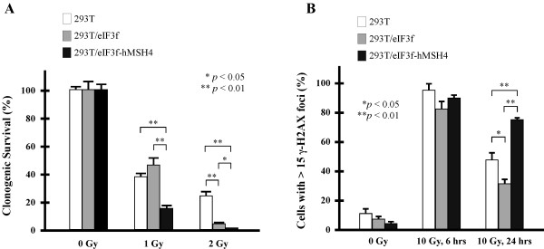
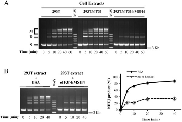
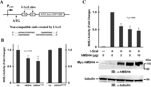
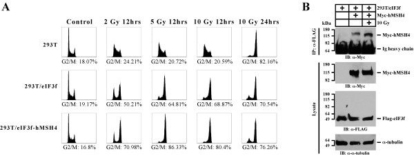
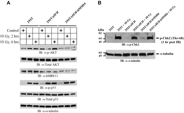
Results: We identified a new hMSH4 interacting protein eIF3f--a protein that functions not only in translation but also in the regulation of apoptosis and tumorigenesis in humans. Our studies have demonstrated that hMSH4-eIF3f interaction is mediated through the N-terminal regions of both proteins. The interaction with eIF3f fosters hMSH4 protein stabilization, which in turn sustains γ-H2AX foci and compromises cell survival in response to ionizing radiation (IR)-induced DNA damage. These effects can be, at least partially, attributed to the down-regulation of NHEJ activity by hMSH4. Furthermore, the interplay between hMSH4 and eIF3f inhibits IR-induced AKT activation, and hMSH4 promotes eIF3f-mediated bypass of S phase arrest, and ultimately enhancing an early G2/M arrest in response to IR treatment.

Conclusion: Our current study has revealed a role for hMSH4 in the maintenance of genomic stability by suppressing NHEJ-mediated DSB repair.


Figures

Figure 1
Figure 1
The interaction between hMSH4 and eIF3f. (A) Yeast-two hybrid analysis of the hMSH4-eIF3f interaction. A series of hMSH4 truncation mutants were utilized to determine the eIF3f-interacting domain on hMSH4. Positive interactions were ascertained by the transcription activation of ADE2 and HIS3 reporter genes. (B) Co-IP analysis of hMSH4-eIF3f interaction in human cells. Immunoblotting with α-tubulin was used as a loading control. (C) Co-IP analysis of the interaction between eIF3f and hMSH4sv. Full-length hMSH4 and hMSH4sv were expressed separately with eIF3f and the respective interactions were analyzed by co-IP. (D) GST pull-down analysis of hMSH4-eIF3f interaction and determination of the hMSH4-interacting domain on eIF3f. The full-length eIF3f and four truncated fragments were expressed as GST-fusion proteins. GST pull-down experiments were performed with Glutathione-Sepharose 4B beads.
Figure 2
Figure 2
eIF3f facilitates hMSH4 stabilization. (A) The effect of RNAi-mediated down-regulation of eIF3f on the levels of endogenous hMSH4 was analyzed in A549 cells. A mixture of eIF3f sh-1 and sh-2 RNAi constructs was used for transient transfection, and cells were collected and analyzed by immunoblotting at 48 hrs post-transfection. Immunoblotting with α-tubulin was used as a loading control. (B) Stable expression of eIF3f and hMSH4 in 293T/eIF3f and 293T/eIF3f-hMSH4 cell lines (from selected single clones). (C) Western blotting analysis of the levels of HDAC3, hRad51, and VBP1 expression in 293T, 293T/eIF3f, and 293T/eIF3f-hMSH4 cells. (D) Effects of reduced eIF3f expression on the levels of hMSH4 in the stable cell line 293T/eIF3f-hMSH4. Reduction of eIF3f expression was achieved by transient transfection of eIF3f RNAi constructs. (E) Nuclear and cytoplasmic distribution of hMSH4 and eIF3f proteins in response to IR. 293T/eIF3f-hMSH4 cells treated with 1 or 10 Gy IR were fractionated at 6 hrs post-treatment and the levels of hMSH4 and eIF3f in the nuclear and cytoplasmic fractions were determined by immunoblotting. α-tubulin was used as a marker for the cytoplasmic fraction.
Figure 3
Figure 3
Effects of eIF3f-hMSH4 on cellular response to IR. (A) Clonogenic survival analysis of 293T, 293T/eIF3f and 293T/eIF3f-hMSH4 cells treated with 1 or 2 Gy IR. Colonies that contained at least 50 cells were counted and the percentage of cell survival was determined in reference to untreated control cells. The means of three individual experiments and the corresponding standard deviations (error bars) are presented. (B) Examination of γ-H2AX foci formation at 6 or 24 hrs post-exposure to 10 Gy IR. Percentages of cells possessing 15 or more foci/nucleus are graphically presented, and statistically significant differences are indicated with asterisks (*p < 0.05 and **p < 0.01; Student’s t-test).
Figure 4
Figure 4
In vitro NHEJ assay. (A) Determination of NHEJ activities in extracts prepared from 293T, 293T/eIF3f and 293T/eIF3f-hMSH4 cells. DNA end joining reactions were performed by incubation of cell extracts with SalI-digested plasmid DNA, and these reactions were terminated at the indicated time points. End joining products were separated by agarose gel electrophoresis. ‘S’ signifies linear DNA substrate, ‘D’ for joint dimer, and ‘M’ indicates all other higher order joint products. (B) Analysis of NHEJ activities in 293T extracts complemented with either 5 μg of BSA or 293T/eIF3f-hMSH4 extracts under identical buffer conditions. A representative gel image was shown on the left, and the relative NHEJ activities were quantified and graphed as a function of time (on the right).
Figure 5
Figure 5
In vivo NHEJ analysis. (A) Schematic illustration of the NHEJ reporter locus. The ATG start codon, located upstream of the I-SceI recognition sites, is not in-frame with the GFP coding sequence. The relative location of the CMV promoter (PCMV) is indicated. (B) Analysis of the effect of hMSH4 on NHEJ. Plasmids encoding I-SceI and the full-length hMSH4 or hMSH4 aa1-183 and aa848-936 fragments were co-transfected into the NHEJ reporter cell line 293T/#8-1. Transfected cells were analyzed by FACS at 48 hrs post-transfection. Average NHEJ activities of three independent experiments were graphed. Error bars are standard deviations from the means. (C) Dose-dependent effect of hMSH4 on NHEJ. Increased amounts of hMSH4 expression construct were transfected into NHEJ reporter cells at 24 hrs prior to I-SceI transfection. FACS analysis was performed at 48 hrs post-I-SceI transfection. Error bars represent standard deviations from the means of triplicate experiments. Western blotting analysis was performed to validate the increased levels of hMSH4 expression in the reporter cells.
Figure 6
Figure 6
Effects of eIF3f-hMSH4 on IR-induced cell cycle arrest. (A) Cell cycle analysis of 293T, 293T/eIF3f, and 293T/eIF3f-hMSH4 cells treated with different doses of IR. Cell cycle analysis was conducted either at 12 hrs or 24 hrs post-IR treatment, and percentages of cells in the G2/M phase are indicated. (B) Effect of IR treatment on eIF3f-hMSH4 interaction. 293T/eIF3f cells were transfected to express Myc-hMSH4, and cells were then irradiated with 10 Gy IR at 48 hrs post-transfection. Cell lysates were prepared, 2 hrs post-IR treatment, for subsequent co-IP analysis.
Figure 7
Figure 7
Immunoblotting analysis of IR-induced AKT (Ser473) activation. (A) The levels of AKT activation were measured by AKT Ser473 phosphorylation in 293T, 293T/eIF3f and 293T/eIF3f-hMSH4 cells treated with 10 Gy IR in comparison to untreated controls. Levels of p53 Ser15 phosphorylation and the total protein levels of AKT, hMRE11 and p53 were also analyzed. α-tubulin was used as a loading control. (B) The levels of Chk2 activation (Chk2 Thr68 phosphorylation) in 293T, 293T/eIF3f and 293T/eIF3f-hMSH4 cells in response to 10 Gy IR. Cell lysates were prepared at 1 hr post IR treatment. Untreated cells were analyzed as controls, while α-tubulin was used as a loading control.

Similar articles

Cited by

References

    1. Moynahan ME, Jasin M. Mitotic homologous recombination maintains genomic stability and suppresses tumorigenesis. Nat Rev Mol Cell Biol. 2010;11:196–207. doi: 10.1038/nrm2851. - DOI - PMC - PubMed
    1. Lieber MR. The mechanism of double-strand DNA break repair by the nonhomologous DNA end-joining pathway. Annu Rev Biochem. 2010;79:181–211. doi: 10.1146/annurev.biochem.052308.093131. - DOI - PMC - PubMed
    1. Deans AJ, West SC. DNA interstrand crosslink repair and cancer. Nat Rev Cancer. 2011;11:467–480. doi: 10.1038/nrc3088. - DOI - PMC - PubMed
    1. Helleday T, Lo J, van Gent DC, Engelward BP. DNA double-strand break repair: from mechanistic understanding to cancer treatment. DNA Repair (Amst) 2007;6:923–935. doi: 10.1016/j.dnarep.2007.02.006. - DOI - PubMed
    1. Surtees JA, Argueso JL, Alani E. Mismatch repair proteins: key regulators of genetic recombination. Cytogenet Genome Res. 2004;107:146–159. doi: 10.1159/000080593. - DOI - PubMed

Publication types

MeSH terms