. 2013 Jun 1:9:26.
doi: 10.1186/1744-8069-9-26.

Differential modulation of nociceptive versus non-nociceptive synapses by endocannabinoids

Affiliations

Differential modulation of nociceptive versus non-nociceptive synapses by endocannabinoids

Alexandra Higgins et al. Mol Pain. .

Abstract

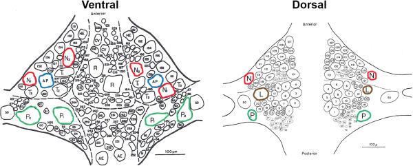
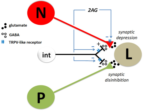
Background: Although a number of clinical and preclinical studies have demonstrated analgesic effects of cannabinoid treatments, there are also instances when cannabinoids have had no effect or even exacerbated pain. The observed pro-nociceptive effects appear to be due to cannabinoid-induced disinhibition of afferent synaptic input to nociceptive circuits. To better understand how cannabinoid-mediated plasticity can have both pro- and anti-nociceptive effects, we examined the possibility that cannabinoids differentially modulate nociceptive vs. non-nociceptive synapses onto a shared postsynaptic target. These experiments were carried out in the central nervous system (CNS) of the medicinal leech, in which it is possible to intracellularly record from presynaptic nociceptive (N-cell) or pressure-sensitive (P-cell) neurons and their shared postsynaptic targets.

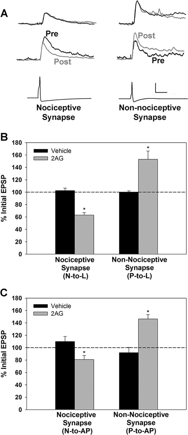
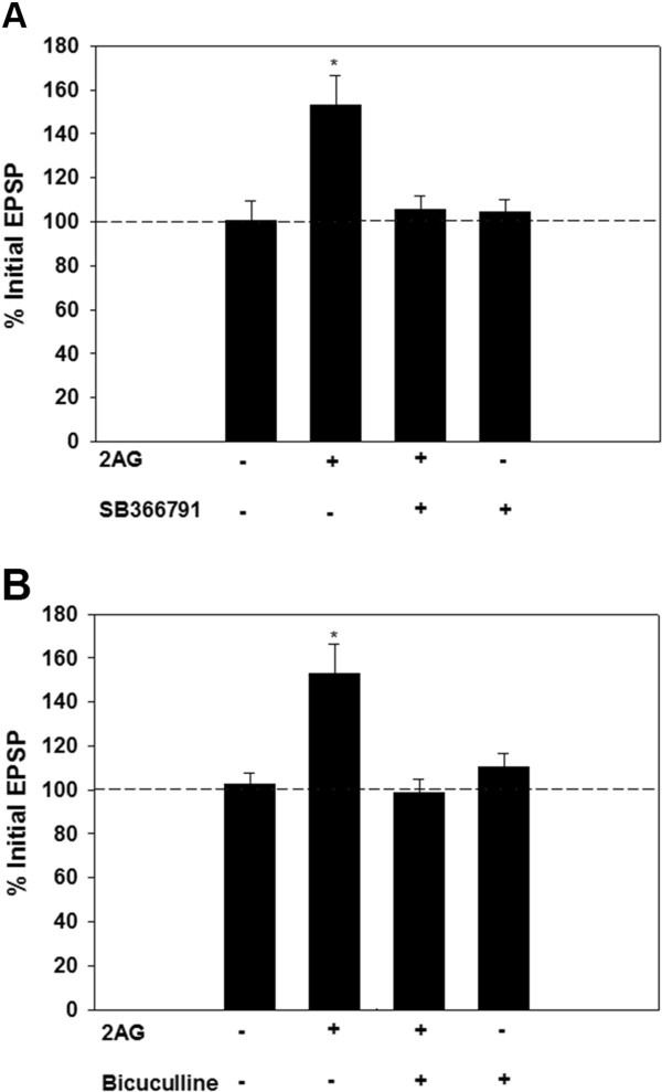
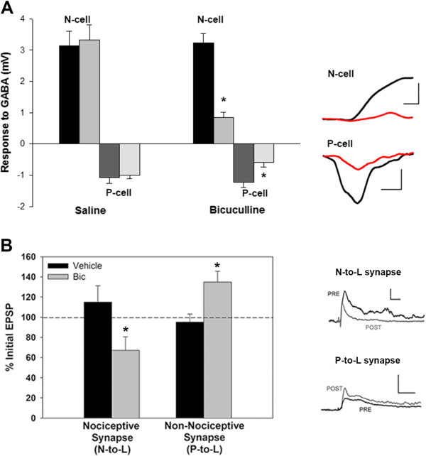
Results: The endocannabinoid 2-arachidonoyl glycerol (2AG) elicited significant long-lasting depression in nociceptive (N-cell) synapses. However, non-nociceptive (P-cell) synapses were potentiated following 2AG treatment. 2AG-induced potentiation of non-nociceptive synapses was blocked by the TRPV antagonist SB366791, suggesting involvement of the same TRPV-like receptor that has already been shown to mediate endocannabinoid-dependent depression in nociceptive inputs. Treatment with the GABA receptor antagonist bicuculline also blocked 2AG-induced potentiation, consistent with the idea that increased synaptic signaling was the result of endocannabinoid-mediated disinhibition. Interestingly, while bicuculline by itself increased non-nociceptive synaptic transmission, nociceptive synapses were depressed by this GABA receptor antagonist indicating that nociceptive synapses were actually excited by GABAergic input. Consistent with these observations, GABA application depolarized the nociceptive afferent and hyperpolarized the non-nociceptive afferent.

Conclusions: These findings show that endocannabinoids can differentially modulate nociceptive vs. non-nociceptive synapses and that GABAergic regulation of these synapses plays an important role in determining whether endocannabinoids have a potentiating or depressing effect.


Figures

Figure 1
Figure 1
Ventral and dorsal aspects of a single ganglion from the leech CNS (modified from [[13]]). Neurons used in this study have been outlined in red (N-cell), green (P-cell), blue (AP-cell) and brown (L motor neuron).
Figure 2
Figure 2
Differential endocannabinoid modulation of nociceptive vs. non-nociceptive synapses. (A) Sample traces from experiments using nociceptive (N-to-L, left) and non-nociceptive (P-to-L, right) synapses. Top EPSP traces are from control experiments in which pre- and post-test recordings (black and grey traces, respectively) were made 75 mins apart without 2AG treatment. Middle EPSP traces are from experiments in which pre- and post-test recordings were made in 2AG-treated ganglia. Bottom traces are action potentials from the presynaptic N-cell (left) or P-cell (right). Vertical calibration bar is 2mV EPSP traces and 50 mV for action potential traces. Horizontal calibration bar is 50 msec for all. (B) Bar graph showing that 2AG depressed the N-to-L synapse, but potentiated the P-to-L synapse. (C) Bar graph showing that 2AG depressed the N-to-AP synapse, but potentiated the P-to-AP synapse. Asterisks indicate statistically significant difference relative to the vehicle control group (see Results section for details).
Figure 3
Figure 3
Role of the leech TRPV-like receptor and GABA during 2AG-mediated synaptic potentiation. (A) The TRPV1 antagonist, SB366791 (10 μM), prevents potentiation of the non-nociceptive P-to-L synapses that is normally observed following 2AG treatment. Asterisks indicate statistically significant difference relative to the vehicle control group (see Results section for details). (B) Role of GABA signaling during 2AG-mediated synaptic potentiation. Pretreatment of synapses with the GABA receptor antagonist bicuculline (100 μM), prevented 2AG-induced potentiation of the non-nociceptive N-to-L synapse. Asterisks indicate statistically significant difference relative to the vehicle control group (see Results section for details).
Figure 4
Figure 4
Opposing effects of GABA on nociceptive vs. non-nociceptive afferents and their synapses. (A) Response of nociceptive (N-cell) and non-nociceptive (P-cell) afferents to GABA. GABA elicited depolarization in the N-cells and hyperpolarization in the P-cells (scale bars are, respectively, 2 mV/500 msec and 0.5 mV/1000 msec). Both responses were inhibited by subsequent treatment 15 mins with bicuculline (100 μM) and this decrease was not observed when a 15 mins saline treatment was used in place of bicuculline. (B) Acute bicuculline treatment depresses the nociceptive (N-to-L) synapse, but enhances the non-nociceptive (P-to-L) synapse. Scale bars are, respectively, 2 mV/50 msec and 5 mV/50 msec. Asterisks indicate statistically significant difference relative to the vehicle control group (see Results section for details).
Figure 5
Figure 5
Hypothetical model for opposing effects of 2AG on nociceptive (N) versus non-nociceptive (P) synapses. Both N- and P-cells have input onto the same postsynaptic targets (in this example, the L motor neuron) via glutamatergic synapses. Based on previous studies [10], 2AG directly depresses the nociceptive synapse via a TRPV-like receptor that reduces presynaptic neurotransmitter release. In the present study, 2AG was observed to potentiate the non-nociceptive synapse via an indirect mechanism in which the eCB reduces inhibitory input from an unknown GABAergic interneuron (int). This 2AG-mediated disinhibition appears to be mediated by the TRPV-like receptor as well. Nociceptive synapses are “protected” from this disinhibitory because they are depolarized by GABA. The same GABAergic interneuron is shown to act on both the N- and P-cells for diagrammatic purposes and it is not known whether these afferents receive GABAergic input from a common source or from distinct interneurons.

Similar articles

Cited by

References

    1. Di Marzo V, Bifulco M, De Petrocellis L. The endocannabinoid system and its therapeutic exploitation. Nat Rev Drug Discov. 2004;3(9):771–784. doi: 10.1038/nrd1495. - DOI - PubMed
    1. Elphick MR. The evolution and comparative neurobiology of endocannabinoid signalling. Philos Trans R Soc Lond B Biol Sci. 2012;367(1607):3201–3215. doi: 10.1098/rstb.2011.0394. - DOI - PMC - PubMed
    1. Heifets BD, Castillo PE. Endocannabinoid signaling and long-term synaptic plasticity. Annu Rev Physiol. 2009;71:283–306. doi: 10.1146/annurev.physiol.010908.163149. - DOI - PMC - PubMed
    1. Pernia-Andrade AJ, Kato A, Witschi R, Nyilas R, Katona I, Freund TF, Watanabe M, Filitz J, Koppert W, Schuttler J. Spinal endocannabinoids and CB1 receptors mediate C-fiber-induced heterosynaptic pain sensitization. Science. 2009;325(5941):760–764. doi: 10.1126/science.1171870. - DOI - PMC - PubMed
    1. Torsney C, MacDermott AB. Disinhibition opens the gate to pathological pain signaling in superficial neurokinin 1 receptor-expressing neurons in rat spinal cord. J Neurosci. 2006;26(6):1833–1843. doi: 10.1523/JNEUROSCI.4584-05.2006. - DOI - PMC - PubMed

Publication types

MeSH terms

LinkOut - more resources