Review
. 2014 Jan 20;20(3):460-73.
doi: 10.1089/ars.2013.5371. Epub 2013 Aug 2.

An overview of autophagy: morphology, mechanism, and regulation

Affiliations
Review

An overview of autophagy: morphology, mechanism, and regulation

Katherine R Parzych et al. Antioxid Redox Signal. .

Abstract

Significance: Autophagy is a highly conserved eukaryotic cellular recycling process. Through the degradation of cytoplasmic organelles, proteins, and macromolecules, and the recycling of the breakdown products, autophagy plays important roles in cell survival and maintenance. Accordingly, dysfunction of this process contributes to the pathologies of many human diseases.

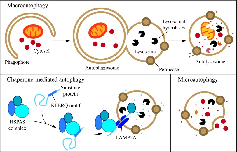
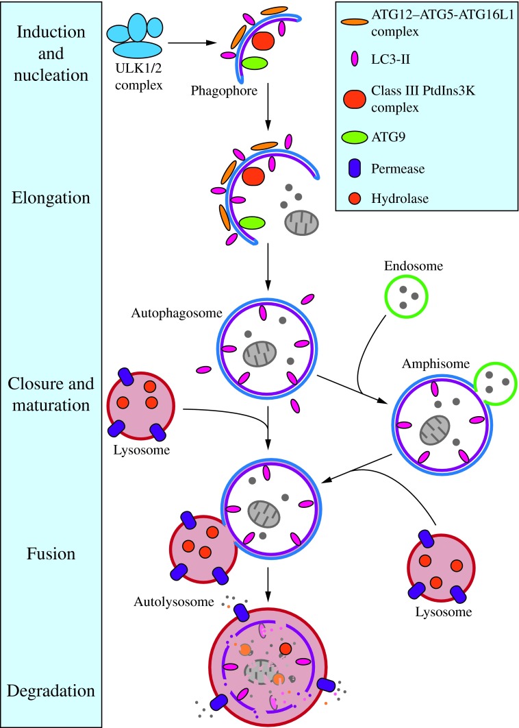
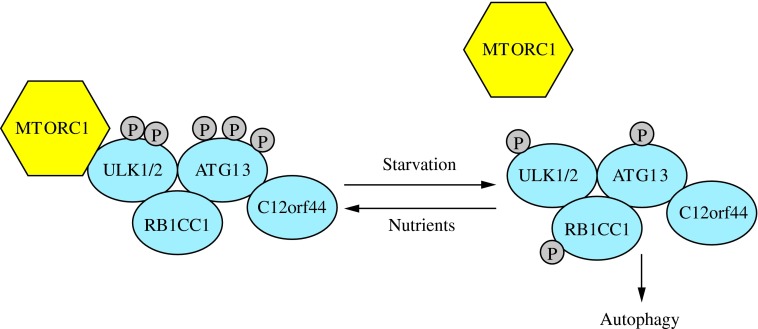
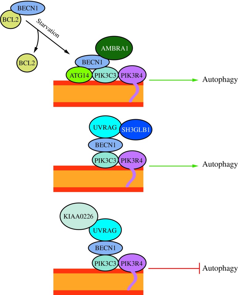
Recent advances: Extensive research is currently being done to better understand the process of autophagy. In this review, we describe current knowledge of the morphology, molecular mechanism, and regulation of mammalian autophagy.

Critical issues: At the mechanistic and regulatory levels, there are still many unanswered questions and points of confusion that have yet to be resolved.

Future directions: Through further research, a more complete and accurate picture of the molecular mechanism and regulation of autophagy will not only strengthen our understanding of this significant cellular process, but will aid in the development of new treatments for human diseases in which autophagy is not functioning properly.


Figures

<b>FIG. 1.</b>
FIG. 1.
Three types of autophagy in mammalian cells. Macroautophagy relies on de novo formation of cytosolic double-membrane vesicles, autophagosomes, to sequester and transport cargo to the lysosome. Chaperone-mediated autophagy transports individual unfolded proteins directly across the lysosomal membrane. Microautophagy involves the direct uptake of cargo through invagination of the lysosomal membrane. All three types of autophagy lead to degradation of cargo and release of the breakdown products back into the cytosol for reuse by the cell. See the text for details. To see this illustration in color, the reader is referred to the web version of this article at www.liebertpub.com/ars
<b>FIG. 2.</b>
FIG. 2.
Morphology of macroautophagy. Nucleation of the phagophore occurs following induction by the ULK1/2 complex. Elongation of the phagophore is aided by the ATG12–ATG5-ATG16L1 complex, the class III PtdIns3K complex, LC3-II, and ATG9. Eventually, the expanding membrane closes around its cargo to form an autophagosome and LC3-II is cleaved from the outer membrane of this structure. The outer membrane of the autophagosome will then fuse with the lysosomal membrane to form an autolysosome. In some instances, the autophagosome may fuse with an endosome, forming an amphisome, before fusing with the lysosome. The contents of the autolysosome are then degraded and exported back into the cytoplasm for reuse by the cell. See the text for details. This figure was modified from Figure 1 in Yang and Klionsky (153). ATG, autophagy-related; PtdIns3K, phosphatidylinositol 3-kinase; ULK, unc-51-like kinase (C. elegans). To see this illustration in color, the reader is referred to the web version of this article at www.liebertpub.com/ars
<b>FIG. 3.</b>
FIG. 3.
The induction complex consists of ULK1/2, ATG13, RB1CC1, and C12orf44. Under nutrient-rich conditions, MTORC1 associates with the complex and inactivates ULK1/2 and ATG13 through phosphorylation. During starvation, MTORC1 dissociates from the complex and ATG13 and ULK1/2 become partially dephosphorylated by as yet unidentified phosphatases, allowing the complex to induce macroautophagy. RB1CC1/FIP200 and C12orf44/ATG101 are also associated with the induction complex and are essential for macroautophagy. RB1CC1/FIP200 may be the ortholog of yeast Atg17, whereas the function of C12orf44/ATG101 is not known. This figure was modified from Figure 1 in Yang and Klionsky (154). MTORC1, mechanistic target of rapamycin complex 1; RB1CC1, RB1-inducible coiled-coil 1. To see this illustration in color, the reader is referred to the web version of this article at www.liebertpub.com/ars
<b>FIG. 4.</b>
FIG. 4.
The activity of the class III PtdIns3K complex is regulated by subunit composition. The ATG14 complex (ATG14-BECN1-PIK3C3-PIK3R4) is required for macroautophagy. It can be positively regulated by AMBRA1 and negatively regulated by BCL2 binding to BECN1 and preventing association with the complex. The UVRAG (UVRAG-BECN1-PIK3C3-PIK3R4) complex is involved in the endocytic pathway and also participates in macroautophagy. SH3GLB1/Bif-1 positively regulates this complex by binding UVRAG. The KIAA0226/Rubicon complex (KIAA0226-UVRAG-BECN1-PIK3C3-PIK3R4) negatively regulates macroautophagy. This figure was modified from Figure 1 in Yang and Klionsky (154). To see this illustration in color, the reader is referred to the web version of this article at www.liebertpub.com/ars
<b>FIG. 5.</b>
FIG. 5.
ATG12–ATG5-ATG16L1 conjugation complex. The ubiquitin-like protein ATG12 is irreversibly conjugated to ATG5 in an ATG7- and ATG10-dependent manner. ATG7 and ATG10 function as E1 activating and E2 conjugating enzymes, respectively. The ATG12–ATG5 conjugate binds ATG16L1 through ATG5. ATG16L1 dimerizes and allows association with the phagophore, promoting membrane expansion. To see this illustration in color, the reader is referred to the web version of this article at www.liebertpub.com/ars
<b>FIG. 6.</b>
FIG. 6.
The LC3 conjugation system. LC3 is processed by ATG4 to reveal a C-terminal glycine (LC3-I). ATG7, an E1-like enzyme, activates LC3-I and transfers it to the E2-like enzyme ATG3. The ATG12–ATG5-ATG16L1 complex may participate as an E3 ligase in the conjugation of PE to LC3-I to create LC3-II, which can associate with the phagophore. LC3-II can subsequently be cleaved by ATG4 to release LC3 (deconjugation). PE, phosphatidylethanolamine. To see this illustration in color, the reader is referred to the web version of this article at www.liebertpub.com/ars
<b>FIG. 7.</b>
FIG. 7.
Regulation of macroautophagy. Three of the major kinases that regulate macroautophagy are PKA, AMPK, and MTORC1. These kinases, along with proteins such as TSC1/2 and CAMKK2/CaMKKβ, respond to a variety of intracellular and extracellular signals to regulate macroautophagy. Green arrows indicate activation of a target and red bars indicate inhibition of a target. See the text for details. This figure was modified from Figure 4 of Chen and Klionsky (14). PKA, cAMP-dependent protein kinase A; AMPK, AMP-activated protein kinase; CAMKK2/CaMKKβ, calcium/calmodulin-dependent protein kinase kinase 2, beta. To see this illustration in color, the reader is referred to the web version of this article at www.liebertpub.com/ars
<b>FIG. 8.</b>
FIG. 8.
Two mechanisms of mitophagy. Mitochondria are cleared from maturing red blood cells through a mechanism involving autophagic recognition of mitochondria through a BNIP3L–LC3 interaction. During removal of damaged mitochondria, PARK2 binds to PINK1 on the mitochondrial surface and ubiquitinates mitochondrial outer membrane proteins, which may then bind SQSTM1, a receptor that interacts with LC3. In either case, the interaction with LC3 leads to sequestration by the phagophore and eventual degradation. This figure was modified from Figure 2 of Youle and Narendra (158). To see this illustration in color, the reader is referred to the web version of this article at www.liebertpub.com/ars

Similar articles

Cited by

References

    1. Agarraberes FA. and Dice JF. A molecular chaperone complex at the lysosomal membrane is required for protein translocation. J Cell Sci 114: 2491–2499, 2001 - PubMed
    1. Alers S, Loffler AS, Wesselborg S, and Stork B. Role of AMPK-mTOR-Ulk1/2 in the regulation of autophagy: cross talk, shortcuts, and feedbacks. Mol Cell Biol 32: 2–11, 2012 - PMC - PubMed
    1. Arias E. and Cuervo AM. Chaperone-mediated autophagy in protein quality control. Curr Opin Cell Biol 23: 184–189, 2011 - PMC - PubMed
    1. Arsham AM, Howell JJ, and Simon MC. A novel hypoxia-inducible factor-independent hypoxic response regulating mammalian target of rapamycin and its targets. J Biol Chem 278: 29655–29660, 2003 - PubMed
    1. Atlashkin V, Kreykenbohm V, Eskelinen E-L, Wenzel D, Fayyazi A, and Fischer von Mollard G. Deletion of the SNARE vti1b in mice results in the loss of a single SNARE partner, syntaxin 8. Mol Cell Biol 23: 5198–5207, 2003 - PMC - PubMed