Review
. 2013 Aug;25(4):495-505.
doi: 10.1016/j.ceb.2013.04.008. Epub 2013 May 29.

Lysosome-related organelles: unusual compartments become mainstream

Affiliations
Review

Lysosome-related organelles: unusual compartments become mainstream

Michael S Marks et al. Curr Opin Cell Biol. 2013 Aug.

Abstract

Lysosome-related organelles (LROs) comprise a group of cell type-specific subcellular compartments with unique composition, morphology and structure that share some features with endosomes and lysosomes and that function in varied processes such as pigmentation, hemostasis, lung plasticity and immunity. In recent years, studies of genetic diseases in which LRO functions are compromised have provided new insights into the mechanisms of LRO biogenesis and the regulated secretion of LRO contents. These insights have revealed previously unappreciated specialized endosomal sorting processes in all cell types, and are expanding our views of the plasticity of the endosomal and secretory systems in adapting to cell type-specific needs.


Figures

Figure 1
Figure 1. Model for biogenesis and docking of four vertebrate LROs
Shown are models for the biogenesis of immature (i) and mature (m) melanosomes (left, orange), platelet α granules (pink), lysosomes (violet), CTL LGs (gray) and WPBs (right, blue) relative to the endocytic and biosynthetic pathways (Golgi, TGN, early endosomes, late endosomes/ MVBs, and lysosomes). Key cargo molecules discussed in the text are noted in the same color as the LRO, and effectors involved in biogenetic steps are labeled in black text. Arrows indicate relevant trafficking pathways. Left, immature melanosomes (iMel) emerge from vacuolar domains of early endosomes, and mature by cargo delivery from tubulovesicular domains of early endosomes through AP-1- or AP-3-coated vesicles; recycling endosomal domains associated with KIF13A and AP-1 migrate along microtubules towards maturing melanosomes for delivery of some cargoes as indicated. BLOC-1 facilitates tubule-mediated transport; BLOC-2, BLOC-3, RAB32 and RAB38 likely function downstream. Platelet α granules derive in an NBEAL2-dependent process from late endosomes/ MVBs within megakaryocytes, and receive both biosynthetic and endocytic cargoes. MVBs in the same cells also fuse with lysosomes to deliver other cargoes. In CTLs and NK cells, immature LGs (iLGs) also derive by fusion of MVBs with dense core structures, and then fuse with recycling endosome-derived structures upon stimulation by target cells to form mature LGs (mLGs). The dense cores of iLGs contain perforin and granzymes that likely aggregate within the TGN. Likewise, vWF forms tubules in the TGN of endothelial cells, that then bud off perhaps together with cargoes such as P-selectin and IGFBP7 to form immature WPBs; other cargoes, such as CD63, are then delivered from early endosomes in an AP-3-dependent manner.
Figure 2
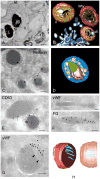
Figure 2. LRO ultrastructure
Shown are images from electron microscopy analyses or three-dimensional (3d) reconstructions of electron tomograms for four model LROs discussed in this review. A, B, melanosomes from MNT-1 human melanoma cells fixed by high pressure freezing and embedded in plastic by freeze substitution. A, a thin section emphasizing stage I and II premelanosomes and stage III and IV mature melanosomes, as indicated. Note the striated appearance in stages II and III. M, mitochondrion. B, 3d reconstruction emphasizing fibrillar structure (yellow) emanating from intralumenal vesicles (green) in stage I/ II melanosomes. Note the accumulation of melanin (brown) on the fibrils in a stage III melanosome. Melanosome limiting membranes are indicated in red; surrounding endosomal tubules are indicated in blue. C, D, cytolytic granules from primary human CTLs. C, an ultrathin cryosection was immunogold labeled for perforin. Note labeling over dense cores (arrowheads), with surrounding ILVs (arrows). D, 3d reconstruction emphasizing dense core (green) surrounded by multilamellar membranes (violet) and ILVs (yellow). The limiting membrane is pseudocolored blue. E, WPBs within an ultrathin cryosection of a human umbilical vein endothelial cell are shown, immunogold labeled for CD63. Note the tubular vWF polymers captured in profile (arrow) or cross-section (*). F, G, H, α granules within human platelets. F, G, ultrathin cryosections were immunogold labeled for vWF or fibrinogen (FG). Note that fibrinogen is present throughout the tubular α granules but vWF is polarized to one side, adjacent to ILVs (arrows in G). H, 3d reconstruction emphasizing vWF tubules (blue) polarized to one side of a spherical α granule domain, shown in two orientations; the limiting membrane is pseudocolored red. Bars: A–D, 200 nm; E–H, 100 nm.
Figure 3
Figure 3. Model for molecular control of LRO secretion
Shown is a schematic diagram of discrete steps in LRO secretion. Tethering (bottom) refers to the attachment of LROs to the subplasmalemmal actin cytoskeleton (royal blue network). RAB27A facilitates tethering of LGs, melanosomes and WPBs, but by distinct effectors as shown, and in several kinetically distinct stages that have not yet been detailed. In CTLs and NK cells, LGs become tethered following centriole polarisation in a process requiring RAB7, its effector RILP, and dynein (not shown). RAB27A and Munc13-4 function independently to regulate fusion of immature LGs with RAB11-positive exocytic vesicles (see Figure 1), and then together at a later step of tethering; RAB27A also functions together with a Slp3/kinesin-1 complex at a yet undefined stage. In endothelial cells, RAB27A, RAB15 and their joint effector, Munc13-4, facilitate WPB tethering; the RAB27A effectors Slp4-a and MyRIP also function in a mutually antagonistic manner at a distinct stage. In melanocytes, a complex of RAB27A, melanophilin and Myosin VA (MYOVA) recruit melanosomes from microtubules to cortical actin. Docking (middle) refers to the engagement of SNAREs on the LRO and target membranes in a pre-fusion complex. In platelets, docking is mediated by VAMP8 on α granule and dense granule membranes, a syntaxin 11 (STX11)/ SNAP-23 complex on the plasma membrane, and accessory proteins Munc13-4 and Munc18b/MUNC18-2. The same components promote docking in CTLs and NK cells for LG secretion, but it is not yet clear whether this is at the plasma membrane or at a pre-secretory step. Fusion/ secretion involves the zippering of opposing SNAREs, applying force to fuse the LRO and plasma membranes. This permits release of the lumenal LRO contents; a few examples of some LRO contents are indicated in the blue box. In endothelial cells, contractile forces from actin and myosin II are required to “squeeze” the elongated vWF tubules from fused WPBs. In skin melanocytes, it is not yet clear whether melanin is secreted like other LRO contents or whether melanin transfer is mediated by a non-secretory process.

Similar articles

Cited by

References

    1. Raposo G, Marks MS, Cutler DF. Lysosome-related organelles: driving post-golgi compartments into specialisation. Curr Opin Cell Biol. 2007;19:394–401. - PMC - PubMed
    1. Hurbain I, Geerts WJC, Boudier T, Marco S, Verkleij A, Marks MS, Raposo G. Electron tomography of early melanosomes: implications for melanogenesis and the generation of fibrillar amyloid sheets. Proc Natl Acad Sci US A. 2008;105:19726–19731. - PMC - PubMed
    1. Watt B, van Niel G, Raposo G, Marks MS. PMEL: A pigment cell-specific model for functional amyloid formation. Pigment Cell Melanoma Res. 2013 doi: 10.1111/pcmr.12067. In press. - DOI - PMC - PubMed
    1. Hellström AR, Watt B, Fard SS, Tenza D, Mannström P, Narfström K, Ekesten B, Ito S, Wakamatsu K, Larsson J, et al. Inactivation of the Pmel gene alters melanosome shape but has only a subtle effect on visible pigmentation. PLoS Genet. 2011;7:e1002285. - PMC - PubMed
    1. Michaux G, Abbitt KB, Collinson LM, Haberichter SL, Norman KE, Cutler DF. The physiological function of von Willebrand’s factor depends on its tubular storage in endothelial Weibel-Palade bodies. Dev Cell. 2006;10:223–232. - PubMed

Publication types

LinkOut - more resources