. 2013 Oct;14(10):1031-44.
doi: 10.1016/j.jpain.2013.03.012. Epub 2013 May 31.

Induction of monocyte chemoattractant protein-1 (MCP-1) and its receptor CCR2 in primary sensory neurons contributes to paclitaxel-induced peripheral neuropathy

Affiliations

Induction of monocyte chemoattractant protein-1 (MCP-1) and its receptor CCR2 in primary sensory neurons contributes to paclitaxel-induced peripheral neuropathy

Haijun Zhang et al. J Pain. 2013 Oct.

Abstract

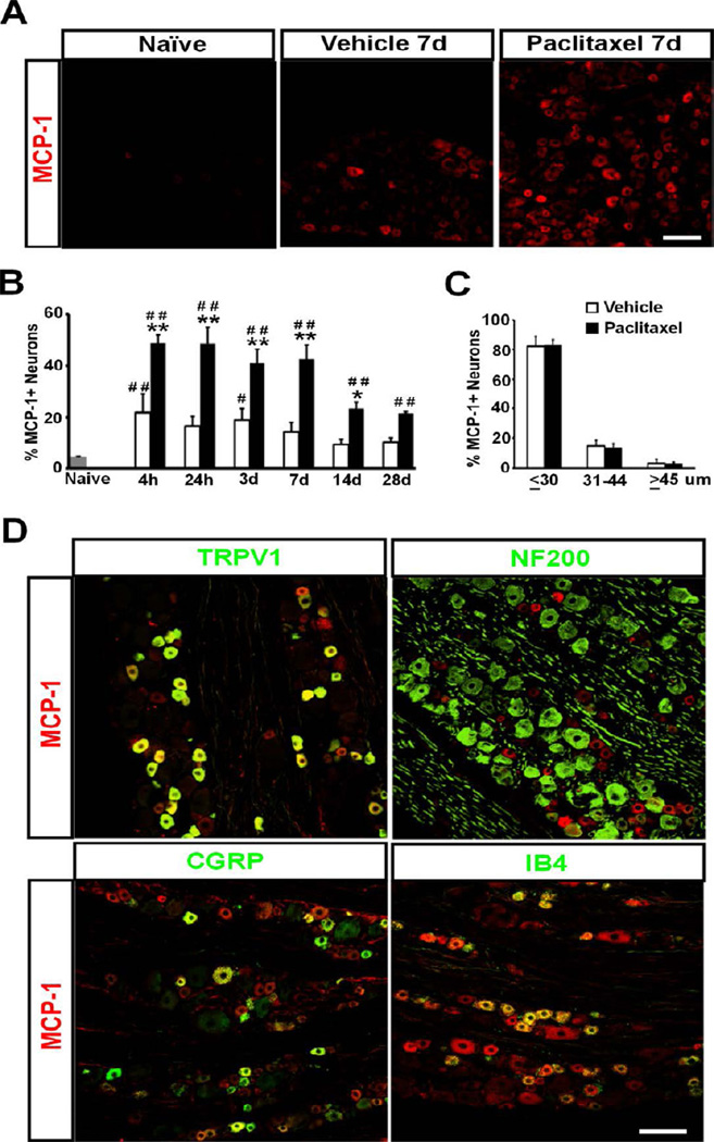
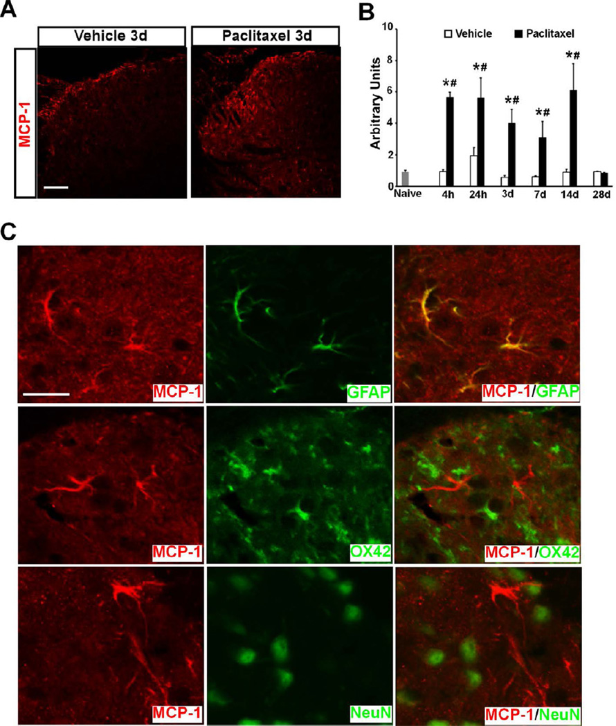
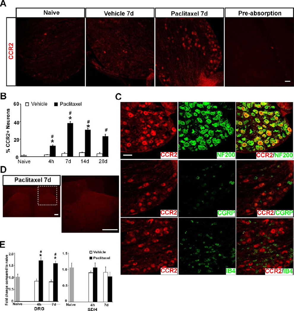
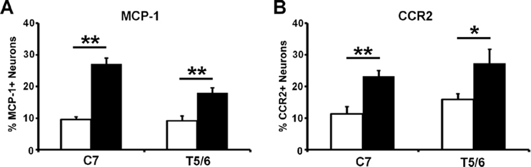
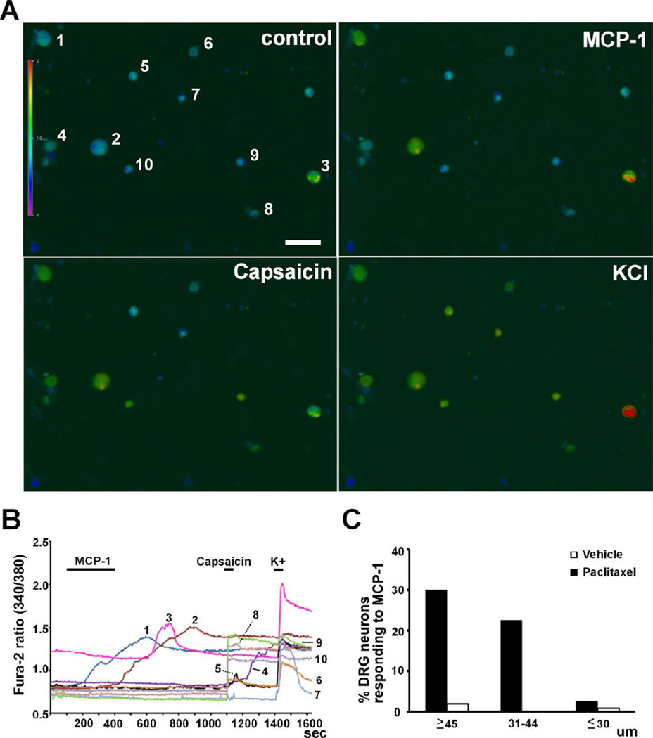
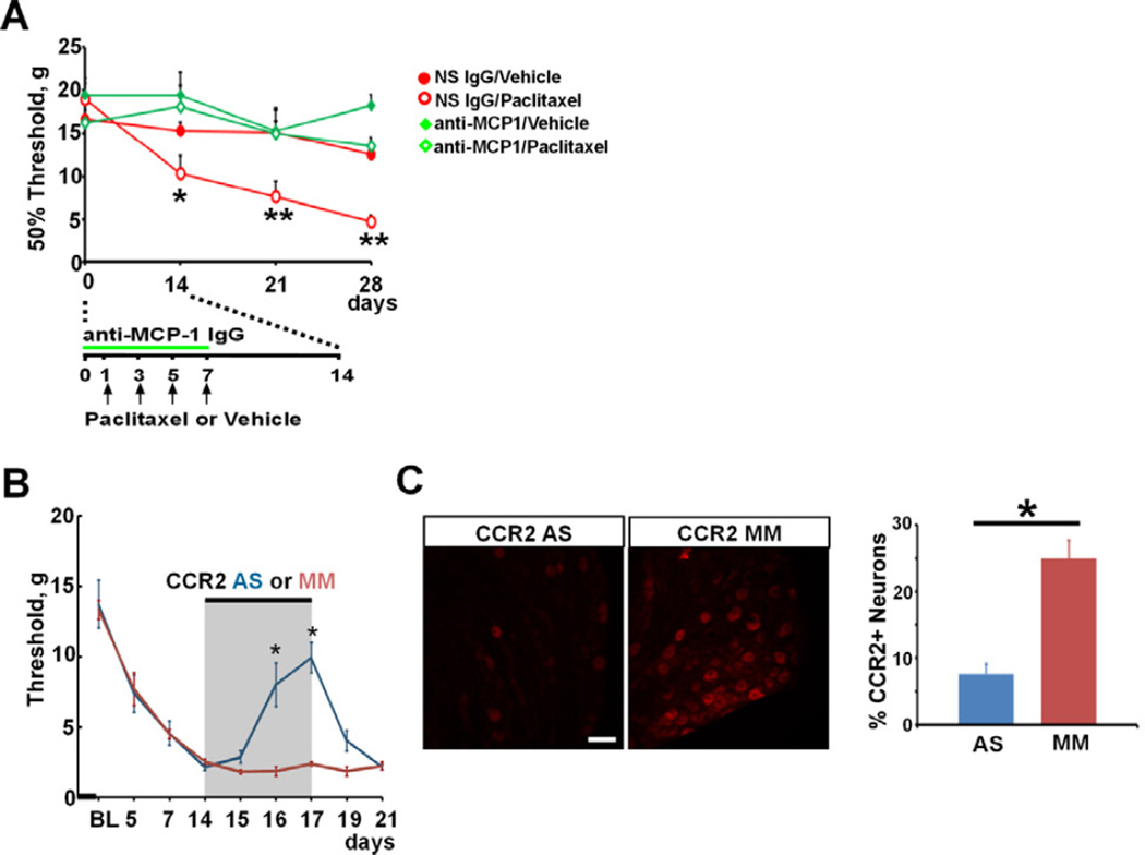
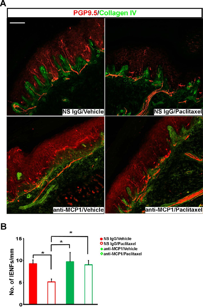
The use of paclitaxel (Taxol), a microtubule stabilizer, for cancer treatment is often limited by its associated peripheral neuropathy (chemotherapy-induced peripheral neuropathy [CIPN]), which predominantly results in sensory dysfunction, including chronic pain. Here we show that paclitaxel CIPN was associated with induction of chemokine monocyte chemoattractant protein-1 (MCP-1) and its cognate receptor CCR2 in primary sensory neurons of dorsal root ganglia. Immunostaining revealed that MCP-1 was mainly expressed in small nociceptive neurons whereas CCR2 was expressed in large and medium-sized myelinated neurons. Direct application of MCP-1 consistently induced intracellular calcium increases in dorsal root ganglia large and medium-sized neurons but not in small neurons mainly dissociated from paclitaxel-treated but not vehicle-treated animals. Paclitaxel also induced increased expression of MCP-1 in spinal astrocytes, but no CCR2 signal was detected in the spinal cord. Local blockade of MCP-1/CCR2 signaling by anti-MCP-1 antibody or CCR2 antisense oligodeoxynucleotides significantly attenuated paclitaxel CIPN phenotypes including mechanical hypersensitivity and loss of intraepidermal nerve fibers in hindpaw glabrous skin. These results suggest that activation of paracrine MCP-1/CCR2 signaling between dorsal root ganglion neurons plays a critical role in the development of paclitaxel CIPN, and targeting MCP-1/CCR2 signaling could be a novel therapeutic approach.

Perspective: CIPN is a severe side effect accompanying paclitaxel chemotherapy and lacks effective treatments. The current study suggests that blocking MCP-1/CCR2 signaling could be a new therapeutic strategy to prevent or reverse paclitaxel CIPN. This preclinical evidence encourages future clinical evaluation of this strategy.

Keywords: CCR2; MCP-1; Paclitaxel; dorsal root ganglion; neuropathy.


Figures

Figure 1
Figure 1
The induction of MCP-1 in DRG by Paclitaxel. A. Immunostaining shows marked upregulation of MCP-1 in DRG neurons from paclitaxel-treated animals compared to vehicletreated or naïve animals. B. The increase of MCP-1 is observed as early as 4 hours and persists through 28 days after treatment. C. The distribution of MCP-1+ neurons in DRG at 4 hours after paclitaxel or vehicle treatment shows the majority of MCP-1+ neurons is small cells. D. MCP-1+ (red) neurons are co-labeled (yellow) with TRPV1 (green), CGRP (green) and IB4 (green), but minimally with NF200 (green). * p < 0.05, ** p < 0.01 versus vehicle; # p < 0.05, ## p < 0.01 versus naïve. Scale bar: 100 um.
Figure 2
Figure 2
The induction of MCP-1 in SDH by Paclitaxel. A. Immunostaining shows marked upregulation of MCP-1 in SDH following paclitaxel treatment. B. The increase of MCP-1 is observed as early as 4 hours and persists through 14 days after treatment. C. Double immunostaining shows MCP-1 (red) is induced in spinal astrocytes (green) but not neurons (green) or microglia (green). * p < 0.01 versus vehicle; # p < 0.01 versus naïve. Scale bar: 100 µm (A) and 20 µm (C).
Figure 3
Figure 3
Paclitaxel induces expression of CCR2 in DRG but not SDH. A. Immunostaining shows marked upregulation of CCR2 in DRG neurons from paclitaxel- but not vehicle-treated or naïve animals. Pre-absorption of CCR2 blocked the immunohistochemical staining of DRG. B. The increase of CCR2 is observed as early as 4 hours and persists through 28 days after treatment. C. CCR2+ (red) neurons are co-labeled (yellow) with NF200 (green) but not with CGRP (green) or IB4 (green). D. No signal of CCR2 is detected by immunostaining in SDH after either paclitaxel or vehicle treatment. E. Paclitaxel induces an increase in mRNA of CCR2 in DRG (F[2,34] = 40.05, p < 0.0001, two-way ANOVA, n = 4, 8 and 8 for naïve, paclitaxel or vehicle at both time points) but not SDH at 4 hours and 7 days following treatment. * p < 0.0001 versus vehicle; # p < 0.001 versus naïve. Scale bar: 100 um (A and C) and 200 um (D).
Figure 4
Figure 4
Paclitaxel induces the prominent expression of MCP-1 (A) and CCR2 (B) in both C7 and T5/6 DRGs. Tissues were examined on day 14 after chemotherapy. * p < 0.05, ** p < 0.01, paclitaxel versus vehicle, t-test, n = 3 each group.
Figure 5
Figure 5
Intracellular calcium response of DRG neurons to MCP-1. A, B. MCP-1 (1 µg/ml) induced increases in intracellular calcium concentration in large (#1 and 2) and medium-sized (#3) neurons which did not respond to capsaicin (0.5 µM) but did respond to a high concentration of K+ (50 mM). Small neurons did not respond to MCP-1 but responded to capsaicin and K+ (#4, 5, 6, 8, 9 and 10). One small neuron did not respond to MCP-1 or capsaicin but responded to K+ (#7). C. Size distribution of DRG neurons responding to MCP-1 from paclitaxel- and vehicle-treated animals. Scale bar: 100 µm.
Figure 6
Figure 6
Blockade of MCP-1 or CCR2 attenuates paclitaxel-induced mechanical hypersensitivity. A. Intrathecal anti-MCP-1 IgG prevents paclitaxel-induced mechanical hypersensitivity (n = 7, 7, 5 and 6 for non-specific (NS) IgG/vehicle, NS IgG/paclitaxel, anti-MCP-1/vehicle and anti-MCP-1/paclitaxel, respectively). * p < 0.05, ** p < 0.01, two-way ANOVA. B. Intrathecal CCR2 antisense (AS) but not mismatch (MM) ODN significantly reduces paclitaxel-induced mechanical hypersensitivity. * p < 0.01, one-way ANOVA. BL: baseline. C. Intrathecal CCR2 AS but not MM ODN significantly reduces the induction of CCR2 in DRG from paclitaxel-treated animals. Tissues are collected on the last day of CCR2 ODNsb treatment (Day 17 following chemotherapy). * p < 0.01, n = 3 each group, t-test. Scale bar: 100 µm.
Figure 7
Figure 7
Blockade MCP-1 prevents the loss of IENFs induced by paclitaxel. A. Representative images of IENFs in glabrous skin of hindpaw foot pad. The density of IENFs stained by PGP9.5 (red) crossing dermal/epidermal border (green) is much lower in NS IgG/paclitaxel group and intrathecal anti-MCP-1 IgG prevents the loss of IENFs. Confocal images are taken with Z-stack of 2µm-step in one view using 20X objective and montaged as a single image. B. Paclitaxel induces a significant decrease in the density of IENFs and anti-MCP-1 treatment prevents the loss of IENFs (n = 6, 7, 4 and 4 for NS IgG/vehicle, NS IgG/paclitaxel, anti-MCP-1/vehicle and anti-MCP-1/paclitaxel, respectively). * p < 0.05, one-way ANOVA. Scale bar: 100 µm.

References

    1. Abbadie C, Lindia JA, Cumiskey AM, Peterson LB, Mudgett JS, Bayne EK, DeMartino JA, MacIntyre DE, Forrest MJ. Impaired neuropathic pain responses in mice lacking the chemokine receptor CCR2. Proc Natl Acad Sci USA. 2003;100:7947–7952. - PMC - PubMed
    1. Alessandri-Haber N, Yeh JJ, Boyd AE, Parada CA, Chen X, Reichling DB, Levine JD. Hypotonicity induces TRPV4-mediated nociception in rat. Neuron. 2003;39:497–511. - PubMed
    1. Amir R, Devor M. Chemically mediated cross-excitation in rat dorsal root ganglia. J Neurosci. 1996;16:4733–4741. - PMC - PubMed
    1. Archer DR, Dahlin LB, McLean WG. Changes in slow axonal transport of tubulin induced by local application of colchicine to rabbit vagus nerve. Acta Physiol Scand. 1994;150:57–65. - PubMed
    1. Bedi SS, Yang Q, Crook RJ, Du J, Wu Z, Fishman HM, Grill RJ, Carlton SM, Walters ET. Chronic spontaneous activity generated in the somata of primary nociceptors is associated with pain-related behavior after spinal cord injury. J Neurosci. 2010;30:14870–14882. - PMC - PubMed

Publication types

MeSH terms