Review
. 2013 Sep 17:248:499-528.
doi: 10.1016/j.neuroscience.2013.05.042. Epub 2013 May 30.

Post-transcriptional regulatory elements and spatiotemporal specification of neocortical stem cells and projection neurons

Affiliations
Review

Post-transcriptional regulatory elements and spatiotemporal specification of neocortical stem cells and projection neurons

E M DeBoer et al. Neuroscience. .

Abstract

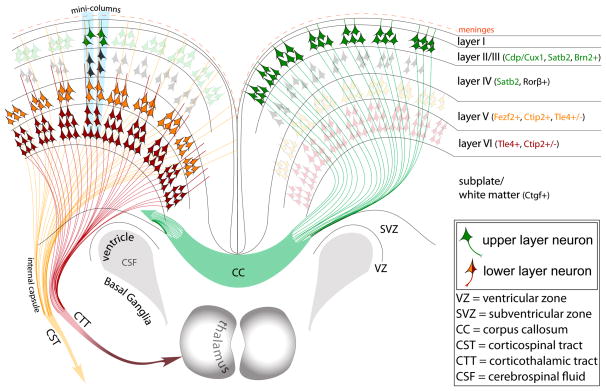
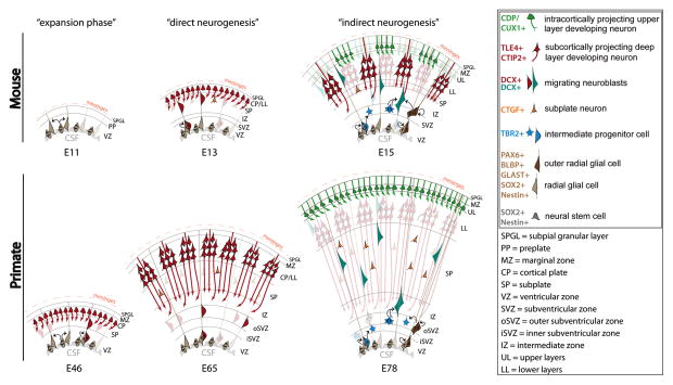
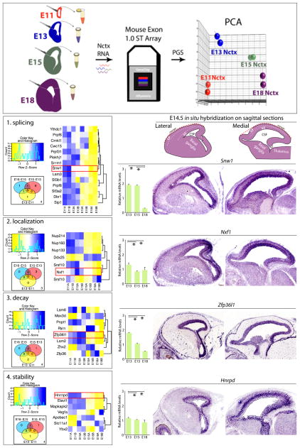
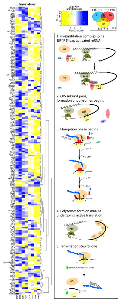
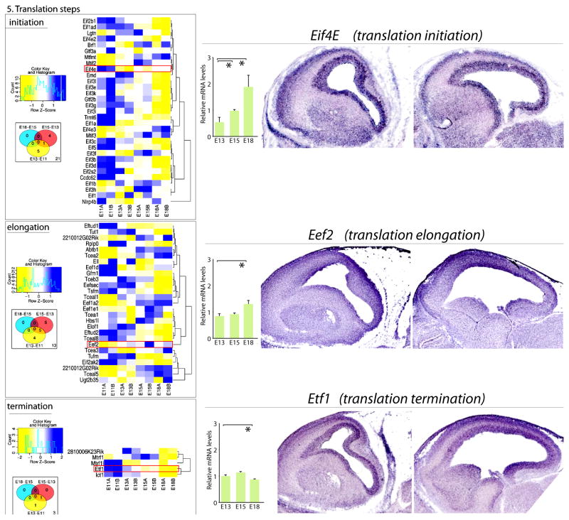
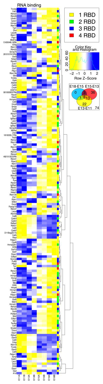
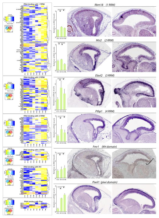
The mature neocortex is a unique six-layered mammalian brain region. It is composed of morphologically and functionally distinct subpopulations of primary projection neurons that form complex circuits across the central nervous system. The precisely-timed generation of projection neurons from neural stem cells governs their differentiation, postmitotic specification, and signaling, and is critical for cognitive and sensorimotor ability. Developmental perturbations to the birthdate, location, and connectivity of neocortical neurons are observed in neurological and psychiatric disorders. These facts are highlighting the importance of the precise spatiotemporal development of the neocortex regulated by intricate transcriptional, but also complex post-transcriptional events. Indeed, mRNA transcripts undergo many post-transcriptional regulatory steps before the production of functional proteins, which specify neocortical neural stem cells and subpopulations of neocortical neurons. Therefore, particular attention is paid to the differential post-transcriptional regulation of key transcripts by RNA-binding proteins, including splicing, localization, stability, and translation. We also present a transcriptome screen of candidate molecules associated with post-transcriptional mRNA processing that are differentially expressed at key developmental time points across neocortical prenatal neurogenesis.

Keywords: RNA-binding protein; neocortex; neural stem cell; post-transcriptional processing; projection pyramidal neuron; translation.


Figures

Figure 1
Figure 1. A simplified schematic of the postnatal organization and projections of neocortical projection neurons
The neocortex is highly organized in both horizontal and vertical dimensions. Horizontally, six layers are defined by highly organized subpopulations of glutamatergic projection neurons, which represent approximately 85% of all neocortical neurons. These subpopulations of projection neurons are characterized by specific molecular identities, dendritic morphologies and terminal targets corresponding to each layer. Projection neurons that are born during later stages of prenatal neurogenesis will be predominantly placed in upper layers II–IV (green neurons). These neurons express specific transcription factors like CDP/Cux1, and project solely intracortically forming the corpus callosum that connects the two hemispheres. However, there is also a smaller portion of intracortically projecting neurons placed in lower layers too (not shown). In contrast, earlier born projection neurons will be placed in lower layers V–VI (orange and red neurons). These subopulations will express transcription factors like FEZF2, and will project subcortically to form long range tracts across the central nervous system like the corticothalamic tract (CTT) originating mainly from layer 6, and somewhat from layer 5, or corticospinal tract (CST) originating solely from layer 5. Within the subventricular zone (SVZ) of the corticostriatal junction, adult progenitors are found giving rise to olfactory cortex neurons.
Figure 2
Figure 2. Schematic of distinct stages of neocortical neurogenesis in developing mouse and primate neocortices
The first “phase” of neocorticogenesis in mouse and primates is characterized by symmetric divisions of neural stem cells called neuroepithelium cells (NEC), which amplifies the number of neocortical progenitors at the ventricular zone (VZ) surface (left panels). This initial phase is accordingly called the “expansion phase”. NECs will then transition into a different lineage of neocortical neural stem cells called radial glia (RG), which divide asymmetrically and first predominantly produce neuronal progeny. This phase was named “direct neurogenesis” (middle panels). As the neurogenic phase progresses, RG continue to undergo a series of asymmetric divisions, but they predominantly produce another progenitor subtype, intermediate progenitor cells (IPCs) and outer radial glia (oRG) (right panels). IPCs and oRG will divide in the subventricular zone (SVZ), which in primates is divided into inner (iSVZ) and outer (oSVZ) portions. Importantly, IPCs terminally divide symmetrically and produce at least two neural progenies. However, oRG will self renew and give rise to neural progenies and IPCs. In this way, both IPCs and oRGs amplify the output of RG, and thus, this later stage of neurogenesis was named “indirect neurogenesis”. These progressive changes in differentiation of neocortical neural stem cells define the birth of distinct subpopulations of projection neurons. Deep layer neurons that will project subcortically into thalamus, brain stem and spinal cord will be born before upper layer intracortically projecting neurons. (Adapted from Angevine and Sidman, 1961, Rakic, 1974, Smart et al., 2002, and Molnár et al., 2006).
Figure 3
Figure 3. Transcriptome analysis of molecules involved in post-transcriptional mRNA processing steps within the developing mouse neocortex
(top) Neocortex (Nctx) from embryonic days 11 (E11), E13, E15, and E18 was dissected, and RNA was isolated then assayed using Mouse Exon 1.0 ST Arrays, and analyzed using Partek Genome Suite (PGS) and R/Bioconductor. Principal component analysis (PCA) revealed clustering among replicates and distinct differentiation among developmental stages. (Left) Gene ontology (GO) analysis of whole developing mouse neocortices during key steps of the neurogenesis for distinct steps of post-transcriptional mRNA processing: splicing (1), localization (2), decay (3), and stability (4). GO analysis is presented as heatmap (blue = higher expression; yellow = lower expression; normalized by gene) and corresponding Venn analyses. Heatmaps include all genes on the Affymetrix Mouse Exon array annotated with the listed GO term, and Venn diagrams depict numbers of genes having significant contrasts between adjacent time points. (Right) qRT-PCR of whole developing neocortices for sample genes annotated with red boxes on the heatmaps. Corresponding in situ hybridization of lateral (middle panels) and medial (right panels) sagittal neocortical sections of E14.5 neocortices were obtained from www.genepaint.com. Remarkably, besides temporally distinct expression levels, the post- transcriptional regulatory elements also show restricted enrichment in different compartments of developing neocortices. For example, expression of the decay regulator Zfp36l1 decreased during neurogenesis, while its expression is highly enriched in the VZ where RG reside. In contrast, expression of a splicing regulator, Snw1, dramatically decreased at E18 when neurogenesis ceases, but is enriched at E14.5 in both progenitor characterized compartment VZ and postmitotic compartment CP. All qRT-PCR values were normalized to four housekeeping genes Gapdh, Mrps6, Rps13, and Rps18, and then scaled to average. * p < 0.05.
Figure 4
Figure 4. Transcriptome analysis of developing neocortices during prenatal neurogenesis for mRNA translation reveals numerous mRNA clusters showing dynamic expression patterns
(Right) Canonical process of translation and points of regulation. 5′-cap activated mRNA carries EIF4F complex (EIF4A+EIF4G+EIF4E) bound to the 5′ untranslated region (UTR) (step 1, top right animation). EIF4G from the complex is associating with PABP bound to the 3′ UTR making the active mRNA into a loop (step 2). On these mRNAs the preinitation complex (eIF2–40S ribosome-Eif3) joins the 5′UTR and screens for the start codon. Then the 60S ribosomal subunit joins the 40S to form the 80S ribosomal monosome. This step is partially regulated by Eif5b, while Eif2 and EIf3 are removed from the 40S (step 3). This initiation phase then transitions into the elongation phase when active translation is being governed by polysome assembly and polypeptide elongation (step 4). Once the polypeptide is finalized by reaching the stop codon, the termination step dissociates the 80S ribosome back into 40S and 60S subunits (step 5, bottom right animation). Each step of translation is regulated by distinct molecules, as shown in the next figure.
Figure 5
Figure 5. Transcriptome analysis of the developing mouse neocortex for mRNAs encoding regulators of distinct steps of mRNA translation
(Left) Remarkably, even mRNAs encoding regulators of mRNA translation show dynamic changes in their expression during neurogenesis. GO analysis is again presented as heatmap (blue = higher expression; yellow = lower expression, normalized by gene) together with corresponding Venn analyses. (Right) qRT-PCR of whole developing neocortices for genes corresponds to relative gene expression in the heatmaps (red boxes). Corresponding in situ hybridization of lateral and medial sagittal neocortical sections of E14.5 neocortices were obtained from www.genepaint.com. Interestingly, initiation factor EIf4E and termination factor, Etf1, are both enriched in the VZ and CP, suggesting highly dynamic regulation of these two regulatory steps in RG progenitors and postmitotic differentiating neurons. All qRT-PCR values were normalized to four housekeeping genes (Gapdh, Mrps6, Rps13, and Rps18), and then scaled to average. * p < 0.05.
Figure 6
Figure 6. Transcriptome analysis of the developing mouse neocortex for RNA binding proteins (RBPs)
(left) GO analysis for RBPs revealed their substantial enrichment in developing neocortices, again with a predominant switch in expression levels occurring around E15. Each RBP has one or more RNA binding domain (RBDs) like KH, piwi or an RNA recognition motif (RRM). Total number of RBDs per RBP are presented with the color key (upper right corner).
Figure 7
Figure 7. Transcriptome analysis of the developing mouse neocortex for RBPs with RRM, KH or piwi domains
(Left) RBPs with distinct RBD signatures show differences in their number and temporal expression. For example, RBPs with 1 RRMs are the most numerous group. Corresponding Venn diagrams are provided below the heatmap key. (Right) qRT-PCR and in situ hybridization from www.genepaint.com revealed dynamic spatiotemporal expression of distinct RBPs. One example for each RBP group from the heatmap on the left is presented. Ptbp1, which is characterized by 4 RRM domains is highly enriched in the VZ, while Rbm18 characterized by 1 RRM seems to be in postmitotic neurons of CP, and some signal is detected in the migratory zone between VZ and CP. Piwil1 shows high signal in the VZ and CP, but is somewhat decreased in the anterolateral VZ. All qRT-PCR values were again normalized to four housekeeping genes Gapdh, Mrps6, Rps13, and Rps18, then scaled to average. * p < 0.05.

References

    1. Brain Museum. http://brainmuseum.org/
    1. Ables JL, Breunig JJ, Eisch AJ, Rakic P. Not(ch) just development: Notch signalling in the adult brain. Nat Rev Neurosci. 2011;12:269–283. - PMC - PubMed
    1. Akamatsu W, Fujihara H, Mitsuhashi T, Yano M, Shibata S, Hayakawa Y, Okano HJ, Sakakibara S, Takano H, Takano T, Takahashi T, Noda T, Okano H. The RNA-binding protein HuD regulates neuronal cell identity and maturation. Proc Natl Acad Sci U S A. 2005;102:4625–4630. - PMC - PubMed
    1. Alcamo EA, Chirivella L, Dautzenberg M, Dobreva G, Farinas I, Grosschedl R, McConnell SK. Satb2 regulates callosal projection neuron identity in the developing cerebral cortex. Neuron. 2008;57:364–377. - PubMed
    1. Alexandre P, Reugels AM, Barker D, Blanc E, Clarke JDW. Neurons derive from the more apical daughter in asymmetric divisions in the zebrafish neural tube. Nat Neurosci. 2010;13:673–679. - PubMed

Publication types

Substances