

Preservation of micro-architecture and angiogenic potential in a pulmonary acellular matrix obtained using intermittent intra-tracheal flow of detergent enzymatic treatment
- PMID: 23727263
- PMCID: PMC3988964
- DOI: 10.1016/j.biomaterials.2013.05.015
Preservation of micro-architecture and angiogenic potential in a pulmonary acellular matrix obtained using intermittent intra-tracheal flow of detergent enzymatic treatment
Abstract
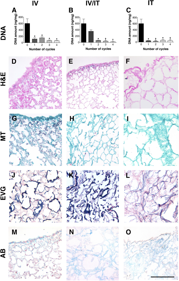
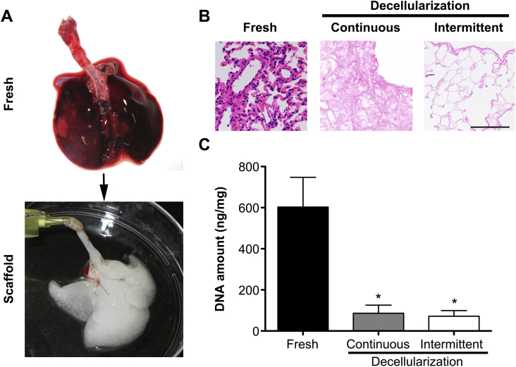
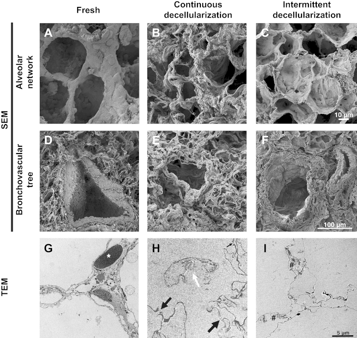
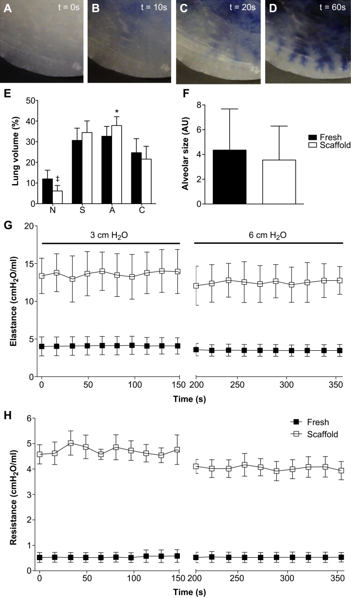
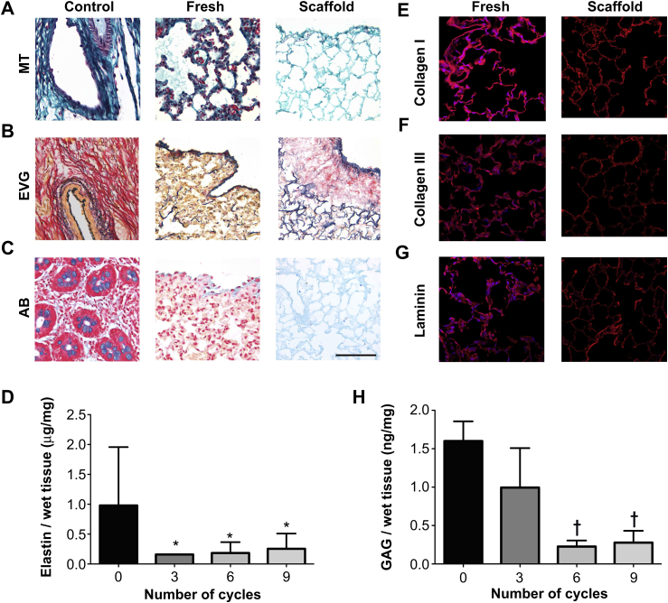
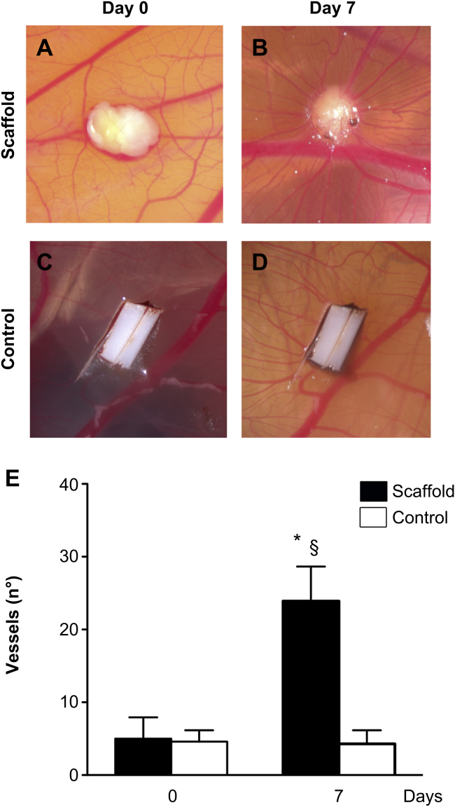
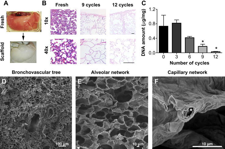
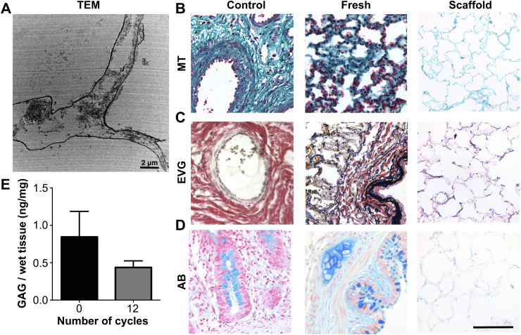
Tissue engineering of autologous lung tissue aims to become a therapeutic alternative to transplantation. Efforts published so far in creating scaffolds have used harsh decellularization techniques that damage the extracellular matrix (ECM), deplete its components and take up to 5 weeks to perform. The aim of this study was to create a lung natural acellular scaffold using a method that will reduce the time of production and better preserve scaffold architecture and ECM components. Decellularization of rat lungs via the intratracheal route removed most of the nuclear material when compared to the other entry points. An intermittent inflation approach that mimics lung respiration yielded an acellular scaffold in a shorter time with an improved preservation of pulmonary micro-architecture. Electron microscopy demonstrated the maintenance of an intact alveolar network, with no evidence of collapse or tearing. Pulsatile dye injection via the vasculature indicated an intact capillary network in the scaffold. Morphometry analysis demonstrated a significant increase in alveolar fractional volume, with alveolar size analysis confirming that alveolar dimensions were maintained. Biomechanical testing of the scaffolds indicated an increase in resistance and elastance when compared to fresh lungs. Staining and quantification for ECM components showed a presence of collagen, elastin, GAG and laminin. The intratracheal intermittent decellularization methodology could be translated to sheep lungs, demonstrating a preservation of ECM components, alveolar and vascular architecture. Decellularization treatment and methodology preserves lung architecture and ECM whilst reducing the production time to 3 h. Cell seeding and in vivo experiments are necessary to proceed towards clinical translation.
Copyright © 2013 Elsevier Ltd. All rights reserved.
Figures
Similar articles
-
Perfusion decellularization of human and porcine lungs: bringing the matrix to clinical scale.J Heart Lung Transplant. 2014 Mar;33(3):298-308. doi: 10.1016/j.healun.2013.10.030. Epub 2013 Oct 26. J Heart Lung Transplant. 2014. PMID: 24365767
-
A nonhuman primate model of lung regeneration: detergent-mediated decellularization and initial in vitro recellularization with mesenchymal stem cells.Tissue Eng Part A. 2012 Dec;18(23-24):2437-52. doi: 10.1089/ten.TEA.2011.0594. Epub 2012 Aug 23. Tissue Eng Part A. 2012. PMID: 22764775 Free PMC article.
-
Hypertensive rat lungs retain hallmarks of vascular disease upon decellularization but support the growth of mesenchymal stem cells.Tissue Eng Part A. 2014 May;20(9-10):1426-43. doi: 10.1089/ten.TEA.2013.0438. Epub 2014 Feb 28. Tissue Eng Part A. 2014. PMID: 24378017 Free PMC article.
-
An overview of the production of tissue extracellular matrix and decellularization process.Cell Tissue Bank. 2024 Mar;25(1):369-387. doi: 10.1007/s10561-023-10112-1. Epub 2023 Oct 9. Cell Tissue Bank. 2024. PMID: 37812368 Review.
-
Methods of tissue decellularization used for preparation of biologic scaffolds and in vivo relevance.Methods. 2015 Aug;84:25-34. doi: 10.1016/j.ymeth.2015.03.005. Epub 2015 Mar 16. Methods. 2015. PMID: 25791470 Review.
Cited by
-
Ovary-derived Decellularized Extracellular Matrix-based Bioink for Fabricating 3D Primary Ovarian Cells-laden Structures for Mouse Ovarian Failure Correction.Int J Bioprint. 2022 Jul 26;8(3):597. doi: 10.18063/ijb.v8i3.597. eCollection 2022. Int J Bioprint. 2022. PMID: 36105140 Free PMC article.
-
Multi-stage bioengineering of a layered oesophagus with in vitro expanded muscle and epithelial adult progenitors.Nat Commun. 2018 Oct 16;9(1):4286. doi: 10.1038/s41467-018-06385-w. Nat Commun. 2018. PMID: 30327457 Free PMC article.
-
Lessons learned from pre-clinical testing of xenogeneic decellularized esophagi in a rabbit model.iScience. 2022 Sep 22;25(10):105174. doi: 10.1016/j.isci.2022.105174. eCollection 2022 Oct 21. iScience. 2022. PMID: 36217545 Free PMC article.
-
Angiogenesis and Re-endothelialization in decellularized scaffolds: Recent advances and current challenges in tissue engineering.Front Bioeng Biotechnol. 2023 Feb 16;11:1103727. doi: 10.3389/fbioe.2023.1103727. eCollection 2023. Front Bioeng Biotechnol. 2023. PMID: 36873356 Free PMC article. Review.
-
Cell-Seeded Biomaterial Scaffolds: The Urgent Need for Unanswered Accelerated Angiogenesis.Int J Nanomedicine. 2022 Mar 12;17:1035-1068. doi: 10.2147/IJN.S353062. eCollection 2022. Int J Nanomedicine. 2022. PMID: 35309965 Free PMC article. Review.
References
-
- Christie J.D., Edwards L.B., Kucheryavaya A.Y., Benden C., Dipchand A.I., Dobbels F. The Registry of the International Society for Heart and Lung Transplantation: 29th adult lung and heart-lung transplant report-2012. J Heart Lung Transplant. 2012 Oct;31(10):1073–1086. - PubMed
-
- Atala A., Bauer S.B., Soker S., Yoo J.J., Retik A.B. Tissue-engineered autologous bladders for patients needing cystoplasty. Lancet. 2006 Apr 15;367(9518):1241–1246. - PubMed
-
- Macchiarini P., Walles T., Biancosino C., Mertsching H. First human transplantation of a bioengineered airway tissue. J Thorac Cardiovasc Surg. 2004 Oct;128(4):638–641. - PubMed
Publication types
MeSH terms
Grants and funding
LinkOut - more resources
Full Text Sources
Other Literature Sources
