. 2014 Jul;73(7):1405-13.
doi: 10.1136/annrheumdis-2012-203146. Epub 2013 Jun 1.

Glucocorticoids induce senescence in primary human tenocytes by inhibition of sirtuin 1 and activation of the p53/p21 pathway: in vivo and in vitro evidence

Affiliations
Free PMC article

Glucocorticoids induce senescence in primary human tenocytes by inhibition of sirtuin 1 and activation of the p53/p21 pathway: in vivo and in vitro evidence

Raewyn C Poulsen et al. Ann Rheum Dis. 2014 Jul.
Free PMC article

Abstract

Cellular senescence is an irreversible side effect of some pharmaceuticals which can contribute to tissue degeneration.

Objective: To determine whether pharmaceutical glucocorticoids induce senescence in tenocytes.

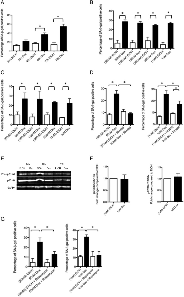
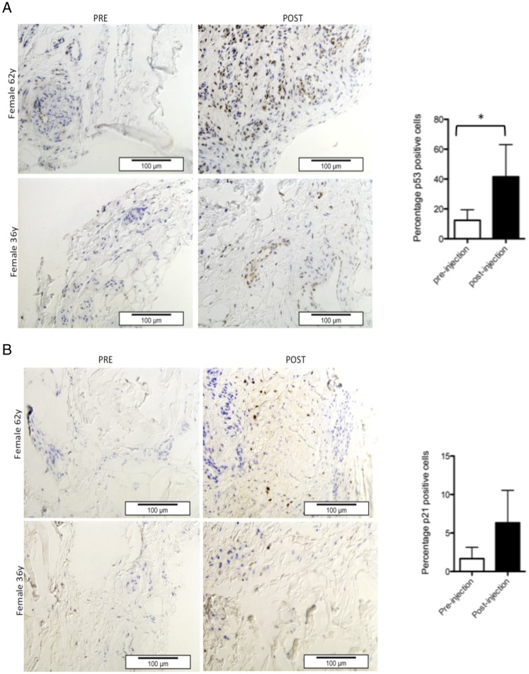
Methods: Features of senescence (β-galactosidase activity at pH 6 (SA-β-gal) and active mammalian/mechanistic target of rapamycin (mTOR) in cell cycle arrest) as well as the activity of the two main pathways leading to cell senescence were examined in glucocorticoid-treated primary human tenocytes. Evidence of senescence-inducing pathway induction in vivo was obtained using immunohistochemistry on tendon biopsy specimens taken before and 7 weeks after subacromial Depo-Medrone injection.

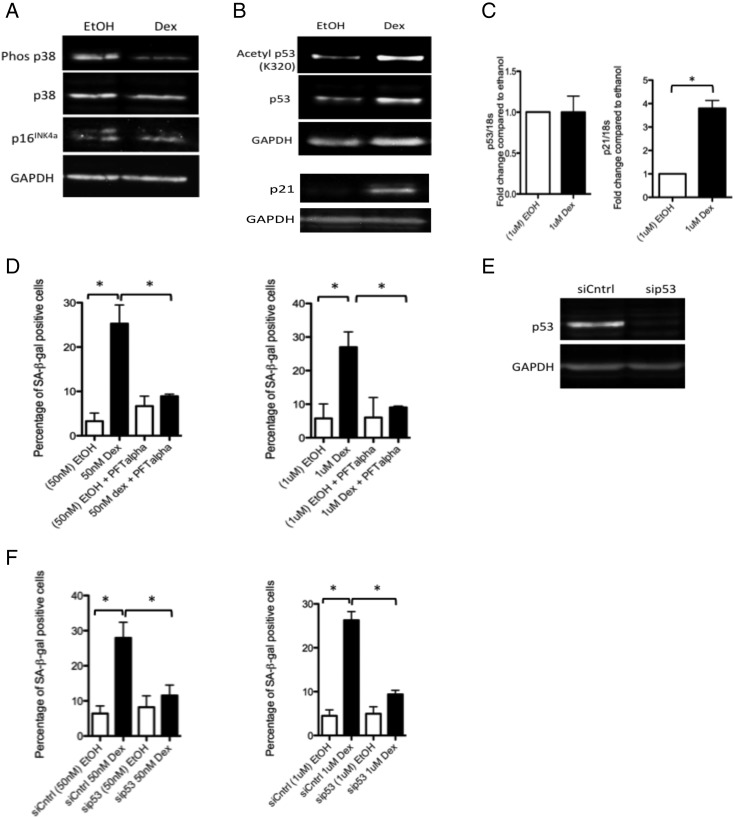
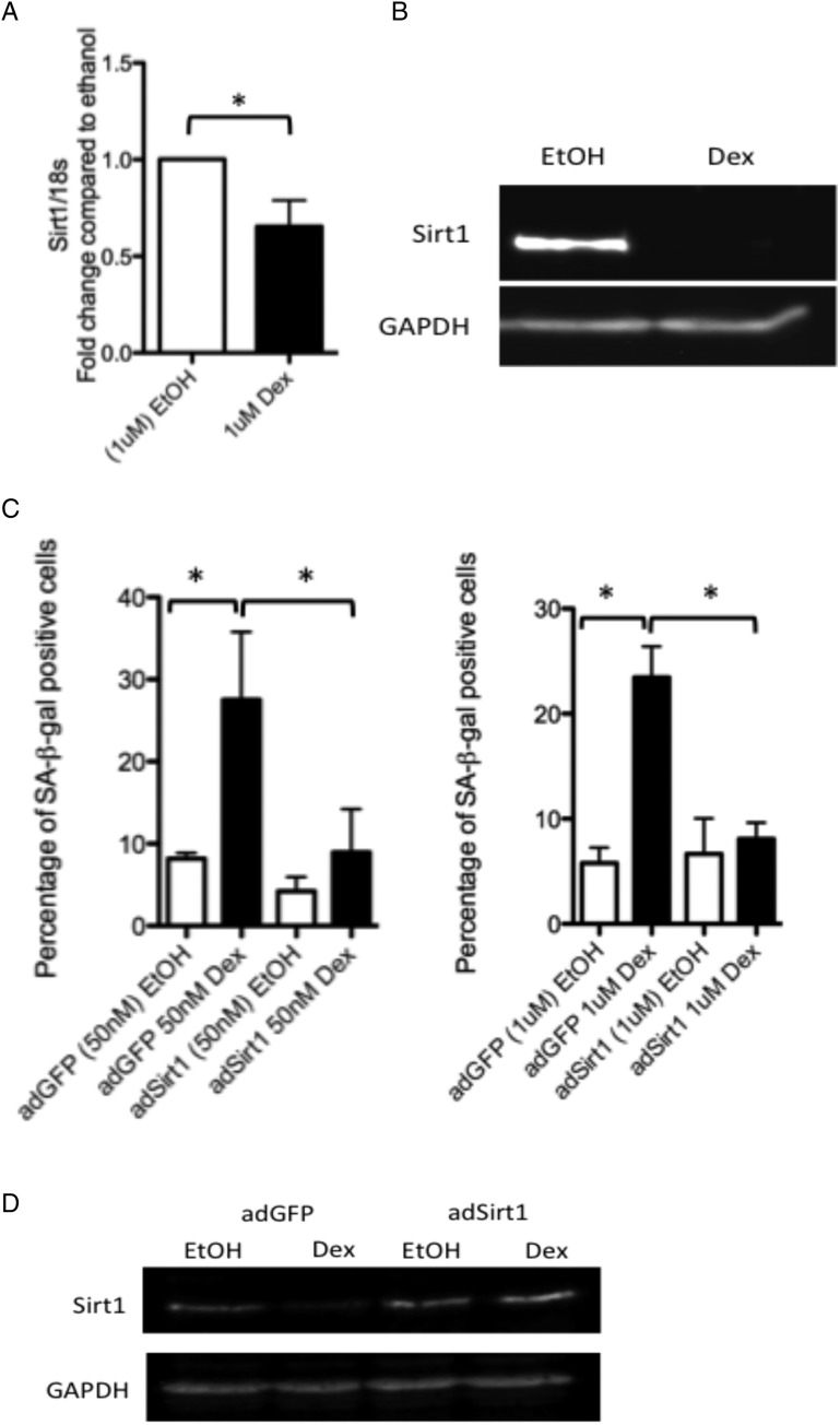
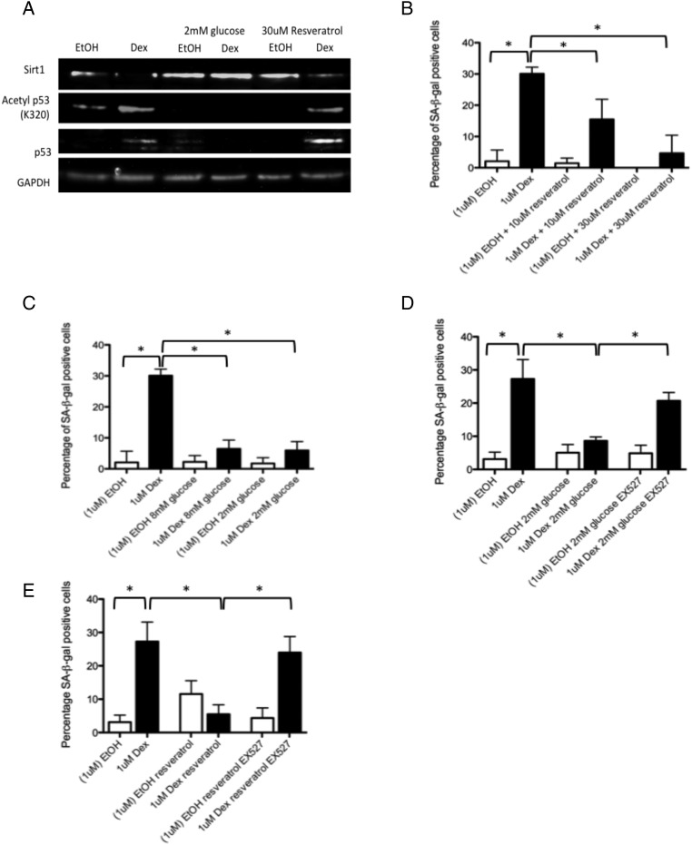
Results: Dexamethasone treatment of tenocytes resulted in an increased percentage of SA-βgal-positive cells. Levels of phosphorylated p70S6K did not decrease with glucocorticoid treatment indicating mTOR remained active. Increased levels of acetylated p53 as well as increased RNA levels of its pro-senescence effector p21 were evident in dexamethasone-treated tenocytes. Levels of the p53 deacetylase sirtuin 1 were lower in dexamethasone-treated cells compared with controls. Knockdown of p53 or inhibition of p53 activity prevented dexamethasone-induced senescence. Activation of sirtuin 1 either by exogenous overexpression or by treatment with resveratrol or low glucose prevented dexamethasone-induced senescence. Immunohistochemical analysis of tendon biopsies taken before and after glucocorticoid injection revealed a significant increase in the percentage of p53-positive cells (p=0.03). The percentage of p21-positive cells also tended to be higher post-injection (p=0.06) suggesting glucocorticoids activate the p53/p21 senescence-inducing pathway in vivo as well as in vitro.

Conclusion: As cell senescence is irreversible in vivo, glucocorticoid-induced senescence may result in long-term degenerative changes in tendon tissue.

Keywords: Chondrocytes; Corticosteroids; Fibroblasts; Inflammation; Tendinitis.


Figures

Figure 1
Figure 1
Glucocorticoids cause morphological and functional changes in tenocytes consistent with a senescent phenotype. (A) The percentage of SA-βgal-positive cells was significantly higher 48 and 72 h post-treatment of primary human tenocytes with 1 μM dexamethasone compared with tenocytes treated with ethanol (carrier) alone. There was no evidence of a dose effect of dexamethasone treatment on the percentage of SA-βgal-positive cells following (B) 3 days or (C) 7 days of treatment (n=3) compared with cells treated with the corresponding amount of ethanol carrier. (D) The percentage of SA-βgal-positive cells was significantly lower in cells treated for 72 h with dexamethasone (50 nM or 1 μM) and the glucocorticoid receptor (GR) antagonist RU486 (80 nM) compared with cells treated with dexamethasone alone, n=3. (E) Western blot showing levels of phosphorylated and total p70 S6 kinase, a substrate of the mTORC1 complex and an indicator of mTORC1 activity 24, 48 and 72 h post-dexamethasone (dex) treatment (1 μM) in primary human tenocytes. Western blots were conducted on protein lysates from cells obtained from three different tissue donors. Blot shown is representative of all experiments. (F) No difference in RNA levels of either of the two p70 S6 kinase subunits (p70S6KB1 and p70S6KB2) was evident in tenocytes treated with dexamethasone (1 μM) compared with ethanol-treated controls. (G) A significantly lower percentage of SA-βgal-positive cells was evident in cells treated for 72 h with dexamethasone (50 nM or 1 μM) and the mammalian/mechanistic target of rapamycin (mTOR) inhibitor rapamycin (rapa) compared with cells treated with dexamethasone alone, n=3. Statistically significant differences between ethanol (EtOH) and dexamethasone (Dex) and between dexamethasone with and without either the GR inhibitor (RU486) or mTOR inhibitor (rapamycin) are indicated by * (p<0.05).
Figure 2
Figure 2
Glucocorticoids activate p53 signalling in tenocytes. Western blots showing levels of (A) phosphorylated and total p38 MAPK and p16INK4a and (B) acetylated (K320) and total p53 and p21cip/waf1 in lysates of primary human tenocytes following treatment with 1 μM dexamethasone or carrier (ethanol) for 48 h. Western blots were conducted on protein lysates from cells obtained from three different tissue donors. Blots shown are representative of all experiments. (C) No significant differences in RNA levels of p53 were apparent in tenocytes treated with 1 μM dexamethasone compared with ethanol-treated controls. However, RNA levels of p21 were significantly greater in tenocytes treated with dexamethasone compared with ethanol-treated controls. (D) The percentage of SA-βgal-positive cells was significantly lower in cells treated with dexamethasone (50 nM or 1 μM) and the p53 inhibitor pifithrin-α (PFT-α, 100 nM) compared with cells treated with dexamethasone alone, n=3. (E) Western blot demonstrating the effectiveness of p53 knockdown obtained using RNAi in primary human tenocytes. (F) The percentage of SA-βgal-positive cells was significantly lower in tenocytes in which p53 expression was knocked down (sip53) compared with dexamethasone-treated controls (siCntrl) (n=3). Statistically significant differences between ethanol (EtOH) and dexamethasone (Dex) and between dexamethasone with and without the p53 inhibitor (PFT-α) or with and without p53 siRNA (sip53) are indicated by * (p<0.05).
Figure 3
Figure 3
Glucocorticoids inhibit sirtuin 1 expression in tenocytes. Both (A) RNA (as measured by real-time qRT-PCR 7 h post-treatment, n=3) and (B) protein (as determined by western blotting) levels of sirtuin 1 were reduced in dexamethasone-treated cells compared with carrier (ethanol)-treated controls. Western blots were conducted on protein lysates from cells obtained from three different tissue donors. Blot shown is representative of all experiments. (C) The percentage of SA-βgal-positive cells was significantly lower in dexamethasone-treated tenocytes infected with an adenoviral vector containing a sirtuin 1 overexpression construct (adSirt1) compared with dexamethasone-treated tenocytes infected with a control adenoviral vector expressing GFP (adGFP). (D) Western blot demonstrating the effectiveness of the adSirt1 virus to prevent dexamethasone-induced inhibition of sirtuin 1 expression. Statistically significant differences between ethanol (EtOH) and dexamethasone (Dex) and between dexamethasone with and without sirt1 overexpression (adSirt1) are indicated by * (p<0.05).
Figure 4
Figure 4
Protective effect of low glucose, low serum and resveratrol on glucocorticoid-induced senescence (A) Protein levels of sirtuin 1 were higher in tenocytes treated with dexamethasone (1 μM) and cultured in low glucose (2 mM) media or co-treated with resveratrol (30 μM) compared with tenocytes treated with dexamethasone and cultured in standard growth media (17.5 mM glucose). Levels of acetylated p53 (lys320) were lower in dexamethasone-treated cells cultured in low glucose (2 mM) media or co-treated with resveratrol compared with tenocytes treated with dexamethasone and cultured in standard growth media (17.5 mM glucose). Western blots were conducted on protein lysates from cells obtained from three different tissue donors. Blots shown are representative of all three experiments. The percentage of SA-βgal-positive cells was significantly lower in dexamethasone-treated cells (B) co-treated with resveratrol (10 or 30 μM) or (C) cultured in low glucose (2 or 8 mM) media; however, no significant difference in the percentage of SA-βgal-positive cells was observed following addition of EX527, a sirtuin 1 inhibitor, in dexamethasone-treated cells (1 μM) cultured in (D) 2 mM glucose or (E) co-treated with resveratrol (30 μM) compared with cells treated with dexamethasone alone and cultured in standard (17.5 mM) growth media (n=3). Statistically significant differences between ethanol (EtOH) and dexamethasone (Dex) and between dexamethasone with and without either resveratrol or 2 mM glucose are indicated by * (p<0.05).
Figure 5
Figure 5
The p53/p21 pathway is activated in vivo following local injection with glucocorticoids. Tendon biopsy samples were taken from five patients with rotator cuff tendinopathy immediately prior to subacromial injection with the synthetic glucocorticoid, Depo-Medrone, and compared with subsequent biopsy specimens taken from the same patients 7 weeks following glucocorticoid injection. Using immunohistochemistry, (A) the percentage of p53-positive cells was significantly greater in tissue samples post-injection compared with pre-injection. (B) A non-statistically significant increase in the percentage of p21-positive cells was observed in the post-injection tissue samples compared with pre-injection tissue samples. Representative photomicrographs are shown for tissue samples from two patients. p53-positive cells or p21-positive cells were detected using horseradish peroxidase-conjugated secondary antibodies and diaminobenzadine. Positive cells appear brown. Tissue was counterstained with haematoxylin (blue) to allow quantification of total cell numbers. Statistically significant differences are indicated by * (p<0.05).

References

    1. NHS Business Services Authority. Electronic Prescribing & Financial Information for Practices (ePFIP). http://wwwnhsbsanhsuk/PrescriptionServices/963aspx, 2009
    1. Garbe E, LeLorier J, Boivin JF, et al. Inhaled and nasal glucocorticoids and the risks of ocular hypertension or open-angle glaucoma. JAMA 1997;277:722–7 - PubMed
    1. Oikarinen A, Autio P. New aspects of the mechanism of corticosteroid-induced dermal atrophy. Clin Exper Dermatol 1991;16:416–19 - PubMed
    1. Butler RC, Davie MWJ, Worsfold M, et al. Bone-mineral content in patients with rheumatoid-arthritis—relationship to low-dose steroid-therapy. Br J Rheumatol 1991;30:86–90 - PubMed
    1. Ford LT, Debender J. Tendon-rupture after local steroid injection. South Med J 1979;72:827–30 - PubMed

Publication types

MeSH terms