. 2013 Oct 15;22(20):4074-83.
doi: 10.1093/hmg/ddt257. Epub 2013 May 31.

The DcpS inhibitor RG3039 improves motor function in SMA mice

Affiliations

The DcpS inhibitor RG3039 improves motor function in SMA mice

James P Van Meerbeke et al. Hum Mol Genet. .

Abstract

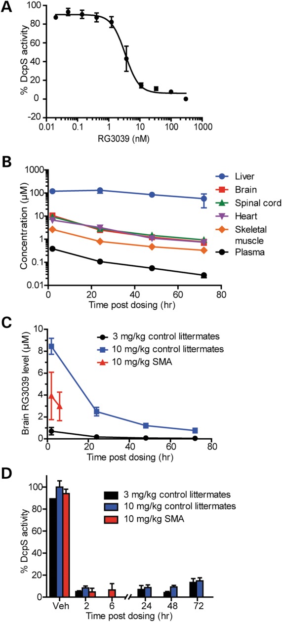
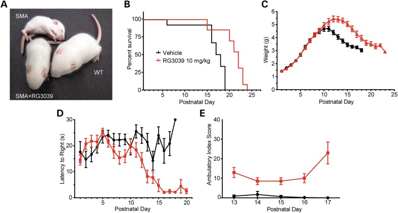
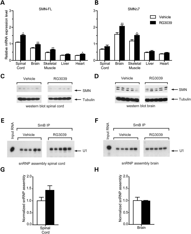
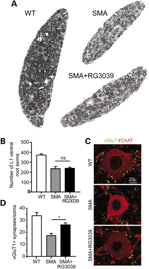
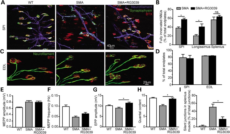
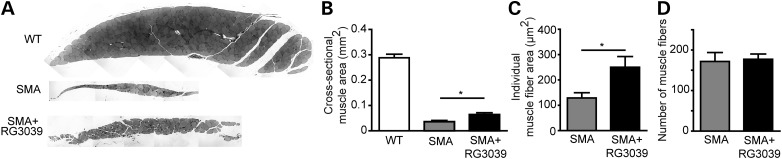
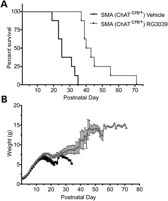
Spinal muscular atrophy (SMA) is caused by mutations of the survival motor neuron 1 (SMN1) gene, retention of the survival motor neuron 2 (SMN2) gene and insufficient expression of full-length survival motor neuron (SMN) protein. Quinazolines increase SMN2 promoter activity and inhibit the ribonucleic acid scavenger enzyme DcpS. The quinazoline derivative RG3039 has advanced to early phase clinical trials. In preparation for efficacy studies in SMA patients, we investigated the effects of RG3039 in severe SMA mice. Here, we show that RG3039 distributed to central nervous system tissues where it robustly inhibited DcpS enzyme activity, but minimally activated SMN expression or the assembly of small nuclear ribonucleoproteins. Nonetheless, treated SMA mice showed a dose-dependent increase in survival, weight and motor function. This was associated with improved motor neuron somal and neuromuscular junction synaptic innervation and function and increased muscle size. RG3039 also enhanced survival of conditional SMA mice in which SMN had been genetically restored to motor neurons. As this systemically delivered drug may have therapeutic benefits that extend beyond motor neurons, it could act additively with SMN-restoring therapies delivered directly to the central nervous system such as antisense oligonucleotides or gene therapy.


Figures

Figure 1.
Figure 1.
RG3039 crosses the blood brain barrier and inhibits the RNA decapping enzyme DcpS in the CNS. (A) DcpS enzyme activity in brain protein extracts treated in vitro with increasing doses of RG3039 (n = 3 per data point). (B) RG3039 drug levels in various tissues harvested from normal littermate mice treated from P1 to P10 with 10 mg/kg of RG3039 and harvested at various times after the last dose (liver, brain, plasma: n = 4 or 5 mice per time point and heart, spinal cord and muscle: n = 4–5 mice pooled). (C) RG3039 drug levels in brain and plasma of control littermate and SMA mice dosed with multiple doses of either 3 or 10 mg/kg of RG3039 (CNTL 3 mg/kg: n = 4 per time point, CNTL 10 mg/kg: n = 4–5 per time point, SMA 10 mg/kg: n = 13–14 per time point). (D) DcpS enzyme activity in the brains of control littermate and SMA mice dosed with 3 or 10 mg/kg of RG3039 (n = at least 4–5 per data point, except vehicle controls n = 1–2).
Figure 2.
Figure 2.
RG3039 improves survival and motor function of SMA mice. (A) Photo of vehicle-treated and 10 mg/kg RG3039-treated SMA mice and an untreated normal littermate at P13. (B) Kaplan–Meier survival curves, (C) weights, (D) righting time latencies, and (E) ambulatory index composite scores of vehicle-treated and 10 mg/kg RG3039-treated SMA mice (vehicle: n = 12, RG3039 10 mg/kg: n = 13, except ambulatory index n = 5–8 per group).
Figure 3.
Figure 3.
RG3039 minimally effects SMN levels in vivo. (A) Full-length (FL) and (B) SMNΔ7 SMN transcript levels in tissues from vehicle and RG3039-treated SMA mice at P10 6 h after the last dose, *P < 0.05, **P < 0.01 (brain: n = 13–15 per group, spinal cord: n = 5 per group, liver, heart, muscle: n = 10 per group). (C and D) Western blot analysis of SMN protein levels in (C) spinal cord and (D) brain extracts from vehicle and RG3039-treated SMA mice (n = 5 per group). (E and F) In vitro snRNP assembly analysis with radioactive U1 snRNA in (E) spinal cord or (F) brain extracts. U1 snRNA was immunoprecipitated with anti-SmB antibodies from each snRNP assembly reaction and analyzed by denaturing gel electrophoresis and autoradiography. (G and H) Quantification of U1 snRNA levels immunoprecipitated from snRNP assembly reactions with (G) spinal cord and (H) brain extracts from the experiments in (E) and (F) (n = 5 per group).
Figure 4.
Figure 4.
RG3039 does not change ventral root axon number, but does improve motor neuron somal synaptic input. (A) Image of L1 ventral roots from control littermate, vehicle-treated SMA and RG3039-treated SMA mice. (B) Quantification of ventral root number. (C) Representative images of motor neurons (red = ChAT) and somal VGLUT1 synapses (green = VGLUT1) from control littermate, vehicle-treated SMA and RG3039-treated SMA mice. (D) Quantification of VGLUT1 motor neuron somal synapses (*P < 0.05, n = 3 per group).
Figure 5.
Figure 5.
RG3039 prevents NMJ denervation and improves NMJ synaptic transmission. (A) Image of NMJs in the SPI muscle of WT, vehicle-treated SMA and RG3039-treated SMA mice. Blue (neurofilament), green (synaptophysin) and red (α-bungarotoxin). (B) Quantification of percentage of fully innervated NMJs in the longissimus, SPI and splenius muscles, *P < 0.05 (n = 7 per group). (C) Image of NMJs demonstrating neurofilament accumulations in presynaptic terminals. Green (neurofilament) and red (α-bungarotoxin). (D) Quantification of NF accumulation in SPI and EDL muscle (n = 4 per group). (E) MEPP amplitudes, (F) MEPP frequencies, (G) EPP amplitudes, and (H) quantal content in the EDL muscle of WT, SMA vehicle-treated and RG3039-treated SMA mice, *P < 0.05 (n = 6 per group). (I) The percentage of silent NMJs in the splenius muscle in WT, SMA vehicle-treated and RG3039-treated SMA mice, *P < 0.05 (n = 3–5 per group).
Figure 6.
Figure 6.
RG3039 increases myofiber size. (A) Image of cross-sections of the longissimus muscle from SMA vehicle-treated, SMA RG3039-treated and WT littermate mice. Quantification of (B) muscle cross sectional area, (C) myofiber size, and (D) myofiber number, *P < 0.05 (n = 6–7 per group).
Figure 7.
Figure 7.
RG3039 further extends survival of SMA mice expressing increased SMN levels in motor neurons. (A) Kaplan–Meier survival and (B) weight curves of ChATCre+ SmnRes SMA mice treated with vehicle or RG3039 (n = 8 per group).

References

    1. Lefebvre S., Burglen L., Reboullet S., Clermont O., Burlet P., Viollet L., Benichou B., Cruaud C., Millasseau P., Zeviani M., et al. Identification and characterization of a spinal muscular atrophy-determining gene. Cell. 1995;80:155–165. - PubMed
    1. Lorson C.L., Strasswimmer J., Yao J.M., Baleja J.D., Hahnen E., Wirth B., Le T., Burghes A.H., Androphy E.J. SMN oligomerization defect correlates with spinal muscular atrophy severity. Nat. Genet. 1998;19:63–66. - PubMed
    1. Monani U.R., Lorson C.L., Parsons D.W., Prior T.W., Androphy E.J., Burghes A.H., McPherson J.D. A single nucleotide difference that alters splicing patterns distinguishes the SMA gene SMN1 from the copy gene SMN2. Hum. Mol. Genet. 1999;8:1177–1183. - PubMed
    1. Feldkotter M., Schwarzer V., Wirth R., Wienker T.F., Wirth B. Quantitative analyses of SMN1 and SMN2 based on real-time lightCycler PCR: fast and highly reliable carrier testing and prediction of severity of spinal muscular atrophy. Am. J. Hum. Genet. 2002;70:358–368. - PMC - PubMed
    1. Prior T.W., Swoboda K.J., Scott H.D., Hejmanowski A.Q. Homozygous SMN1 deletions in unaffected family members and modification of the phenotype by SMN2. Am. J. Med. Genet. A. 2004;130:307–310. - PMC - PubMed

Publication types

MeSH terms

Substances