. 2013 Jun;123(6):2434-46.
doi: 10.1172/JCI68280.

C3 glomerulopathy-associated CFHR1 mutation alters FHR oligomerization and complement regulation

Affiliations

C3 glomerulopathy-associated CFHR1 mutation alters FHR oligomerization and complement regulation

Agustín Tortajada et al. J Clin Invest. 2013 Jun.

Abstract

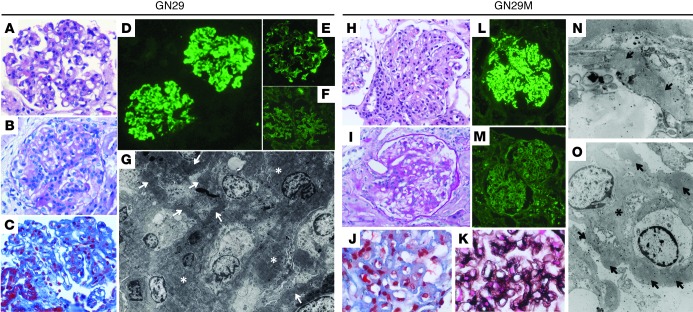
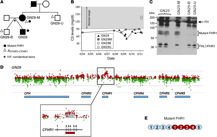
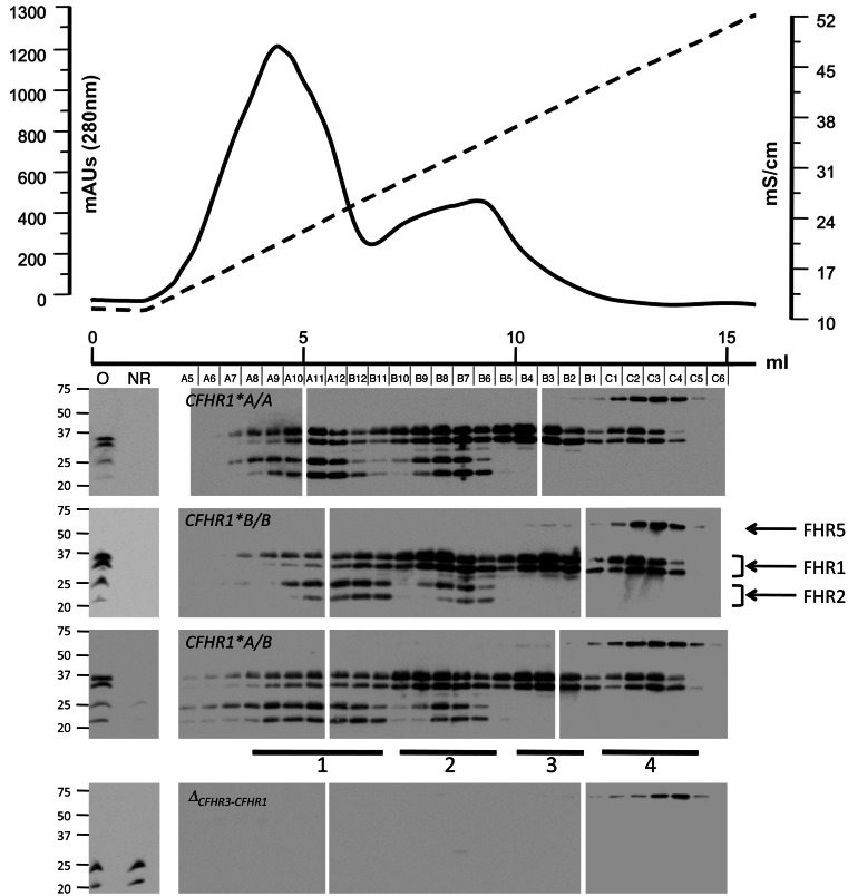
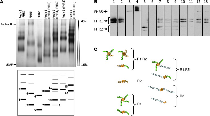
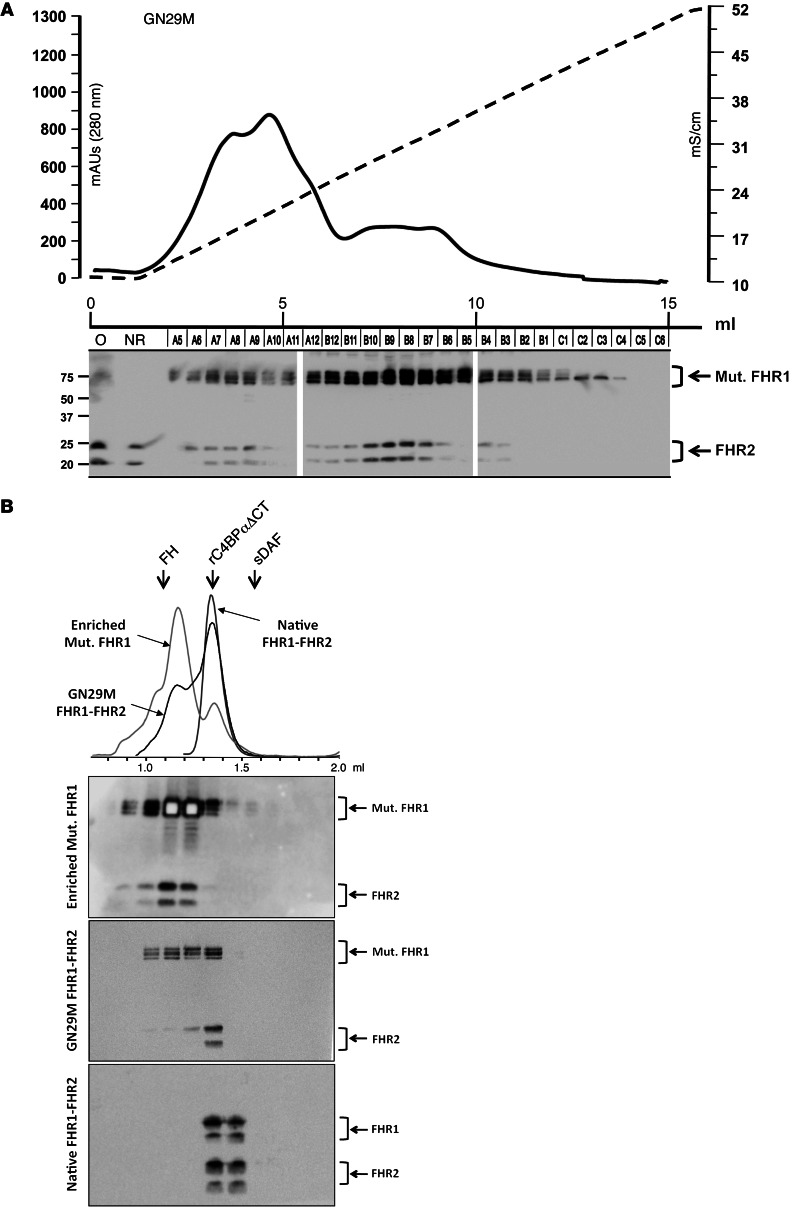
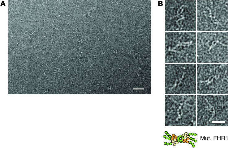
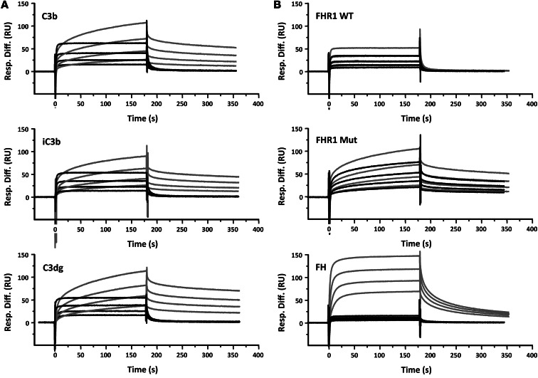
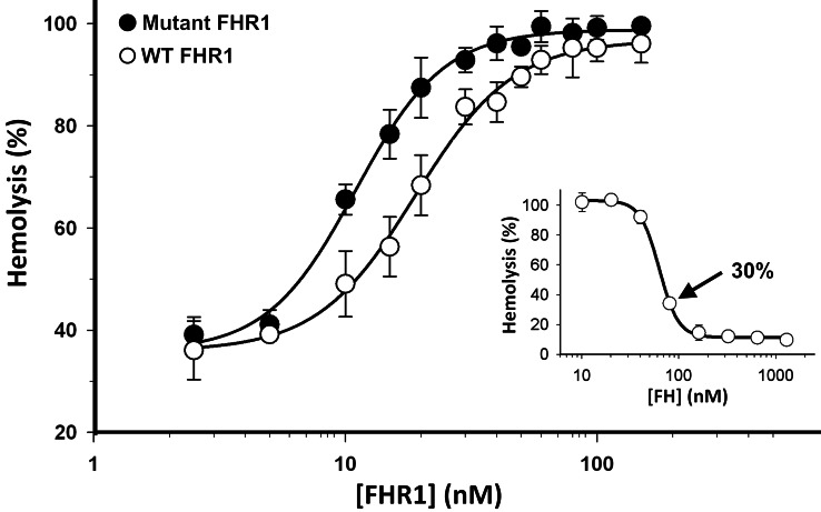
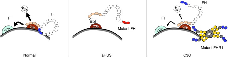
C3 glomerulopathies (C3G) are a group of severe renal diseases with distinct patterns of glomerular inflammation and C3 deposition caused by complement dysregulation. Here we report the identification of a familial C3G-associated genomic mutation in the gene complement factor H–related 1 (CFHR1), which encodes FHR1. The mutation resulted in the duplication of the N-terminal short consensus repeats (SCRs) that are conserved in FHR2 and FHR5. We determined that native FHR1, FHR2, and FHR5 circulate in plasma as homo- and hetero-oligomeric complexes, the formation of which is likely mediated by the conserved N-terminal domain. In mutant FHR1, duplication of the N-terminal domain resulted in the formation of unusually large multimeric FHR complexes that exhibited increased avidity for the FHR1 ligands C3b, iC3b, and C3dg and enhanced competition with complement factor H (FH) in surface plasmon resonance (SPR) studies and hemolytic assays. These data revealed that FHR1, FHR2, and FHR5 organize a combinatorial repertoire of oligomeric complexes and demonstrated that changes in FHR oligomerization influence the regulation of complement activation. In summary, our identification and characterization of a unique CFHR1 mutation provides insights into the biology of the FHRs and contributes to our understanding of the pathogenic mechanisms underlying C3G.


Figures

Figure 1
Figure 1. Histology, immunofluorescence, and EM findings.
Kidney biopsies from probate GN29 (AG) and his mother, GN29M (HO), showed remarkably similar light, immunofluorescence, and ultrastructural findings. The characteristic histological lesion consisted of mesangial hypercellularity with thickened, eosinophil-rich segments of GBM (A and H). The affected glomerular segments were PAS positive and reacted to trichrome and Jones methenamine-silver stain (B, C, and IK). The main immunofluorescence findings were prominent and diffuse C3 deposits, which were granular in some glomerular areas (D and L). IgG was absent from these deposits (F and M), although local deposits of IgM were observed in GN29 (E). Both biopsies showed similar ultrastructural alterations (G, N, and O) consisting of the presence of ribbon-like, osmiophilic deposits in the GBM (arrows); these electron-dense deposits were also evident in the mesangial matrix (asterisks). Original magnification, ×400 (AC, H, and I); ×200 (DF); ×1,600 (G); ×600 (J and K); ×100 (L and M); ×2,950 (N); ×8,900 (O).
Figure 2
Figure 2. Internal duplication of CFHR1 is associated with C3G in pedigree GN29.
(A) The GN29 pedigree. Affected individuals are indicated with solid symbols; carriers of the mutant FHR1 and of ΔCFHR3-CFHR1 are indicated with solid circles and triangles, respectively. (B) Levels of C3 in affected and healthy individuals in the GN29 pedigree. (C) Western blot of whole human plasma identifying FH, FHL1, and FHR1 with an in-house rabbit polyclonal anti-FH. An anomalous band of approximately 70 kDa was identified in the proband and his mother. Proteomic analysis of material from this band demonstrated that this band was the product of a mutant CFHR1 gene (Supplemental Figure 2). (D) High-resolution CGH array of the CFH-CFHR1 locus, illustrating an internal duplication of CFHR1 in the genomic DNA from GN29, encompassing exons 2–5. (E) Putative structure of a 9-SCR protein encoded by the mutant CFHR1. Red denotes duplicated SCRs.
Figure 3
Figure 3. Heparin chromatography of FHR1, FHR2, and FHR5 proteins.
Elution profiles of FHR1, FHR2, and FHR5 from individuals of the 3 CFHR1*A/B genotypes illustrated that FHR1 eluted in 4 distinct peaks at different NaCl concentrations. Top: Representative case of how plasma proteins retained in the heparin column were eluted with the NaCl gradient. Bottom: Western blots used the MBC125 mAb to determine the elution position of the different FHRs. Peaks 1–4 are indicated. FHR1 coeluted with FHR2 in the first 2 peaks: in peak 1, FHR1 and FHR2 showed equimolar quantities (FHR1-FHR2); in peak 2, FHR1 was approximately 3 times more abundant than FHR2 (FHR13-FHR2). In peak 3, FHR1 eluted alone, and in peak 4, FHR1 and FHR5 coeluted at approximately equimolar quantities (FHR1-FHR5). FHRs were identified simultaneously by Western blot using MBC125 mAb, which recognizes an epitope common to the 3 FHRs. Protein bands corresponding to FHR1, FHR2, and FHR5 are indicated at right. Also shown is Western blot of the elution profile corresponding to an individual homozygote for the ΔCFHR3-CFHR1 allele, illustrating that FHR2 was not retained in the column in the absence of FHR1. Samples from the elution profiles were run in 3 separate gels. O, original; NR, nonretained.
Figure 4
Figure 4. FHR1, FHR2, and FHR5 assemble into homo- and hetero-oligomers.
(A) Purified FHRs corresponding to the 4 FHR1-containing peaks from heparin chromatography, and FHR2 and FHR5 proteins purified from a FHR1-deficient individual, were analyzed in 4%–16% polyacrylamide native gels. sDAF (4 SCRs) and FH (20 SCRs) were used as molecular weight markers. Gels were silver stained. Protein complexes were obtained for each sample (bottom). (B) FHR composition of the numbered protein complexes in A was analyzed by SDS-PAGE. Comparison of the relative mobility of the bands obtained for each sample, together with the Western blot analysis, demonstrated the presence of different homo- and hetero-oligomeric complexes. For example, lanes 1, 4, and 5 were interpreted as FHR1, FHR5, and FHR2 homodimers, respectively. A faint and diffuse band (lane 3) running below the band of the FHR1 dimer could correspond to a FHR5 monomer. Lane 6 did not contain FHR2 (likely a protein contamination). Therefore, there was only 1 protein complex in plasma containing FHR2 (likely a dimer). Lane 7 was a FHR1-FHR2 heterodimer: it contained both FHR1 and FHR2 and presented mobility intermediate between the FHR2 and FHR1 homodimers. Lanes 12 and 13 corresponded to different FHR1-FHR5 hetero-oligomers. Lanes were run on the same gel but were noncontiguous (black lines). (C) Putative structure of these complexes, based on structural data demonstrating that the first 2 N-terminal SCRs (orange) of these proteins formed dimers in a head-to-tail orientation (13).
Figure 5
Figure 5. Mutant FHR1 protein shows an abnormal heparin chromatography elution profile and assembles into high–molecular weight multimers.
(A) Elution profiles of the FHR1, FHR2, and FHR5 proteins from GN29M illustrated that the mutant FHR1 coeluted with FHR2 in a major peak extending through most of the NaCl gradient and a minor peak at low NaCl concentration. Heparin fractions were characterized by Western blot using MBC125. Samples from the elution profiles were run in 3 separate gels. (B) EDTA plasma from GN29M was passed through the MBC125 affinity column. The retained FHRs were eluted in 100 mM glycine, pH 2.5; dialyzed against PBS; and analyzed by size-exclusion Superdex 200 PC 3.2/30 chromatography in 20 mM Tris-HCl (pH 7.5) and 350 mM NaCl (GN29M FHR1-FHR2). Purified proteins corresponding to the FHRs heparin chromatography elution peak 1 from a normal individual (Native FHR1-FHR2; corresponding to FHR1-FHR2 dimers) is also shown for size comparison. Also included is a preparation enriched of the high–molecular size species of the mutant FHR1 protein (Enriched Mut. FHR1). Proteins in the gel filtration fractions were characterized by Western blot and are shown below.
Figure 6
Figure 6. EM analyses of the mutant FHR1 high–molecular weight forms.
(A) Typical field for a diluted sample of a negative-staining EM analysis of the purified mutant FHR1 enriched in high–molecular weight oligomers. Isolated complexes were detected as filaments of white density on the background of the micrograph. Scale bar: 50 nm. (B) Gallery of selected images for single complexes, illustrating that each was composed of at least 2 elongated and flexible chains, presumably mutant FHR1 monomers. The length of individual chains was measured (about 30 nm) and found to be in agreement with the estimated length of an elongated mutant FHR1 molecule composed of 9 SCR domains in tandem (approximately 3 nm per SCR domain). Scale bar: 25 nm.
Figure 7
Figure 7. Mutant FHR1 shows increased binding to surface-bound C3b, iC3b, and C3dg.
(A) SPR analysis of native (black lines) and mutant (gray lines) FHR1 binding to C3b, iC3b, and C3dg. C3b (150 RU) was amine-coupled to a CM5 Biacore chip and used as a nidus for convertase formation. Further C3b (1,240 RU) was deposited on the chip surface by flowing FB and FD to form C3bBb, followed by C3 as convertase substrate. The surface C3b was converted to iC3b by incubating with FH and FI until the surface no longer supported convertase formation. Conversion to C3dg was achieved by incubating the iC3b surface with soluble CR1 and FI until no further C3c was released. 1:2 serial dilutions from 28 μg/ml native and mutant FHR1 were flowed across the surface for 3 minutes, then allowed to dissociate for 3 minutes prior to regenerating the surface. Avidity effects affecting dissociation of mutant FHR1 are clear. (B) Comparative binding of native and mutant FHR1 and FH to C3b and iC3b. Serial dilutions of native FHR1 (WT; 28 μg/ml), mutant FHR1 (Mut; 28 μg/ml), and FH (bottom; 26 μg/ml) were flowed across the C3b surface (gray lines) as described in A. The surface C3b was converted to iC3b by incubating with FH and FI until the surface no longer supported convertase formation. Native and mutant FHR1 as well as FH were flowed across iC3b (black lines) at identical concentrations as in A. Binding of native or mutant FHR1 was little affected by conversion of C3b to iC3b, whereas binding of FH was almost eliminated.
Figure 8
Figure 8. Mutant FHR1 shows enhanced competition with FH in a FH-dependent hemolytic assay.
Inset shows the preliminary experiment to determine the amount of FH to give 30% lysis in the guinea pig hemolytic assay. Main plot shows the competition assay between native FHR1 (open circles) or mutant FHR1 (filled circles) and FH amount. Percent hemolysis was determined as mean ± SD of 3 independent experiments. The EC50 values — 18.97 nM for native FHR1 and 10.97 nM for mutant FHR1 — were significantly different (P = 0,001, 2-tailed t test).
Figure 9
Figure 9. Proposed model for a novel pathogenic mechanism in C3G.
FH is an elongated glycoprotein of 155 kDa composed of 20 SCRs (small circles). FH presents C3b binding sites at each end of the molecule. The N-terminal C3b binding site mediates the accelerated decay of the AP C3 convertase (C3bBb) and the cofactor activity for the FI-dependent proteolytic inactivation of C3b. The C-terminal region binds both C3b and polyanions normally present in the cell surfaces (e.g., sialic acid, heparan sulfates, and glycosaminoglycans). This region is essential for the complement regulatory activity of FH on surfaces and to discriminate between self and pathogens, which normally lack these polyanions on their surfaces. Extensive experimental data generated during the last 10 years has provided conclusive evidence that mutations disrupting the functional activity of the C-terminal region, like those associated with aHUS, decrease the avidity of FH for cell surfaces and impair complement regulation (–36). The data reported here suggest similarities between the established model for the aHUS-associated FH mutations and the FHR1 mutant described here. We therefore propose that multimerization of the FHRs as a consequence of duplication of the oligomerization domain in mutant FHR1, FHR2, or FHR5 proteins increases binding to surface-bound C3b, iC3b, C3dg, and carbohydrates, resulting in enhanced competition with FH that decreases its complement regulatory capacity and causes different degrees of cell surface complement dysregulation.

Comment in

References

    1. Jozsi M, Zipfel PF. Factor H family proteins and human diseases. Trends Immunol. 2008;29(8):380–387. doi: 10.1016/j.it.2008.04.008. - DOI - PubMed
    1. Perez-Caballero D, et al. Clustering of missense mutations in the C-terminal region of factor H in atypical hemolytic uremic syndrome. Am J Hum Genet. 2001;68(2):478–484. doi: 10.1086/318201. - DOI - PMC - PubMed
    1. Fritsche LG, et al. An imbalance of human complement regulatory proteins CFHR1, CFHR3 and factor H influences risk for age-related macular degeneration (AMD). Hum Mol Genet. 2010;19(23):4694–4704. doi: 10.1093/hmg/ddq399. - DOI - PubMed
    1. Heinen S, et al. Factor H-related protein 1 (CFHR-1) inhibits complement C5 convertase activity and terminal complex formation. Blood. 2009;114(12):2439–2447. doi: 10.1182/blood-2009-02-205641. - DOI - PubMed
    1. Hebecker M, Jozsi M. Factor H-related protein 4 activates complement by serving as a platform for the assembly of alternative pathway C3 convertase via its interaction with C3b protein. J Biol Chem. 2012;287(23):19528–19536. doi: 10.1074/jbc.M112.364471. - DOI - PMC - PubMed

Publication types

MeSH terms