. 2013 Jul;20(7):836-42.
doi: 10.1038/nsmb.2585. Epub 2013 Jun 2.

Nucleosome dynamics regulates DNA processing

Affiliations

Nucleosome dynamics regulates DNA processing

Nicholas L Adkins et al. Nat Struct Mol Biol. 2013 Jul.

Abstract

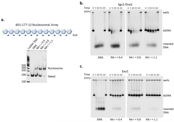
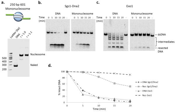
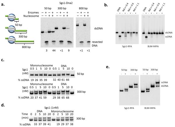
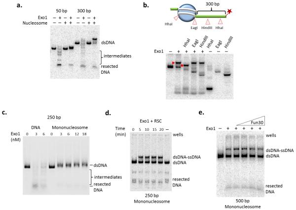
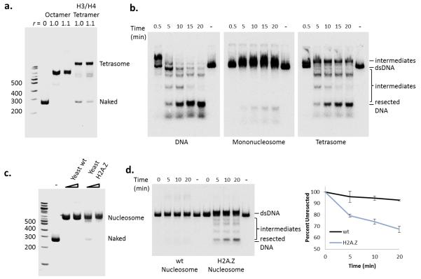
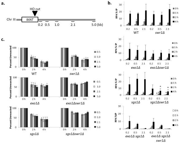
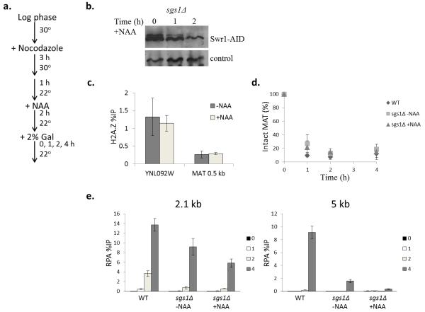
The repair of DNA double-strand breaks (DSBs) is critical for the maintenance of genome integrity. The first step in DSB repair by homologous recombination is the processing of the ends by one of two resection pathways, executed by the Saccharomyces cerevisiae Exo1 and Sgs1-Dna2 machineries. Here we report in vitro and in vivo studies that characterize the impact of chromatin on each resection pathway. We find that efficient resection by the Sgs1-Dna2-dependent machinery requires a nucleosome-free gap adjacent to the DSB. Resection by Exo1 is blocked by nucleosomes, and processing activity can be partially restored by removal of the H2A-H2B dimers. Our study also supports a role for the dynamic incorporation of the H2A.Z histone variant in Exo1 processing, and it further suggests that the two resection pathways require distinct chromatin remodeling events to navigate chromatin structure.


Figures

Figure 1
Figure 1
Increasing nucleosome density inhibits resection. (a) Top: schematic of the 601-177-12 nucleosomal array (NA). Bottom: Native 4% PAGE of nucleosomal array ScaI digests after reconstitution by salt step dialysis. DNA template utilized for chromatin reconstitution comprised of 12 repeats of 177 bp (each flanked by ScaI restriction site) containing the “601” nucleosome positioning sequence. (b, c). Resection assays with 3′-radiolabelled naked DNA and chromatin at increasing ratios (r) of histone octamer to repeat sequence (0.4, 0.6, and 1.1). Time course of resection for both pathways, Sgs1-Dna2 (10 nM Mre11-Rad50-Xrs2 complex, 10 nM Sgs1, 10 nM Top3-Rmi1 complex, 20 nM Dna2 and 100 nM RPA) (b) and Exo1 (6 nM) (c), resulting in intact (dsDNA) or digested (resected) substrates.
Figure 2
Figure 2
Mononucleosomes inhibit resection pathways differentially (a) Top: schematic of the 601-250 mononucleosome. Bottom: Native PAGE analysis of chromatin reconstitutions with Xenopus histone octamers. Template DNA consisted of one 601 positioning sequence in the middle of a 250 bp DNA fragment.(b) Resection assay using 601-250 naked DNA and mononucleosomes (3′ radiolabel on one end). Resection reaction conditions on mononucleosome are identical to those in Fig. 1b, c. (c) Quantification of signal remaining was calculated and graphed as percent of intact radiolabel DNA at indicated times compared to the 0 time point of each assay.
Figure 3
Figure 3
Increasing free DNA adjacent to a nucleosome enhances the helicase activity of Sgs1. (a) Left: schematic of mononucleosome substrates depicting varying amounts of nucleosome-adjacent free DNA (50 bp, 300 bp, and 800 bp). Right: resection assay for the Sgs1-Dna2 pathway using the 50-, 300-, and 800-bp free DNA mononucleosomal substrates after 20 minutes at 30° C. Percent remaining after resection is indicated for each reaction. (b) Sgs1 (10 nM) and BLM (20 nM) helicase activity in the presence of RPA on identical NAs to Fig. 1a after 20 min at 30° C. (c, d) Helicase assay of Sgs1 on DNA and mononucleosomes of indicated sizes. Substrates were incubated with 100 nM RPA and the indicated concentrations of Sgs1 at 30° C for 20 minutes (c) or indicated times (d). (e) The helicase activity of Sgs1 (left panel) and human homolog BLM (right panel) in the presence of RPA on chromatin substrates described in Fig. 2a.
Figure 4
Figure 4
Nucleosomes act as a barrier to Exo1 DNA resection. (a) Resection assay with increasing concentration of Exo1 on 250 bp DNA or mononucleosome (50 bp end) after 20 minutes at 30° C. (b) Exo1 resection assay using naked DNA or mononucleosome containing either 50- or 300-bp of nucleosome-adjacent free DNA after 20 minutes at 30°C. (c) Top: schematic of restriction enzyme site locations on the 500 bp mononucleosome in relation to nucleosome positioning. Star indicates location of the 3′-radiolabel. Bottom: restriction enzyme (RE) digests following Exo1 resection and Proteinase K treatment to map ssDNA formation on the 601-500 mononucleosome. Exo1 resection intermediate and the RE cleaved resection intermediate are indicated by a closed triangle and closed circle respectively. (d) 250 bp mononucleosome incubated with Exo1 (6 nM) and RSC (1 nM). The lane labeled (-) received no Exo1 or RSC. Note that the appearance of the slowly migrating species following RSC treatment is likely due to sliding of the additional free DNA to form a ssDNA-dsDNA intermediate similar to Exo1 product in (b). (e) 500 bp mononucleosome incubated with Exo1 (6 nM) with increasing amounts of Fun30 (0.025 nM to 0.5 nM) after 20 minutes at 30°C.
Figure 5
Figure 5
Loss of H2A-H2B dimers and incorporation of H2A.Z dimers promotes chromatin resection by Exo1. (a) Native PAGE analysis of chromatin reconstitutions with Xenopus histone octamers and H3/H4 tetramers on a 250 bp DNA template. (b) Time-course of Exo1 resection analysis on naked DNA, mononucleosomes, and tetrasomes. (c) Reconstitution of yeast mononucleosomes with either wild type (wt) H2A or H2A.Z-containing octamers. (d) Left: time-course of Exo1 resection analysis using either wild type (H2A) or H2A.Z-containing mononucleosomes. Right: Quantification of dsDNA signal after Exo1 resection provided as mean values +/− SEM (n=3).
Figure 6
Figure 6
Swr1 stimulates DSB repair through the Exo1 pathway. (a) Schematic of MAT locus with sites of HO cut and primer location indicated. (b) ChIP analysis of RPA occupancy at indicated distances and times in indicated yeast strains. RPA levels were measured at increasing distances from the DSB (0.2, 0.5, and 2.1 kb) and normalized to the percentage of DSB in each strain (WT = wild type, JKM139). Graph values reflect the percent of precipitated DNA relative to the input at each region +/− SEM (n=3). Bars with * indicate P value < 0.02 compared to sgs1 single mutant.(c) DNA levels at indicated distances to the right of a galactose-inducible DSB were monitored by quantitative real-time PCR. DNA levels at indicated times were normalized to actin and the 0 hour time point signals were set at 100% with subsequent hours signal indicated as percent with +/− SEM (n=3).
Figure 7
Figure 7
Depletion of Swr1 inhibits Exo1 resection. (a) Schematic of yeast culture growth and treatments. (b) Western blotting of Swr1 depletion during addition of 1 mM 1-naphthaleneacetic acid (NAA), a synthetic auxin hormone. (c) ChIP analysis of H2A.Z occupancy after 2 hrs. of auxin treatment at the YNL092W promoter and +0.5 kb at MAT HO cut site before Gal induction. Error bars represent +/− SEM (n=3). (d) DSB induction kinetics after auxin treatment. Error bars represent +/− SEM (n=3). (e) ChIP analysis of RPA occupancy in the indicated yeast strains (WT = wild type, CY2049) arrested in G2/M with nocodazole. RPA levels were measured at increasing distances from the DSB (2.1 and 5 kb) and normalized to the percentage of DSB in each strain. Graph values reflect the percent of precipitated DNA relative to the input at each region +/− SEM (n=3).

References

    1. Khanna KK, Jackson SP. DNA double-strand breaks: signaling, repair and the cancer connection. Nat Genet. 2001;27:247–254. - PubMed
    1. Zou L, Elledge SJ. Sensing DNA damage through ATRIP recognition of RPA-ssDNA complexes. Science. 2003;300:1542–1548. - PubMed
    1. Krogh BO, Symington LS. Recombination proteins in yeast. Annu Rev Genet. 2004;38:233–271. - PubMed
    1. Zhu Z, Chung WH, Shim EY, Lee SE, Ira G. Sgs1 helicase and two nucleases Dna2 and Exo1 resect DNA double-strand break ends. Cell. 2008;134:981–994. - PMC - PubMed
    1. Mimitou EP, Symington LS. Sae2, Exo1 and Sgs1 collaborate in DNA double-strand break processing. Nature. 2008;455:770–774. - PMC - PubMed

Methods-only reference

    1. Sigurdsson S, Trujillo K, Song B, Stratton S, Sung P. Basis for avid homologous DNA strand exchange by human Rad51 and RPA. J Biol Chem. 2001;276:8798–8806. - PubMed
    1. Raynard S, Bussen W, Sung PA. A double Holliday junction dissolvasome comprising BLM, topoisomerase III alpha, and BLAP75. J Biol Chem. 2006;281:13861–13864. - PubMed
    1. Luger K, Rechsteiner TJ, Richmond TJ. Preparation of nucleosome core particle from recombinant histones. Methods Enzymol. 1999;304:3–19. - PubMed
    1. Smith CL, Horowitz-Scherer R, Flanagan JF, Woodcock CL, Peterson CL. Structural analysis of the yeast SWI/SNF chromatin remodeling complex. Nat Struct Biol. 2003;10:141–145. - PubMed
    1. Goldstein AL, McCusker JH. Three new dominant drug resistance cassettes for gene disruption in Saccharomyces cerevisiae. Yeast. 1999;15:1541–1553. - PubMed

Publication types

MeSH terms