. 2013 Jul;15(7):839-45.
doi: 10.1038/ncb2761. Epub 2013 Jun 2.

A spindle-independent cleavage pathway controls germ cell formation in Drosophila

Affiliations

A spindle-independent cleavage pathway controls germ cell formation in Drosophila

Ryan M Cinalli et al. Nat Cell Biol. 2013 Jul.

Abstract

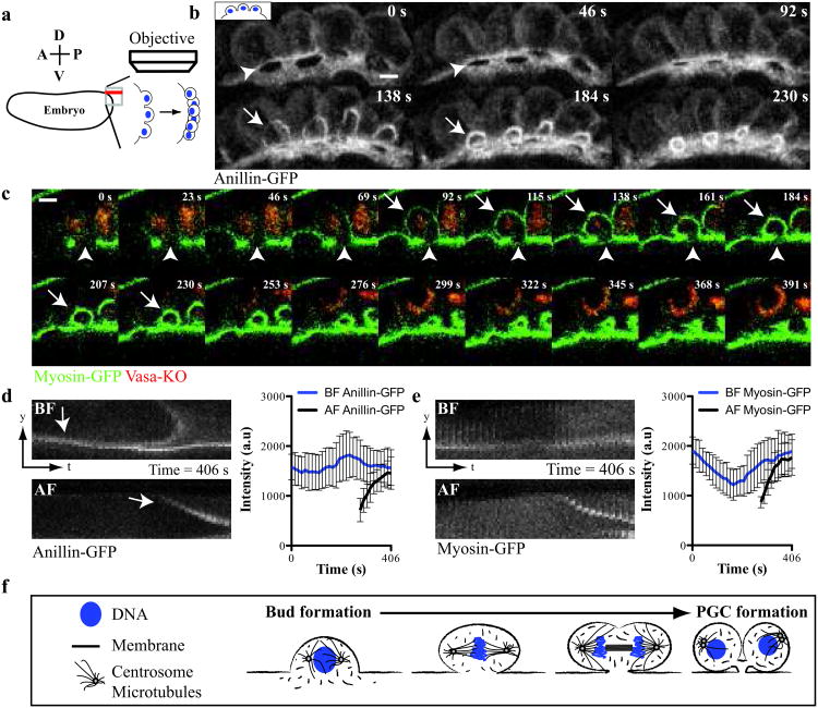
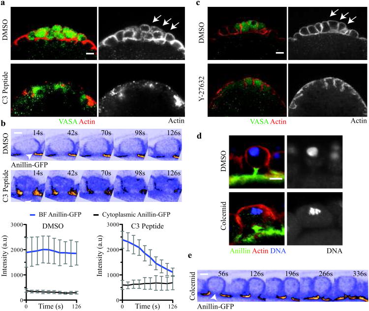
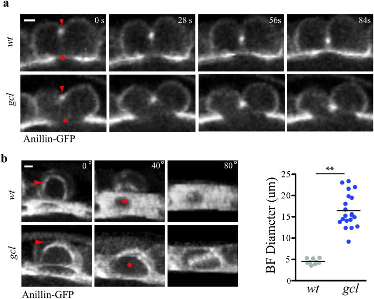
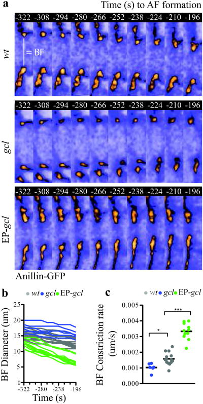
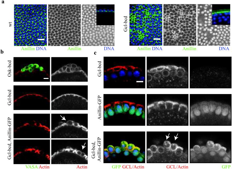
The primordial germ cells (PGCs) are the first cells to form during Drosophila melanogaster embryogenesis. Whereas the process of somatic cell formation has been studied in detail, the mechanics of PGC formation are poorly understood. Here, using four-dimensional multi-photon imaging combined with genetic and pharmacological manipulations, we find that PGC formation requires an anaphase spindle-independent cleavage pathway. In addition to using core regulators of cleavage, including the small GTPase RhoA (Drosophila rho1) and the Rho-associated kinase, ROCK (Drosophila drok), we show that this pathway requires Germ cell-less (GCL), a conserved BTB-domain protein not previously implicated in cleavage mechanics. This alternative form of cell formation suggests that organisms have evolved multiple molecular strategies for regulating the cytoskeleton during cleavage.


Figures

Figure 1
Figure 1. Anillin-GFP and Myosin-GFP localize to paired cleavage furrows during Drosophila PGC formation
(a) Diagram of 4D-imaging strategy used to capture PGC formation at the posterior of the Drosophila embryo. The embryo is positioned with its dorsal surface closest to the cover slip. 40 Z-slices (red line) spaced 1 μm apart along the dorsal-ventral axis were acquired per time point at the apex of the posterior pole. (b) Micrographs of time-lapse maximum intensity projections (MIP) of paired furrows during PGC formation revealed with Anillin-GFP (see Supplementary Video 1). (c) Time-lapse micrographs (single optical sections) of a single bud during PGC formation revealed by Myosin-GFP and Vasa-KO (see Supplementary Video 2). (b, c) Arrows and arrowheads mark the anaphase furrow (AF) and bud furrow (BF) respectively. (d and e) Kymographs showing the localization and quantification of (d) Anillin-GFP and (e) Myosin-GFP at a single paired furrow during PGC formation. We consistently observed biphasic enrichment of Myosin-GFP at the BF suggesting regulation by the cell cycle as previously reported. Data shows the mean of quantifications done in (d) = 4 embryos and (e) = 4 embryos, with 3 buds measured in each embryo. Error bars: S.D. (f) Graphical description of PGC formation showing the remodeling of one bud into two cells. Scale bars = 5 μm
Figure 2
Figure 2. A spindle-independent cleavage pathway directs bud furrow cleavage
(a) Micrographs of vehicle- and C3 peptide-injected embryos (Vasa (green) and F-actin (red)). Arrows mark the PGCs in vehicle-injected embryos. Total number of embryos injected and scored in (a): vehicle-injected = 15, C3 peptide-injected = 12. (b) Time–lapse micrographs and below quantification of Anillin-GFP at the BF in vehicle- and C3 peptide-injected embryos (see Supplementary Video 3 and 4). Arrowheads mark the BF. Quantification shows the mean of 4 embryos, with 3 buds measured in each embryo. Error bars: S.D. (c) Micrographs of vehicle- and Y27682-injected embryos (Vasa (green) and F-actin (red)). Arrows mark the PGCs in vehicle-injected embryos. Total number of embryos injected and scored in c: vehicle-injected = 21, Y27682-injected = 26. (d) MIP micrographs of single PGCs from vehicle- and colcemid-injected embryos (Anillin (green), F-actin (red) and DNA (blue)). Total number of embryos injected and scored in d: vehicle-injected = 10, colcemid-injected = 18. (e) Time-lapse micrographs (single optical sections) of a bud from a colcemid-injected Anillin-GFP expressing embryo showing complete constriction of BF (see Supplementary Video 5). Arrowhead marks the BF. Note the absence of the AF in the time series. Total number of embryos observed in e = 4. Scale bars = 5 μm
Figure 3
Figure 3. Germ cell-less is required for bud furrow constriction
(a) Time-lapse micrographs (single optical sections) of a single wt and gcl mutant bud during PGC formation revealed by Anillin-GFP. Red arrow marks the AF while a red dot marks the BF. Note BF constriction fails in the gcl mutant buds while AF forms and constricts. (b) MIP micrographs and quantification of the BF diameter shortly after AF formation in wt and gcl mutant buds. Red arrows mark the AF while red dots mark the BF. Each paired furrow is rotated around the X-axis by 40° and 80° to better reveal the BF. BF diameter was measured at t = AF formation in wt and gcl mutants. The total number of embryos filmed for b: wt = 4 and gcl = 7, where 1-3 BF diameters were measured per embryo. Total number of buds measured in b: wt = 9, gcl = 20. The black bar represents the mean BF diameter. ** represent a two-tailed t-test with p < .001.Scale bar = 5um.
Figure 4
Figure 4. Germ cell-less is a rate-limiting component of bud furrow constriction
(a) Time-lapse micrographs (single optical sections) showing the BF diameter revealed with Anillin-GFP in wt, gcl mutant and Gcl-overexpressing embryos (EP-gcl) between t=-322s and t=-196s prior to AF formation (see Supplementary Video 6, 7 and 8). (b) Quantification of the BF diameter between t=-322s and t=-196s in wt, gcl mutant and EP-gcl embryos. c, BF rate of constriction between t=-322s and t=-196s in wt, gcl mutant and EP-gcl buds calculated from the slopes of the lines shown in (b). Total number of embryos analyzed for c, d, e: wt = 7, gcl = 6, EP-gcl = 8, where 1 or 2 buds were measured in each embryo. One-way Anova: p < .05 for wt vs gcl and p < .001 for wt vs EP-gcl. Scale bars = 5 μm
Figure 5
Figure 5. Mis-expression of Germ cell-less together with Anillin is sufficient for ectopic cell formation
(a) Surface micrographs of wt and Gcl mis-expressing embryos (gcl-bcd) during somatic cellularization (Anillin (green), and DNA (blue)). Note mis-expression disrupts somatic cell formation by inducing the premature constriction of the cellularization furrow. Insets are representative micrographs depicting lateral views of the cellularization furrow in wt and Gcl mis-expressing embryos.(b) Micrographs of the anterior pole of Oskar mis-expressing (osk-bcd), gcl-bcd, and gcl-bcd, anillin-GFP embryos (Vasa, germ plasm marker (green) and Actin (red)). Note Oskar is sufficient to ectopically recruit germ plasm (Vasa) and instruct cell formation, while gcl-bcd, anillin-GFP embryos instruct cell formation but do not recruit germ plasm. Arrows mark ectopic cells in gcl-bcd, anillin-GFP embryos prior to somatic cell formation. (c) Micrographs of the anterior pole of embryos from females expressing gcl-bcd, anillin-GFP and gcl-bcd, anillin-GFP transgenes (Anillin-GFP (green) GCL-Actin (red) and DNA (blue)). Arrows mark ectopic cells in gcl-bcd, anillin-GFP embryos at the start of somatic cell formation. Scale bars = 5 μm

References

    1. Sullivan W, Theurkauf WE. The cytoskeleton and morphogenesis of the early Drosophila embryo. Current opinion in cell biology. 1995;7:18–22. - PubMed
    1. Glover DM. Mitosis in the Drosophila embryo--in and out of control. Trends Genet. 1991;7:125–132. - PubMed
    1. Edgar BA, Kiehle CP, Schubiger G. Cell cycle control by the nucleo-cytoplasmic ratio in early Drosophila development. Cell. 1986;44:365–372. - PubMed
    1. Foe VE, Field CM, Odell GM. Microtubules and mitotic cycle phase modulate spatiotemporal distributions of F-actin and myosin II in Drosophila syncytial blastoderm embryos. Development. 2000;127:1767–1787. - PubMed
    1. Karr TL, Alberts BM. Organization of the cytoskeleton in early Drosophila embryos. The Journal of Cell Biology. 1986;102:1494–1509. - PMC - PubMed

Publication types

MeSH terms

LinkOut - more resources