. 2013 Nov;158(11):2273-84.
doi: 10.1007/s00705-013-1741-4. Epub 2013 Jun 2.

Characterization of Dak Nong virus, an insect nidovirus isolated from Culex mosquitoes in Vietnam

Affiliations

Characterization of Dak Nong virus, an insect nidovirus isolated from Culex mosquitoes in Vietnam

Ryusei Kuwata et al. Arch Virol. 2013 Nov.

Abstract

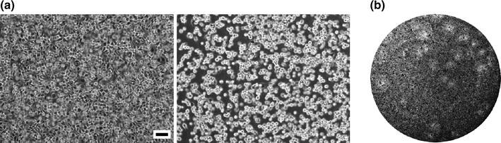
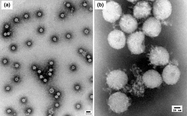
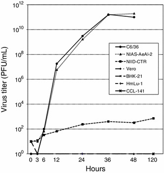
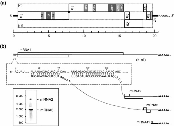
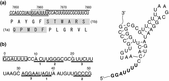
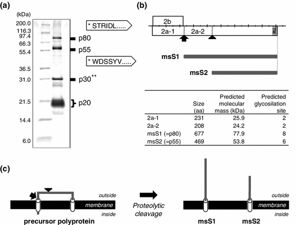
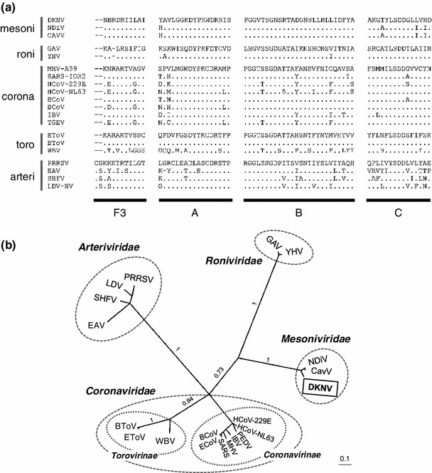
In this study, we isolated and characterized an insect nidovirus from the mosquito Culex tritaeniorhynchus Giles (Diptera: Culicidae) in Vietnam, as an additional member of the new family Mesoniviridae in the order Nidovirales. The virus, designated "Dak Nong virus (DKNV)," shared many characteristics with Cavally virus and Nam Dinh virus, which have also been discovered recently in mosquitoes, and these viruses should be considered members of a single virus species, Alphamesonivirus 1. DKNV grew in cultured mosquito cells but could not replicate in the cultured vertebrate cells tested. N-terminal sequencing of the DKNV structural proteins revealed two posttranslational cleavage sites in the spike glycoprotein precursor. DKNV is assumed to be a new member of the species Alphamesonivirus 1, and the current study provides further understanding of viruses belonging to the new family Mesoniviridae.


Figures

Fig. 1
Fig. 1
(a) Phase-contrast micrographs of mock-infected (left) and DKNV-infected (right) C6/36 cells at 2 days postinfection. Scale bars indicate 100 μm. (b) Plaque morphology induced by DKNV infection of a C6/36 cell monolayer in a 6-well plate (Corning Inc., NY, USA)
Fig. 2
Fig. 2
Negative-contrast electron micrographs of DKNV particles
Fig. 3
Fig. 3
Growth kinetics of DKNV in four vertebrate cell lines (Vero, BHK-21, HmLu-1 and CCL-141) and three mosquito cell lines (C6/36, NIAS-AeAl-2, and NIID-CTR)
Fig. 4
Fig. 4
Genome organization and gene expression of DKNV. (a) Schematic diagram of the organization of the DKNV genome. Horizontal lines and rectangles indicate the viral genomic RNA and the locations of ORFs arranged in three reading frames (−1, 0, +1), respectively, relative to that of ORF1a. Putative protein domains encoded by ORF1a/b are highlighted in gray. The predicted site of RFS is indicated by a black dot. (b) Schematic representation of DKNV gene expression. Horizontal lines and open boxes on the lines indicate the DKNV mRNAs (mRNA1–4) and ORFs corresponding to the positions shown in panel a, respectively. Two different leader TRSs that regulate the expression of DKNV subgenomic mRNA2 and 3 are shown in a gray-dotted box. Northern hybridization analysis (bottom left) identified at least two bands that correspond to the predicted sizes of subgenomic mRNA2 (upper) and mRNA3 (lower)
Fig. 5
Fig. 5
Predicted RFS element and RNA prseudoknot structure in the DKNV genome. (a) The genomic nucleotide sequences (upper) and deduced amino acid sequences (lower) of ORF1a and ORF1b around their overlapping site. The deduced amino acid sequence of DKNV pp1ab is shaded. (b) A putative RNA pseudoknot structure immediately downstream of a “slippery sequence (GGAUUUU)” predicted using the DotKnot program [18, 19]
Fig. 6
Fig. 6
(a) SDS-PAGE and N-terminal sequencing analyses of DKNV virion proteins. *The determined N-terminal aa sequences of p80 and p55 are shown in arrowhead-shaped boxes. **The N terminus of p30 could not be directly sequenced because of the presence of a blocked N-terminal residue. (b) Cleavage sites in the precursor ORF2a polypeptide. An arrow and an arrowhead represent the cleavage sites of signal peptidase and furin-like proteases, respectively. (c) Predicted topology of the two forms of DKNV spike glycoprotein, msS1 and msS2. Cylinders and solid lines represent predicted transmembrane domains and ectodomains, respectively. The precursor ORF2a polypeptide is posttranslationally processed by signal peptidase to yield msS1 and further processed by a furin-like protease to yield msS2
Fig. 7
Fig. 7
Phylogenetic relationships between DKNV and other members of the order Nidovirales. (a) Alignment of the deduced amino acid sequences of the RdRp motif domains (F3, A, B, and C) [27, 28] from DKNV and 19 nidoviruses. Family or subfamily names of the viruses are indicated on the left side of the alignment. (b) The tree was constructed from the posterior distribution of trees generated by Bayesian MCMC coalescent analysis. Family or subfamily names are indicated in boldface type, and the viruses in the same families are circled. Posterior probabilities (on a scale from 0 to 1) are indicated above the branches. The scale bar represents the number of substitutions per site. Abbreviations of nidoviruses and sequence accession numbers are as follows: BCoV, bovine coronavirus ENT (NC_003045); BToV, bovine torovirus Breda1 (AY427798); EAV, equine arteritis virus Bucyrus (NC_002532); ECoV, equine coronavirus NC99 (NC_010327); EToV, equine torovirus Berne (X52374); GAV, gill-associated virus (NC_010306); HCoV-229E, human coronavirus 229E (NC_002645); HCoV-NL63, human coronavirus NL63 (NC_005831); IBV, infectious bronchitis virus Arkansas Vaccine (GQ504721); LDV, lactate dehydrogenase-elevating virus Plagemann (NC_001639); HHV, murine hepatitis virus A59 (AY700211); PEDV, porcine epidemic diarrhea virus CV777 (NC_003436); PRRSV, porcine respiratory and reproductive syndrome virus (NC_001961); SARS, SARS coronavirus Tor2 (NC_004718); SHFV, simian hemorrhagic fever virus LVR 42-0/M6941 (AF180391); WBV, white bream virus DF24/00 (NC_008516); YHV, yellow head virus Chachoengsao 1998 (EU487200)

References

    1. de Groot RJ, Cowley JA, Enjuanes L, Faaberg KS, Perlman S, Rottier PJM, Snijder EJ, Ziebuhr J, Gorbalenya AE. Order Nidovirales. In: King AMQ, Adams MJ, Carstens EB, Lefkowitz EJ, editors. Virus taxonomy; Ninth Report of the International Committee on Taxonomy of Viruses. London: Elsevier; 2012. pp. 785–795.
    1. Adams MJ, Carstens EB. Ratification vote on taxonomic proposals to the International Committee on Taxonomy of Viruses. Arch Virol. 2012;157:1411–1422. doi: 10.1007/s00705-012-1299-6. - DOI - PMC - PubMed
    1. Schütze H, Ulferts R, Schelle B, Bayer S, Granzow H, Hoffmann B, Mettenleiter TC, Ziebuhr J. Characterization of White bream virus reveals a novel genetic cluster of nidoviruses. J Virol. 2006;80:11598–11609. doi: 10.1128/JVI.01758-06. - DOI - PMC - PubMed
    1. Batts WN, Goodwin AE, Winton JR. Genetic analysis of a novel nidovirus from fathead minnows. J Gen Virol. 2012;93:1247–1252. doi: 10.1099/vir.0.041210-0. - DOI - PubMed
    1. Gorbalenya AE, Enjuanes L, Ziebuhr J, Snijder EJ. Nidovirales: Evolving the largest RNA virus genome. Virus Res. 2006;117:17–37. doi: 10.1016/j.virusres.2006.01.017. - DOI - PMC - PubMed

Publication types

MeSH terms

Associated data

LinkOut - more resources