. 2013 Jun:Chapter 4:4.11.1-4.11.24.
doi: 10.1002/0471143030.cb0411s59.

Two-photon excitation microscopy for the study of living cells and tissues

Affiliations

Two-photon excitation microscopy for the study of living cells and tissues

Richard K P Benninger et al. Curr Protoc Cell Biol. 2013 Jun.

Abstract

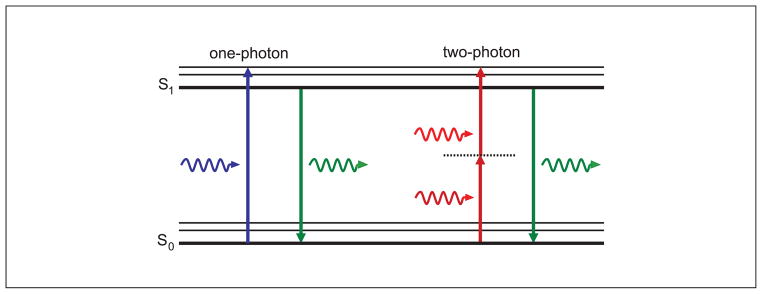
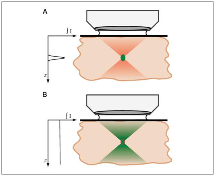
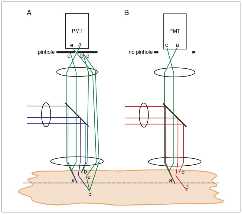
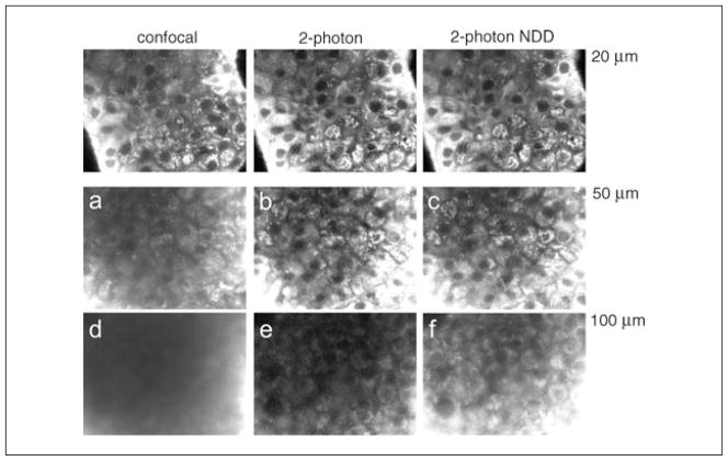
Two-photon excitation microscopy is an alternative to confocal microscopy that provides advantages for three-dimensional and deep tissue imaging. This unit will describe the basic physical principles behind two-photon excitation and discuss the advantages and limitations of its use in laser-scanning microscopy. The principal advantages of two-photon microscopy are reduced phototoxicity, increased imaging depth, and the ability to initiate highly localized photochemistry in thick samples. Practical considerations for the application of two-photon microscopy will then be discussed, including recent technological advances. This unit will conclude with some recent applications of two-photon microscopy that highlight the key advantages over confocal microscopy and the types of experiments which would benefit most from its application.


Figures

Figure 4.11.1
Figure 4.11.1
Jabłoński (energy-level) diagram of conventional one-photon excitation (left) and nonlinear two-photon excitation (right) of fluorescence. In each case, the absorption of photon(s) generates an excited state from which the molecule can relax by emitting a fluorescent photon. Thus, the path to the excited state follows a different path under either one- or two-photon absorption, leading to different absorption spectra. However, fluorescence is emitted from the same excited state producing identical emission spectra.
Figure 4.11.2
Figure 4.11.2
Description of how two-photon absorption occurs only at the focal plane in a microscope. (A) Upon focusing of mode-locked pulsed infrared illumination, the density of photons becomes increasingly greater until at the focal plane. At the focal plane, due to the quadratic dependence of two-photon absorption on intensity, the photon density is increased sufficiently for two-photon absorption to occur. Outside of the focal plane, negligible two-photon absorption occurs, and thus no fluorescence is generated. Pulsed illumination is required to temporally crowd the photons in time, such that the peak photon density is much greater, to achieve two-photon absorption, whereas the time-averaged power is still low. (B) Single-photon absorption depends linearly on the excitation intensity, and thus occurs throughout the focus, requiring a confocal pinhole to achieve optical sectioning (red dotted line). Graph shows total fluorescence generated at each axial position throughout the focus, illustrating the intrinsic optical sectioning provided by two-photon excited fluorescence. For the color version of this figure, go to http://www.currentprotocols.com/protocol/cb0411.
Figure 4.11.3
Figure 4.11.3
Profile (x-z) of photobleaching caused by one-photon and two-photon excitation. A thick fluorescent object is repeatedly imaged at a single focal position either in a confocal microscope (left) or a two-photon microscope (right) until the focal plane is substantially photobleached. The red box indicates the position of the focal plane from which fluorescence is collected. In the confocal microscope, photobleaching can be seen to extend above and below the focal plane, and additional photobleaching throughout the object. Under two-photon excitation, photobleaching has occurred solely at the focal plane, with no photobleaching outside of the region in which fluorescence was collected. For the color version of this figure, go to http://www.currentprotocols.com/protocol/cb0411.
Figure 4.11.4
Figure 4.11.4
Schematic of descanned and nondescanned detection geometries used with a two-photon microscope. Excitation light is raster scanned (x and y scan mirrors) and focused onto the sample by the objective lens. (A) Under descanned detection, the fluorescence emission follows a path returning back along the excitation beam path, first collected by the objective, passing by the scan mirrors, and reflected by a dichroic mirror and focused onto the confocal pinhole, to an ‘internal’ PMT detector. Under two-photon excitation, this pinhole will generally be open. (B) Under non-descanned detection (NDD), the fluorescence emission is collected by the objective and reflected by a dichroic mirror through a transfer lens, which projects the light from the back aperture of the objective onto an ‘internal’ PMT. Under NDD, the position of the dichroic/lens/PMT varies by microscope model, but can be immediately behind the objective in the nose piece. (C) Alternatively, fluorescence emission can be directly detected using an external detector before it reaches the objective.
Figure 4.11.5
Figure 4.11.5
Schematic showing the reduced effect of scattering in reducing signal-to-background in a two-photon microscope compared to a confocal microscope. (A) In a confocal microscope, excitation light is focused on the object, and fluorescence generated at the focal plane is collected and passes through the pinhole to be detected (a). Above or below the focal plane, excitation light generates fluorescence, but when collected, this is rejected by the confocal pinhole and not detected (b). In a turbid object, fluorescence originating from the focal plane can scatter (appearing to originate from outside of the focus), and is thus rejected by the confocal pinhole and not detected (c). Excitation light can also scatter and not reach the focus (d). Both c and d will reduce the detected fluorescent signal. The scattered excitation light will also generate out-of-focus fluorescence, which normally will be rejected by the confocal pinhole, as in b. However, there is a small probability that it can scatter and pass through the pinhole and be detected, increasing the background (e). Similarly, there is a small probability that fluorescence originating from outside of the focus can scatter and pass though the pinhole and be detected, also increasing the background. As the amount of scattering increases, the signal will decrease and the background will increase. (B) In a two-photon excitation microscope, fluorescence generated at the focal plane is collected and detected (a). Above or below the focal plane, no fluorescence is generated (b). In a turbid object, fluorescence originating from the focal plane can scatter, but due to the absence of the confocal pinhole it is still collected. The scattered excitation light will also not generate out-of-focus fluorescence (d). Thus as the amount of scattering increases, the signal will reduce to a lesser degree and the background will not increase.
Figure 4.11.6
Figure 4.11.6
A comparison of confocal microscopy and two-photon microscopy with imaging depth, together with descanned and nondescanned detection. A thick, highly scattering pancreatic islet sample was first imaged at ~20 μm under confocal microscopy and two-photon microscopy with descanned and non-descanned detection, with settings maintained such that the peak fluorescence signal is the same in each case. The sample was then imaged at a greater depth of 50 μm, with the same settings retained. Under confocal microscopy, a decrease in image signal and a substantial background reduces the image contrast (a), which is not present under two-photon excitation (b,c). At an even greater imaging depth (~100 μm) under constant settings, no image contrast is visible under confocal microscopy (d). Two-photon descanned detection shows a reduction in signal but maintains image contrast (e), whereas non-descanned detection retains a good signal and image contrast (f).
Figure 4.11.7
Figure 4.11.7
Optical section of Rhodamine123 fluorescence (A) and NAD(P)H autofluorescence (B) from an intact pancreatic islet. A close-up of a single cell for each channel can be seen in (C). NAD(P)H signal arises from the cytoplasm (indicated ‘c’) and mitochondria (indicated ‘m’), the latter being brighter, somewhat punctate, and overlapping with Rhodamine123 fluorescence. Cell nuclei (indicated ‘n’)—where there is little or no NAD(P)H—appear dark.
Figure 4.11.8
Figure 4.11.8
Well localized photoactivation of a photoconvertible fluorescent protein using two-photon excitation. A single neural growth cone in a Drosophila embryo labeled with PA-GFP is highlighted utilizing two-photon excitation. PA-GFP is efficiently converted to bright green fluorescence using two-photon excitation at 800 nm, and is well differentiated from background autofluorescence (red). For the color version of this figure, go to http://www.currentprotocols.com/protocol/cb0411.

References

    1. Albota MA, Xu C, Webb WW. Two-photon fluorescence excitation cross sections of biomolecular probes from 690 to 960 nm. Appl Optics. 1998;37:7352–7356. - PubMed
    1. Araya R, Eisenthal KB, Yuste R. Dendritic spines linearize the summation of excitatory potentials. Proc Natl Acad Sci USA. 2006;103:18799–18804. - PMC - PubMed
    1. Barretto RP, Ko TH, Jung JC, Wang TJ, Capps G, Waters AC, Ziv Y, Attardo A, Recht L, Schnitzer MJ. Time-lapse imaging of disease progression in deep brain areas using fluorescence microendoscopy. Nat Med. 2011;17:223–228. - PMC - PubMed
    1. Beaurepaire E, Oheim M, Mertz J. Ultra-deep two-photon fluorescence excitation in turbid media. Opt Comm. 2001;188:25–29.
    1. Bennett BD, Jetton TL, Ying G, Magnuson MA, Piston DW. Quantitative sub-cellular imaging of glucose metabolism within intact pancreatic islets. J Biol Chem. 1996;271:3647–3651. - PubMed