

Arthroscopic excision of separated ossicles of the lateral malleolus
- PMID: 23728893
- PMCID: PMC3778221
- DOI: 10.1007/s00776-013-0412-3
Arthroscopic excision of separated ossicles of the lateral malleolus
Abstract
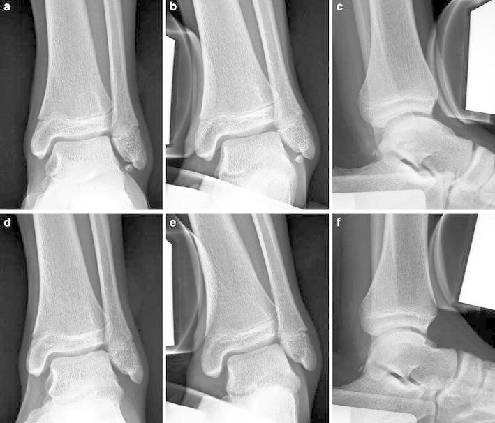
Background: We have conducted a retrospective review of 19 patients for whom 20 separated ossicles of the lateral malleolus were excised arthroscopically. We examined the operating methods, findings, and overall results.
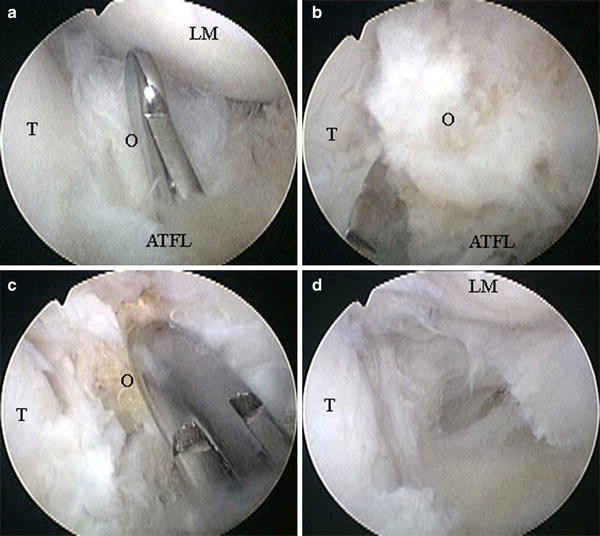
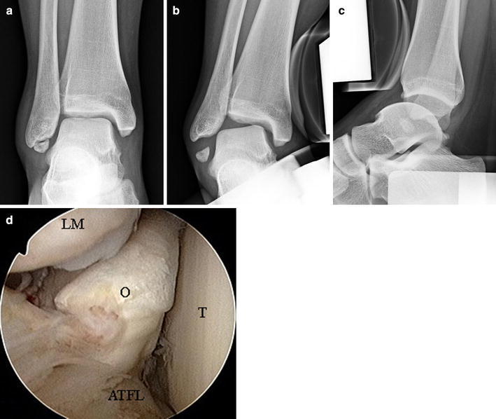
Methods: The patients' indications for this procedure were as follows. The main complaints were pain alone; ossicle sizes were small and ankle instability was minimal. There were 12 ankles of 12 males and eight ankles of seven females. The patients' average age was 17.6 years. A 2.7-mm, 30° arthroscope was inserted into the ankle joint through the anterolateral portal. Instruments were inserted through the accessory anterolateral portal, and ossicles were removed piece by piece. Talar tilt angles and anterior displacements were examined and compared before and after surgery by use of stress radiographs. Japanese Society for Surgery of the Foot (JSSF) ankle/hindfoot scales were assessed pre and postoperatively.
Results: All patients recovered their original levels of activity. The mean talar tilt angle changed from 6.1° ± 2.4° preoperatively to 6.0° ± 1.8° postoperatively (p = 0.93), and the mean anterior displacement changed from 5.9 ± 1.7 mm preoperatively to 6.1 ± 2.0 mm postoperatively (p = 0.42). Average JSSF ankle/hindfoot scale improved from 77.6 ± 2.6 points preoperatively to 97.2 ± 5.2 points postoperatively (p < 0.01).
Conclusions: Arthroscopic excision of separated ossicles of the lateral malleolus achieved good results with minimum incisions, and relatively early resumption of daily and sports activity was possible. However, when the ossicles were embedded within the fibers of the anterior talofibular ligament, it was impossible to avoid cutting of ligament fibers. To reduce the possibility of ligament dysfunction, we believe postoperative treatment should conform to the accepted method for treatment of acute ankle sprains.
Figures
References
-
- Powell HDW. Extra center of ossification for the medial malleolus in children: incidence and significance. J Bone Joint Surg Br. 1961;43:107–113.
-
- Griffiths JD, Menelaus MB. Symptomatic ossicles of the lateral malleolus in children. J Bone Joint Surg Br. 1987;69:317–319. - PubMed
-
- Berg EE. The symptomatic os subfibulare. Avulsion fracture of the fibula associated with recurrent instability of the ankle. J Bone Joint Surg Am. 1991;73:1251–1254. - PubMed
MeSH terms
LinkOut - more resources
Full Text Sources
Other Literature Sources
Medical
