. 2014 Mar;36(3):385-92.
doi: 10.1002/hed.23312. Epub 2013 Jun 1.

Enhancement of head and neck squamous cell carcinoma proliferation, invasion, and metastasis by tumor-associated fibroblasts in preclinical models

Affiliations

Enhancement of head and neck squamous cell carcinoma proliferation, invasion, and metastasis by tumor-associated fibroblasts in preclinical models

Sarah Elizabeth Wheeler et al. Head Neck. 2014 Mar.

Abstract

Background: Head and neck squamous cell carcinoma (HNSCC) has had little improvement in mortality rates in decades. A clearer understanding of the HNSCC tumor microenvironment will aid in finding more effective targeted therapies for this disease. Tumor-associated fibroblasts (TAFs) are the largest stromal cellular components of the tumor microenvironment in HNSCC.

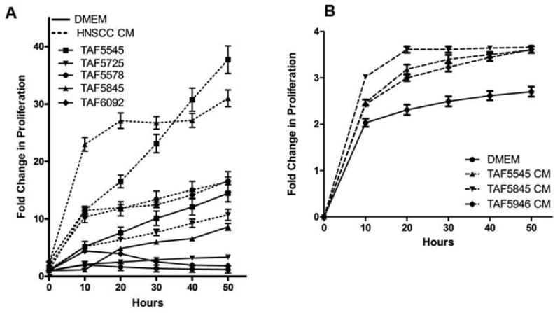
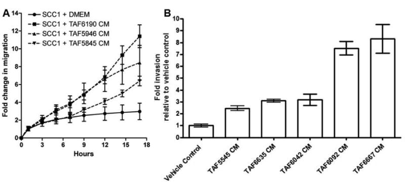
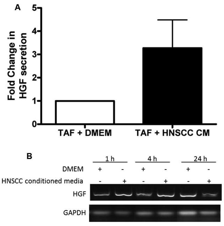
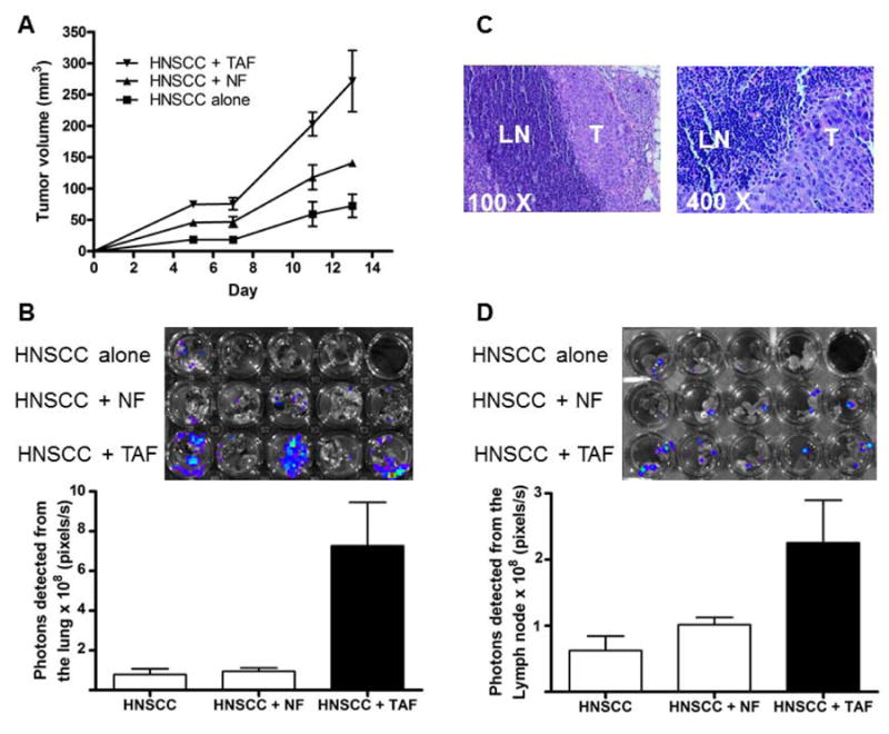
Methods: We isolated TAFs from clinical HNSCC cases and propagated in vitro. The effects of TAF-secreted paracrine factors on in vitro HNSCC migration, invasion, and proliferation was assessed. The effect of TAFs on HNSCC growth and metastases was determined in an orthotopic floor-of-the-mouth tumor model.

Results: TAF-conditioned media increased HNSCC cell migration, invasion, and proliferation. TAFs increased HNSCC tumor growth and metastases in vivo.

Conclusion: TAFs play a major role in increasing tumor growth and metastasis in HNSCC. Targeting the tumor stroma may be important to reduce the rate of HNSCC metastasis.

Keywords: head and neck cancer; invasion; metastasis; tumor microenvironment; tumor-associated fibroblasts.


Figures

Figure 1
Figure 1. Conditioned media from each cell type induces proliferation in the opposite cell type
(A) Five TAF lines were treated with DMEM alone (solid lines) or SCC1 conditioned media (HNSCC conditioned media, dashed lines) and assayed for proliferation using the xCELLigence System. HNSCC conditioned media increased proliferation in all five TAF lines tested. n=5 p=0.004 at 50 hours. (B) UMSCC1 cells were treated with DMEM alone or conditioned media from one of three TAF lines and assayed for proliferation over 60 hours. Data is presented as fold change in proliferation compared to the vehicle control p = 0.029 at 50 hours.
Figure 2
Figure 2. TAF conditioned media increases migration and invasion in HNSCC cells
(A) UMSCC1 cells were treated in quadruplicates with TAF conditioned media (TAF CM) from three different TAF lines and assayed in real-time for migration on the xCELLigence System. TAF conditioned media significantly increased HNSCC migration (p= 0.029). The experiment was repeated three times with similar results. (B) UMSCC1 cells were plated in Boyden invasion chambers, treated in triplicates with conditioned media from 5-TAF lines or DMEM and assayed for invasion after 24 hours. TAF conditioned media significantly increased HNSCC invasion in vitro (p<0.0004). The experiment was repeated 4 times with similar results.
Figure 3
Figure 3. HNSCC conditioned media induces HGF secretion from TAFs
(A) Six TAF lines (TAF5545, TAF5633, TAF5655, TAF5715, TAF5746, TAF5747) were treated with HNSCC cell line UMSCC1 conditioned media or DMEM alone for 48 h. Supernatants were collected and clarified by centrifugation for analysis by HGF ELISA. The average of all 6 TAF lines is shown with error bars (n=6, p=0.001). (B) TAFs (4 × 10⁵ cells/well) were treated with DMEM or HNSCC conditioned medium (CM) for 1h, 4h or 24h. RT-PCR for HGF demonstrates an increase in HGF mRNA at 1 and 4h post treatment with HNSCC CM. The experiment was repeated 3 times with similar results.
Figure 4
Figure 4. TAFs increase HNSCC tumor volume and metastasis in vivo
Nude mice were inoculated in the floor-of-the-mouth with 3 × 10⁶ cells luciferase-expressing OSC19 HNSCC cells alone (n = 4) or combined with 0.5 × 10⁶ TAF cells (TAF5545) or 0.5 × 10⁶ normal fibroblasts (NF) from cancer-free patients (n = 5). (A) The graph depicts growth of primary tumor volume measured using vernier calipers. The volume of HNSCC+TAF tumors at day 13 was significantly higher than the HNSCC alone (p = 0.008) and the HNSCC+NF (p = 0.004) tumors. (B) Lungs were isolated and imaged ex-vivo. Photons emitted by lungs were measured and plotted (±SEM). Light emitted from tumors in the HNSCC +TAF group was significantly higher compared to HNSCC alone (p = 0.016) and HNSCC + NF (p = 0.047) groups. (C) Cervical lymph nodes were paraffin embedded and imaged after hematoxylin and eosin staining. HNSCC metastases (T) to the lymph nodes (LN) are depicted. (D) Cervical lymph nodes were imaged ex-vivo. Photons emitted by lymph nodes were measured and plotted (±SEM). Light emitted from tumors in the HNSCC +TAF group was significantly higher compared to HNSCC alone (p = 0.008) and HNSCC + NF (p = 0.01) groups.

References

    1. Haddad RI, Shin DM. Recent advances in head and neck cancer. N Engl J Med. 2008;359(11):1143–54. - PubMed
    1. Ozdek A, Sarac S, Akyol MU, Unal OF, Sungur A. Histopathological predictors of occult lymph node metastases in supraglottic squamous cell carcinomas. Eur Arch Otorhinolaryngol. 2000;257(7):389–92. - PubMed
    1. Ferlito A, Shaha AR, Silver CE, Rinaldo A, Mondin V. Incidence and sites of distant metastases from head and neck cancer. ORL J Otorhinolaryngol Relat Spec. 2001;63(4):202–7. - PubMed
    1. Jemal A, Siegel R, Ward E, et al. Cancer statistics, 2008. CA Cancer J Clin. 2008;58(2):71–96. - PubMed
    1. Gonda TA, Varro A, Wang TC, Tycko B. Molecular biology of cancer-associated fibroblasts: can these cells be targeted in anti-cancer therapy? Semin Cell Dev Biol. 2010;21(1):2–10. - PMC - PubMed

Publication types

MeSH terms