Review
. 2012 Jan;2(1):1-29.
doi: 10.1002/cphy.c100090.

Mechanobiology in lung epithelial cells: measurements, perturbations, and responses

Affiliations
Review

Mechanobiology in lung epithelial cells: measurements, perturbations, and responses

Christopher M Waters et al. Compr Physiol. 2012 Jan.

Abstract

Epithelial cells of the lung are located at the interface between the environment and the organism and serve many important functions including barrier protection, fluid balance, clearance of particulate, initiation of immune responses, mucus and surfactant production, and repair following injury. Because of the complex structure of the lung and its cyclic deformation during the respiratory cycle, epithelial cells are exposed to continuously varying levels of mechanical stresses. While normal lung function is maintained under these conditions, changes in mechanical stresses can have profound effects on the function of epithelial cells and therefore the function of the organ. In this review, we will describe the types of stresses and strains in the lungs, how these are transmitted, and how these may vary in human disease or animal models. Many approaches have been developed to better understand how cells sense and respond to mechanical stresses, and we will discuss these approaches and how they have been used to study lung epithelial cells in culture. Understanding how cells sense and respond to changes in mechanical stresses will contribute to our understanding of the role of lung epithelial cells during normal function and development and how their function may change in diseases such as acute lung injury, asthma, emphysema, and fibrosis.


Figures

Figure 1
Figure 1
Alveolar distension in lung inflation. An alveolus was imaged at baseline (Palv = 5 cmH2O, green pseudocolor) and hyperinflation (Palv = 20 cmH2O, red pseudocolor). Numbers in baseline image label two perimeter segments. An overlay of the images demonstrates inflation-induced alveolar expansion, which increased perimeter length and alveolar diameter by 13% and 15%, respectively. Adapted (with permission) from reference (196).
Figure 2
Figure 2
Diagram of pressures and forces acting on region of lung isolated by a plane transecting lung. A is the area of the transaction and ΣFi is the sum of all tensile forces in the tissue elements transecting the plane. Adapted (with permission) from reference (168).
Figure 3
Figure 3
Model of the disposition of axial, septal, and peripheral fibers in an acinus showing the effect of surface forces (arrows). From reference (283). Reprinted with permission of the publisher. Copyright © 1984 by the President and Fellows of Harvard College.
Figure 4
Figure 4
Force balance in the epithelial cell monolayer subjected to in-plane stretch. Inward tension (red arrows) produced by active contractile tension generated by actomyosin motors and passive elastic recoil exerted by the actin meshwork is counterbalanced by outward adhesive forces (blue arrows) exerted by the adjacent cells and the extracellular matrix.
Figure 5
Figure 5
Alveolar edema results in the expansion of the air-filled alveolus and the contraction of the fluid-filled alveolus. From reference (197). Reprinted with permission of the American Thoracic Society. Copyright © American Thoracic Society.
Figure 6
Figure 6
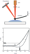
Measurement of cell mechanics with atomic force microscopy.(Top) The atomic force microscope indents the surface of the cell with a flexible cantilever with a sharp tip placed at its end. The cantilever is displaced with a piezoactuator. Force is computed from the lateral displacement on a segmented photodetector of a laser beam reflected on the cantilever. (Bottom) Force-displacement (F-z) curve recorded in an alveolar epithelial cell showing the force measured while the cantilever was approached (solid line) toward the cell and retracted (dashed line) at constant velocity (6 μm/s). The curve exhibits hysteresis indicative of viscoelasticity. The retracted limb exhibits unspecific adhesion before the tip-cell contact was lost (F < 0). The arrow indicates the estimated contact point. Sinusoidal oscillations were applied at a given indentation to compute the complex shear modulus. Reprinted from reference (4) with permission from Elsevier.
Figure 7
Figure 7
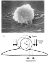
Magnetic twisting cytometry (MTC). (Top) Scanning electron microscopy of a bead bound to the surface of a human airway smooth muscle (HASM) cell. (Bottom) A magnetic field introduces a torque which causes the bead to rotate and to displace. M denotes the direction of the bead's magnetic moment. Images reproduced (with permission) from reference (76).
Figure 8
Figure 8
Optical tweezers. (A) A sketch of optical tweezers-based cytorheometer. Optical tweezers were used to manipulate an intracellular granular structure (lamellar body, left circle), or an extracellular antibody coated glass bead (right circle). (B) A bright-field image of lamellar bodies that abundantly exist in alveolar epithelial type II cells. Reprinted (with permission) from reference (282).
Figure 9
Figure 9
Mice myoblast stretched with microplates. Reprinted from reference (59) with permission from Elsevier.
Figure 10
Figure 10
Microaspiration of adhered cells. The micropipette is gently pressed against the glass slide and slid into contact with the adherent cell (A). A vacuum is applied; aspiration of the cell arrow into the pipette bore (B and C). Reprinted from reference (32) with permission from Elsevier.
Figure 11
Figure 11
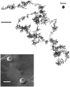
Spontaneous displacement fluctuations of a microbead attached to the surface of human airway smooth muscle cells. Scale bar = 5 μm. Reprinted (with permission) from reference (30).
Figure 12
Figure 12
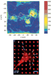
Measurements of cell tractions exerted to the substrate. (Top) Traction microscopy. Cell tractions exerted by a human airway smooth cell on a polyacrylamide gel coated with collagen. Colors show the magnitude and direction of the traction vectors in pascal. Adapted (with permission) from reference (31). (Bottom) Micropost array. Confocal image of immunofluorescence staining of a smooth muscle cell on posts. Position of fibronectin (red) on the tips of the posts was used to calculate force exerted by cells (white arrows). Reprinted (with permission) from reference (240). Copyright (2003) National Academy of Sciences, USA.
Figure 13
Figure 13
Uniaxial stretching device. A strip of silicone is held between the two clamps within the dish. Reprinted from reference (176) with kind permission from Springer Science+Business Media.
Figure 14
Figure 14
Cell stretcher based on the inflation of a clamped elastic diaphragm. Human epidermal keratinocytes (NHEKs) plated on a thin polydimethylsiloxane (PDMS) membrane are subjected to biaxial strain when subject to transdiaphragm pressure. Reprinted from reference (223), Copyright (2007), with permission, from IOS Press.
Figure 15
Figure 15
In-plane biaxial cell membrane stretcher coupled to MTC. A flexible-bottomed well is positioned on a sample holder based on a hollow cylindrical loading post, concentric with the objective of the microscope. The application of a negative pressure underneath the annular outer region of the sample results in a homogeneous and equibiaxial strain of the central area. Two pairs of coaxial coils are coupled to the stretching device to perform MTC. The pair transverse to the sample plane was used to magnetize the beads with a short and strong magnetic pulse. The pair coaxial to the optical axis applied oscillatory twisting fields. Adapted (with permission) from reference (255).
Figure 16
Figure 16
Device to produce homogenous strains in a cruciform silicone membrane. Adapted (with permission) from reference (277).
Figure 17
Figure 17
Biologically inspired design of a human breathing lung-on-a-chip microdevice. Application of vacuum to the side chambers causes mechanical stretching of membrane forming the alveolar-capillary barrier. Adapted (with permission) from reference (125). Reprinted (with permission) from AAAS.
Figure 18
Figure 18
Map of selected in vitro experiments utilizing alveolar epithelial cells, immortalized or primary. To distinguish the type of cells utilized in these studies, that is, AEII, A549, and AEI-like, color coding is utilized as shown in the map. The AEI-like cells are primary AEII cells that are cultured to four or more days. The reference numbers are given in the boxes and are listed in Table 1.
Figure 19
Figure 19
Illustration indicating four ways in which the plasma membrane can respond to stretch. Adapted (with permission) from reference (265).
Figure 20
Figure 20
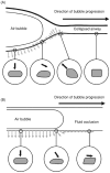
Progression of an air bubble within the collapsed small airways results in large normal and shear stresses that deform the cells significantly and result in plasma membrane injury. Adapted (with permission) from reference (24).
Figure 21
Figure 21
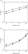
Frequency dependence of G′ (A) and G″(B) under baseline conditions (filled symbols) and during application of a single stretch of 14.1% (open symbols). Data are plotted as means ± SEM. Adapted (with permission) from reference (255).
Figure 22
Figure 22
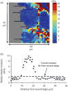
Elastic modulus of cells is dependent on the distance from the wound edge in 16HBE cells. (A) Representative elastic modulus map of migrating 16HBE cells at a wound edge 2 h after wounding. Arrows indicate the wound edge as cells were migrating from right to left. Grey regions indicate plastic substrate. (B) Median elastic modulus values as a function of distance from the wound edge were summarized from four different fields. The dashed line indicates the median value from control cells far away from the wound edge (2.4 kPa), and the asterisks indicate a significant difference from this value (P < 0.05). Adapted (with permission) from reference (270).

Similar articles

Cited by

References

    1. Ventilation with lower tidal volumes as compared with traditional tidal volumes for acute lung injury and the acute respiratory distress syndrome. The acute respiratory distress syndrome network. N Engl J Med. 2000;342:1301–1308. - PubMed
    1. Adkins WK, Hernandez LA, Coker PJ, Buchanan B, Parker JC. Age effects susceptibility to pulmonary barotrauma in rabbits. Crit Care Med. 1991;19:390–393. - PubMed
    1. Agostoni E, Hyatt RE. Static behaviour of the respiratory system. In: Macklem PT, Mead J, editors. Handbook of Physiology Mechanics of Breathing. Bethesda, MD: American Physiological Society; 1986. pp. 113–130. sect. 3, pt. 1.
    1. Alcaraz J, Buscemi L, Grabulosa M, Trepat X, Fabry B, Farre R, Navajas D. Microrheology of human lung epithelial cells measured by atomic force microscopy. Biophys J. 2003;84:2071–2079. - PMC - PubMed
    1. Alcaraz J, Buscemi L, Puig-de-Morales M, Colchero J, Baro A, Navajas D. Correction of microrheological measurements of soft samples with atomic force microscopy for the hydrodynamic drag on the cantilever. Langmuir. 2002;18:716–721.

Publication types

LinkOut - more resources