Review
. 2012 Jan;2(1):255-81.
doi: 10.1002/cphy.c110018.

Motility, survival, and proliferation

Affiliations
Review

Motility, survival, and proliferation

William T Gerthoffer et al. Compr Physiol. 2012 Jan.

Abstract

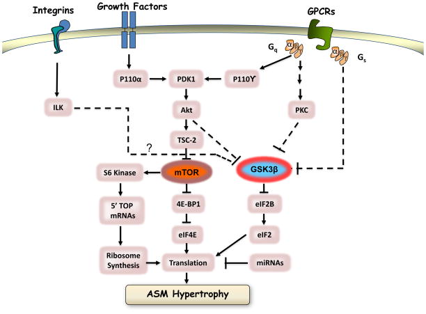
Airway smooth muscle has classically been of interest for its contractile response linked to bronchoconstriction. However, terminally differentiated smooth muscle cells are phenotypically plastic and have multifunctional capacity for proliferation, cellular hypertrophy, migration, and the synthesis of extracellular matrix and inflammatory mediators. These latter properties of airway smooth muscle are important in airway remodeling which is a structural alteration that compounds the impact of contractile responses on limiting airway conductance. In this overview, we describe the important signaling components and the functional evidence supporting a view of smooth muscle cells at the core of fibroproliferative remodeling of hollow organs. Signal transduction components and events are summarized that control the basic cellular processes of proliferation, cell survival, apoptosis, and cellular migration. We delineate known intracellular control mechanisms and suggest future areas of interest to pursue to more fully understand factors that regulate normal myocyte function and airway remodeling in obstructive lung diseases.


Figures

Figure 1
Figure 1. Schematic representation of the role of airway smooth muscle cell proliferation, cellular hypertrophy, apoptosis and migration if development of airway remodeling in asthma
A key local driving force for airway remodeling are cytokines, chemokines, and growth factors released by the epithelium that act on the underlying airway wall (myo)fibroblasts and airway smooth muscle cells. Airway smooth muscle and fibroblasts also release trophic and pro-fibrotic factors that contribute to local inflammation and tissue repair. Central to the initiation and modulation of inflammation, tissue damage and repair is recruitment of active inflammatory cells including Th-2 and Th-1 polarized lymphocytes, eosinophils, neutrophils and mast cells.
Figure 2
Figure 2. Schematic representation of key signaling mechanisms associated with control of airway smooth muscle cell proliferation
See text for details and Table 1 for list of factors that control activation of these pathways.
Figure 3
Figure 3. Schematic representation of key signaling mechanisms associated with control of hypertrophic cell growth
See text for details.
Figure 4
Figure 4. Simplified schematic representation of essential pathways for caspase-dependent apoptotic cell death
Apoptosis is triggered by internal cellular stress (intrinsic pathway) or extracellular signals (extrinsic pathway) that mediate effects via the binding of ligands (eg. Fas, TNFR1, DR5) to cell surface death receptors. Extrinsic pathways directly activate executioner caspases (caspase-3) through initiator caspases (eg. caspase-8 and -9) ultimately leading to cell death. In intrinsic pathways, death signals are conducted through mitochondria, increasing permeability that leads to the release of cytochrome c. Cytosolic cytochrome c binds Apaf-1 to activate the apoptosome and caspase-9 which ultimately leads to downstream activation of executioner caspase-3 [218].
Figure 5
Figure 5. Schematic model illustrating the prominent features of a migrating cell
The leading edge of the cell is represented by the cross hatched region on the right. Inset A: The actin polymerization module located at the leading edge is a site of rapid actin polymerization, depolymerization and filament branching. Actin nucleating proteins (mDia1, mDia2, VASP) promote filament formation at the plus (barbed) end. G-actin monomers are added by the action of profilin. Actin filaments are severed by gelsolin and depolymerized by cofilin. Actin branching is regulated by small G proteins acting on WAVE, WASP and proteins of the ARP2/3 complex. The stiffness of the actin gel and traction forces on the matrix are controlled in part myosin II motor proteins that are regulated by activation of multiple kinases (MLCK, PAK, ROCK) and myosin light chain phosphatase (MLCP). Inset B: Signaling and actin attachment modules in the leading edge promote formation of nascent focal contacts (red bars) that rapidly assemble to transiently attach the cell to the matrix. Actin attachment components include integrins, adaptor proteins (talin, vinculin, tensin, paxillin). Signaling module components control assembly and maturation of the focal contact. These include regulatory proteins (Src, CAS, FAK) and proteins controlling actomyosin assembly and myosin II activation and (MLCK, PAK, MLCP and ROCK). As the cell migrates, nascent focal contacts mature and move towards the rear of cell. Focal contacts at the rear of the cell (red bars on the left) are disassembled as the cell advances. Disassembly requires the action of multiprotein complexes that depend on microtubules (gray filaments) emanating from the microtubule organizing center (MTOC). Reprinted from Gerthoffer [270] by permission of the American Thoracic Society.
Figure 6
Figure 6. Signaling pathways that regulate actin polymerization and myosin II motors in smooth muscle cell migration
Activation of G protein coupled receptors (GPCR) and receptor tyrosine kinases (RTK) initiates activation of parallel signaling cascades that culminate in actin filament remodeling, changes matrix adhesiveness and regulation of myosin II motors that generate traction force. Immediate post-receptor events include activation of trimeric G proteins, Src family tyrosine kinases, phospholipase C (PLC) and PIP2, PI3-kinases (PI3-K) and increased Ca2+. Multiple small G proteins (RhoA, Rac, Cdc42) and calmodulin (CaM) then activate downstream targets that are shown here in darker shades of red. Some targets are effector proteins that regulate actin polymerization including the formins (mDIA1 and mDIA2), WAVE and WASP and the ARP2/3 complex. Other targets include members of the MAP kinase family (p38 MAPK and ERK), Rho kinases (ROCK) and p21-activated protein kinases (PAK). The signaling kinases phosphorylate other protein kinases (MAPKAPK, LIMK) or phosphatases (MLCP) to regulate effector proteins (dark blue ovals) that control actin polymerization and traction forces generated by myosin II. Most of the schematic is organized as sets of parallel linear signaling cascades, which is an oversimplification for the sake of clarity. Pathway convergence and crosstalk are known to occur between the pathways shown. Regulation of MLCK is a good example where both positive and negative inputs are integrated to determine the level of myosin II regulatory light chain phosphorylation and traction force. Reprinted from Gerthoffer271 by permission of the American Heart Association.

References

    1. Wissler RW. The arterial medial cell, smooth muscle, or multifunctional mesenchyme? Circulation. 1967;36:1–4. - PubMed
    1. Jeffery PK. Remodeling in asthma and chronic obstructive lung disease. Am J Respir Crit Care Med. 2001;164:S28–S38. - PubMed
    1. Holgate ST, Davies DE, Lackie PM, Wilson SJ, Puddicombe SM, Lordan JL. Epithelial-mesenchymal-interactions in the pathogenesis of asthma. J Allergy Clin Immunol. 2000;105:193–204. - PubMed
    1. Van Eerdewegh P, Little RD, Dupuis J, Del Mastro RG, Falls K, Simon J, et al. Association of the ADAM33 gene with asthma and bronchial hyperresponsiveness. Nature. 2002;418:426–430. - PubMed
    1. Holgate ST. Airway inflammation and remodeling in asthma: current concepts. Mol Biotechnol. 2002;22:179–189. - PubMed

Publication types

MeSH terms

LinkOut - more resources