. 2013 Jul;136(Pt 7):2063-76.
doi: 10.1093/brain/awt107. Epub 2013 May 31.

Storage of a naturally acquired conditioned response is impaired in patients with cerebellar degeneration

Affiliations

Storage of a naturally acquired conditioned response is impaired in patients with cerebellar degeneration

Andreas Thieme et al. Brain. 2013 Jul.

Abstract

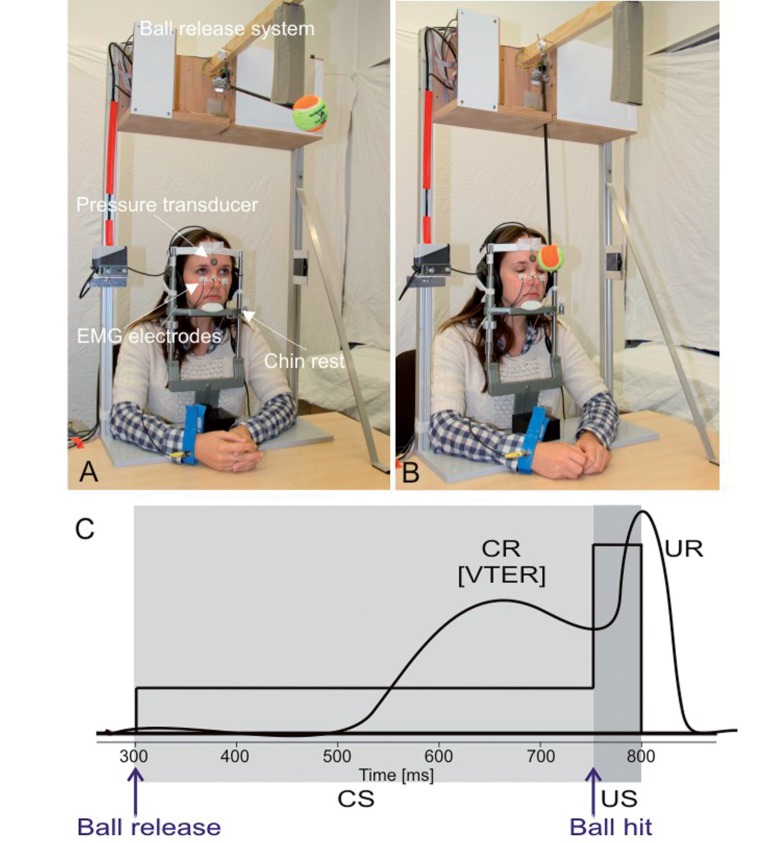
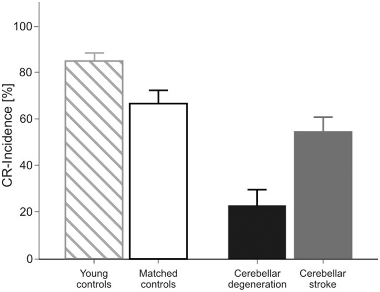
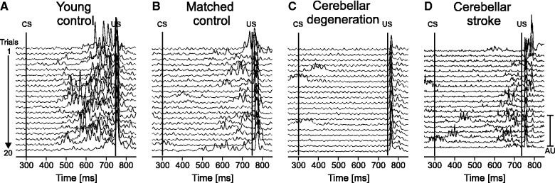
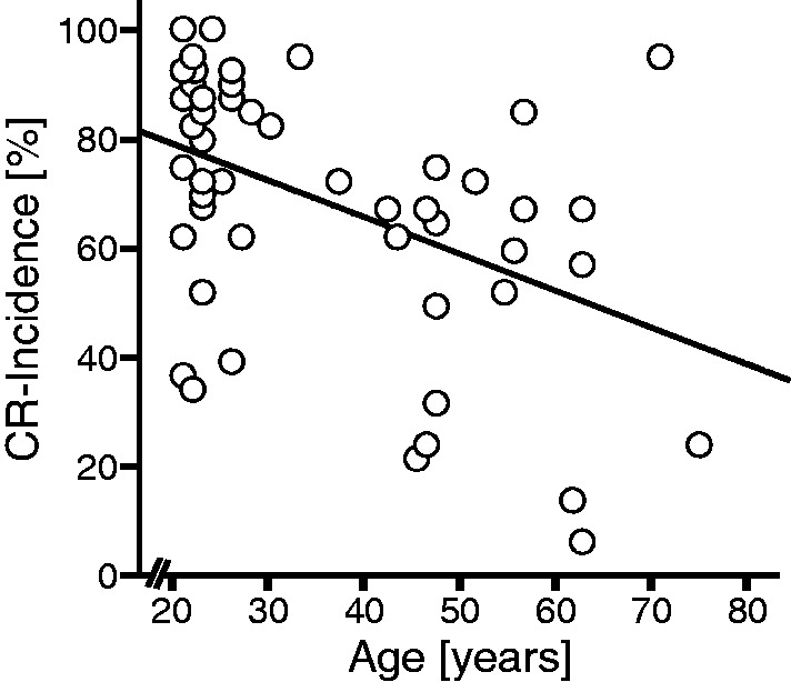
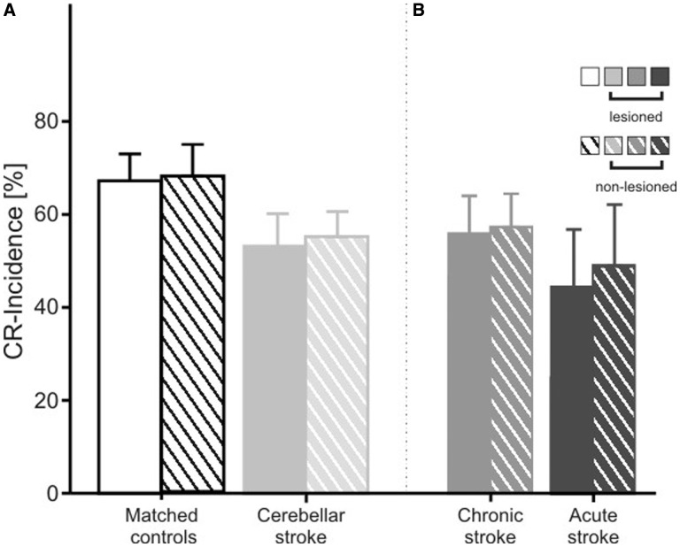
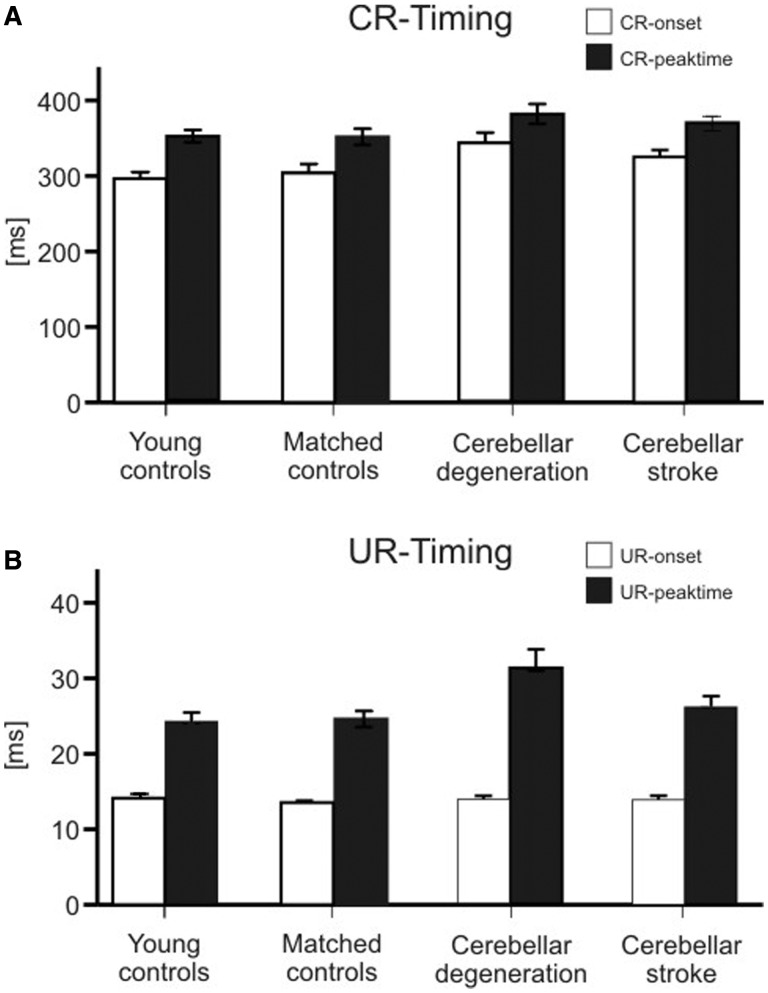
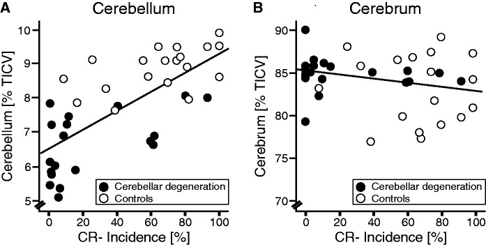
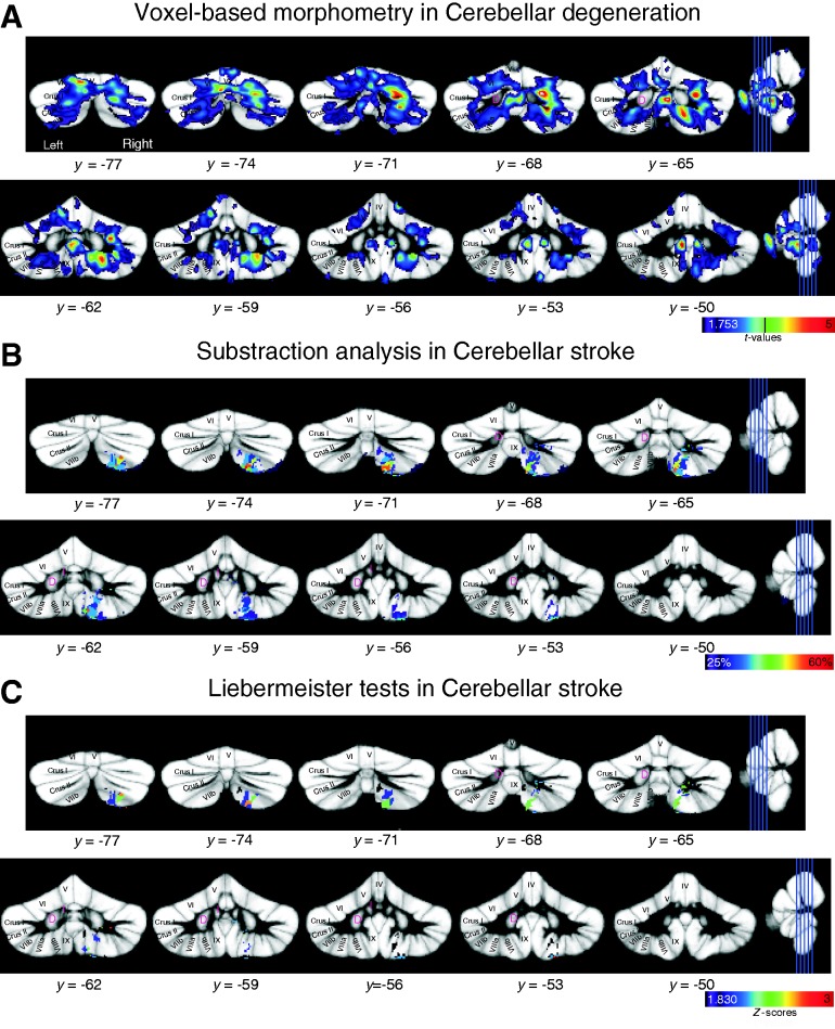
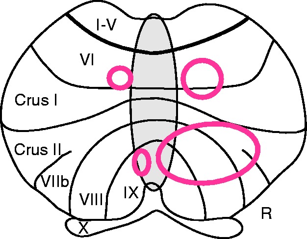
Previous findings suggested that the human cerebellum is involved in the acquisition but not the long-term storage of motor associations. The finding of preserved retention in cerebellar patients was fundamentally different from animal studies which show that both acquisition and retention depends on the integrity of the cerebellum. The present study investigated whether retention had been preserved because critical regions of the cerebellum were spared. Visual threat eye-blink responses, that is, the anticipatory closure of the eyes to visual threats, have previously been found to be naturally acquired conditioned responses. Because acquisition is known to take place in very early childhood, visual threat eye-blink responses can be used to test retention in patients with adult onset cerebellar disease. Visual threat eye-blink responses were tested in 19 adult patients with cerebellar degeneration, 27 adult patients with focal cerebellar lesions due to stroke, 24 age-matched control subjects, and 31 younger control subjects. High-resolution structural magnetic resonance images were acquired in patients to perform lesion-symptom mapping. Voxel-based morphometry was performed in patients with cerebellar degeneration, and voxel-based lesion-symptom mapping in patients with focal disease. Visual threat eye-blink responses were found to be significantly reduced in patients with cerebellar degeneration. Visual threat eye-blink responses were also reduced in patients with focal disease, but to a lesser extent. Visual threat eye-blink responses declined with age. In patients with cerebellar degeneration the degree of cerebellar atrophy was positively correlated with the reduction of conditioned responses. Voxel-based morphometry showed that two main regions within the superior and inferior parts of the posterior cerebellar cortex contributed to expression of visual threat eye-blink responses bilaterally. Involvement of the more inferior parts of the posterior lobe was further supported by voxel-based lesion symptom mapping in focal cerebellar patients. The present findings show that the human cerebellar cortex is involved in long-term storage of learned responses.

Keywords: ataxia; cerebellum; conditioning; human brain mapping; learning.


Figures

Figure 1
Figure 1
Visual threat eye-blink response paradigm. (A) Picture of the experimental set-up before ball release. Ball is in its starting position. (B) Picture after ball release, but before ball hit on the forehead. Eyes are already closed representing the visual threat eye-blink response (VTER). (C) Schematic drawing of the paradigm. Conditioned stimulus (CS, ball moving towards the face) is indicated in light grey, unconditioned stimulus (US, ball hitting the forehead) in dark grey. For further details see text. CR = conditioned eye-blink response; UR = unconditioned response.
Figure 2
Figure 2
Mean conditioned eye-blink response (CR) incidence (plus standard error) of the visual threat eye-blink response for younger-aged controls (hatched columns), age-matched controls (open column), patients with cerebellar degeneration (black column) and patients with focal lesions due to stroke (grey column). Conditioned eye-blink response incidence represents the mean of both eyes.
Figure 3
Figure 3
Rectified EMG recordings of orbicularis oculi muscle in (A) a young control (24 years old, male), (B) a matched control (59 years old, male), (C) a patient with cerebellar degeneration (Patient cer-deg-8 in Table 1) and (D) a patient with cerebellar stroke (Patient cer-foc-11 in Table 1). Recordings of the right eye are shown in A–C, of the left eye in D (ipsilateral to the lesion). Each line represents one trial. All 20 trials are shown with the first on top, and the last on bottom of each stack plot. The first line indicates the onset of the conditioned stimulus (CS, ball begins to move) and the second line the onset of the unconditioned stimulus (US, ball touches the forehead).
Figure 4
Figure 4
Scatterplot comparing conditioned eye-blink response (CR) incidence and age within the group of all control subjects (n = 55). Linear regression line is also shown (R = 3.74, P = 0.005).
Figure 5
Figure 5
Mean conditioned eye-blink response (CR) incidence (plus standard error) of the visual threat eye-blink response (A) in matched control subjects (n = 24), all patients with focal lesions due to stroke (n = 26), and (B) the subgroups of patients with chronic (n = 18) and acute/subacute stroke (n = 8). In focal patients, mean conditioned eye-blink response incidence is shown for the eye ipsilateral to the lesion and the eye contralateral to the lesion. Sides were matched in the controls. Filled columns refer to the lesioned eye and hatched columns to the non-lesioned eye. Only data from the 26 patients with a unilateral lesion are shown.
Figure 6
Figure 6
Conditioned eye-blink response (CR, A) and unconditioned response (UR, B) timing parameters (mean and standard error) in the group of younger-aged control subjects, age-matched control subjects, patients with cerebellar degeneration and cerebellar stroke. Conditioned eye-blink response onset and peak time are expressed as time (ms) after conditioned stimulus onset. Unconditioned response onset and peak time are expressed as time (ms) after unconditioned stimulus onset. White columns = onset latency; filled columns = peak time.
Figure 7
Figure 7
Scatterplots comparing conditioned eye-blink response (CR) incidence and total cerebellar volume (A), and cerebral volume (B) in patients with cerebellar degeneration (filled circles) and matched control subjects (open circles). All volumes are expressed in percentage of total intracranial volume (% total intracranial volume, TICV). Linear regression lines are shown considering both patients and control subjects (cerebellar volume: R = 0.718, P < 0.001; cerebral volume: R = 0.246, P = 0.136).
Figure 8
Figure 8
Results of lesion–symptom mapping superimposed on the SUIT probabilistic atlas template (Diedrichsen et al., 2009). Y-values indicate the coordinate in SUIT space. (A) Voxel-based morphometry in patients with cerebellar degeneration. Cerebellar areas are shown with a positive correlation between conditioned eye-blink response incidence and grey matter values. Results are thresholded at P < 0.05, uncorrected. Colour code indicates t-values, and t-value corresponding to P < 0.001 uncorrected is indicated by a vertical line (t = 3.73). (B) Subtraction analysis in patients with focal lesions due to stroke. Cerebellar areas are shown that are more likely to be lesioned in patients with an abnormally low conditioned eye-blink response incidence. Colour code represents % consistency with a threshold of 25%. All lesions were flipped to the same side. (C) Liebermeister tests in patients with focal lesions due to stroke. Cerebellar areas are shown that are more likely to be lesioned in patients with an abnormally low conditioned eye-blink response incidence. Results are thresholded at P < 0.05 FDR corrected, Z-score = 1.83. Colour code indicates Z-scores. All lesions were flipped to the same side.
Figure 9
Figure 9
Summary diagram based on Fig. 8 superimposed on a flat map of the cerebellar cortex. Two main areas in the cerebellum are related to visual threat eye-blink response storage: one in lobule VI bordering Crus I, and one extending from lobule Crus II to VIII and IX. R = right.

References

    1. Ashburner J, Friston KJ. Unified segmentation. NeuroImage. 2005;26:839–51. - PubMed
    1. Attwell PJ, Cooke SF, Yeo CH. Cerebellar function in consolidation of a motor memory. Neuron. 2002;34:1011–20. - PubMed
    1. Baillieux H, De Smet HJ, Dobbeleir A, Paquier PF, De Deyn PP, Mariën P. Cognitive and affective disturbances following focal cerebellar damage in adults: a neuropsychological and SPECT study. Cortex. 2010;46:869–79. - PubMed
    1. Bastian AJ. Moving, sensing and learning with cerebellar damage. Curr Opin Neurobiol. 2011;21:596–601. - PMC - PubMed
    1. Blaxton TA, Zeffiro TA, Gabrieli JD, Bookheimer SY, Carrillo MC, Theodore WH, et al. Functional mapping of human learning: a positron emission tomography activation study of eyeblink conditioning. J Neurosci. 1996;16:4032–40. - PMC - PubMed

Publication types

MeSH terms