. 2013 Aug 1;305(3):L240-55.
doi: 10.1152/ajplung.00355.2012. Epub 2013 May 31.

Ezrin/radixin/moesin proteins differentially regulate endothelial hyperpermeability after thrombin

Affiliations

Ezrin/radixin/moesin proteins differentially regulate endothelial hyperpermeability after thrombin

Djanybek M Adyshev et al. Am J Physiol Lung Cell Mol Physiol. .

Abstract

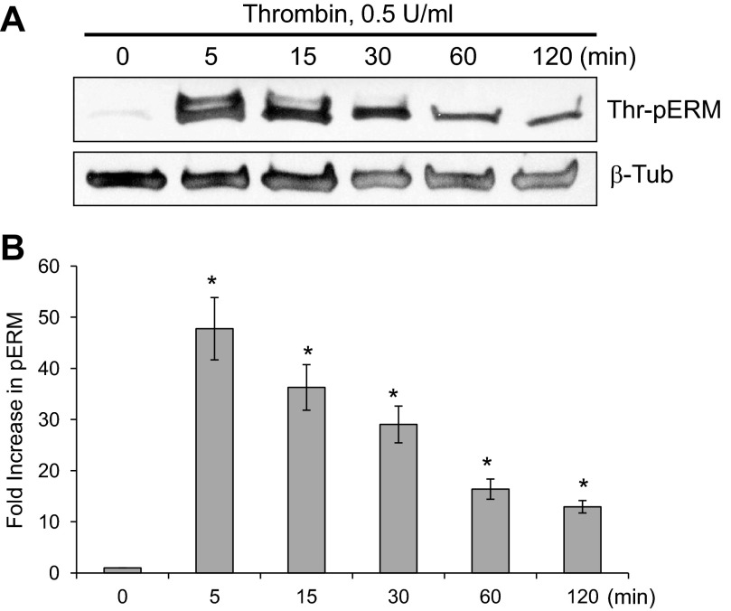
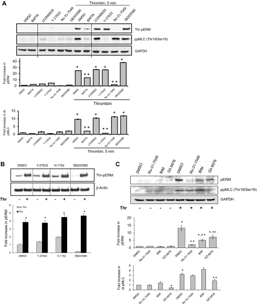
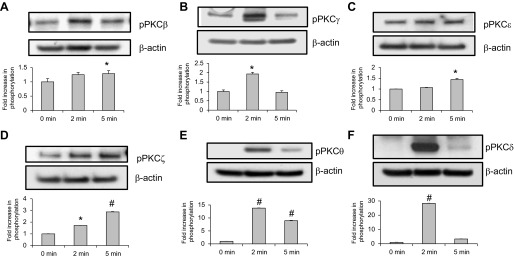
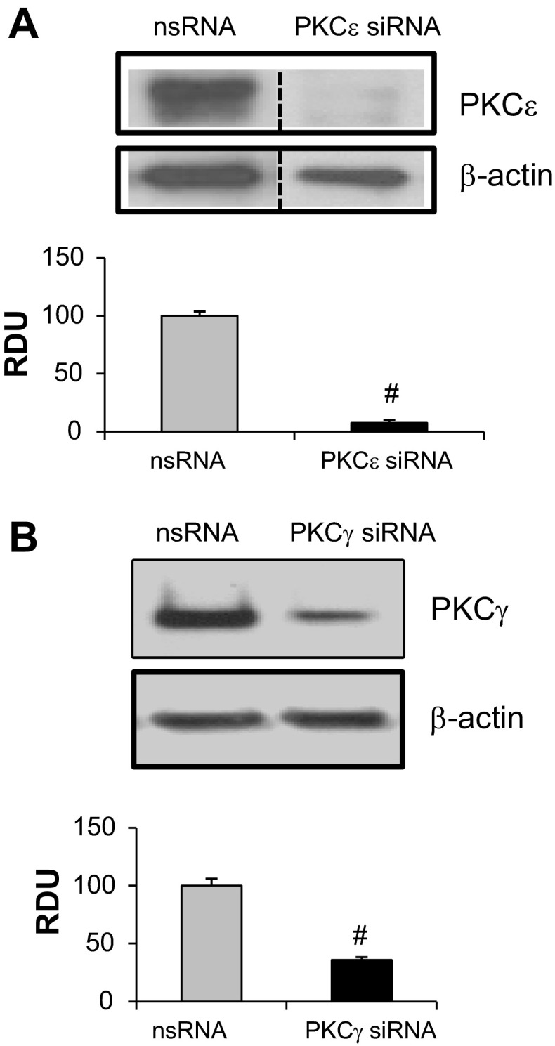
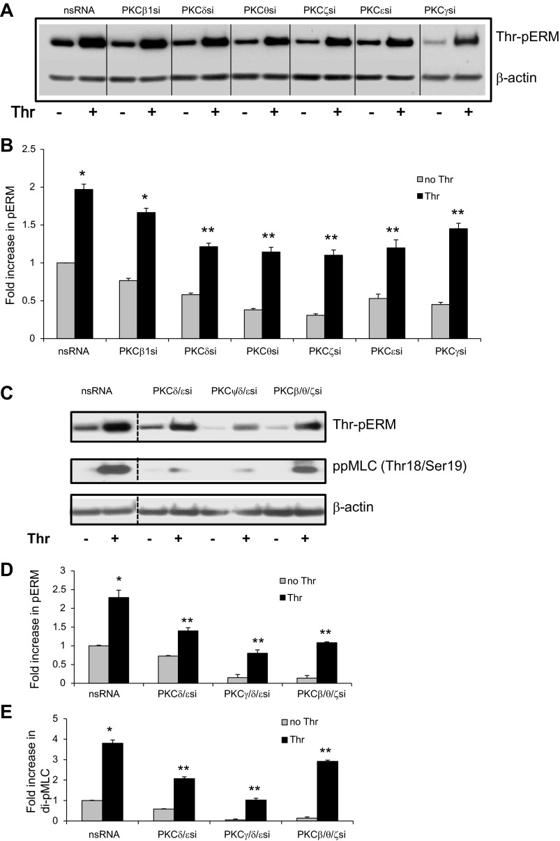
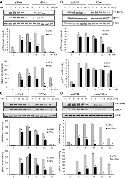
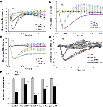
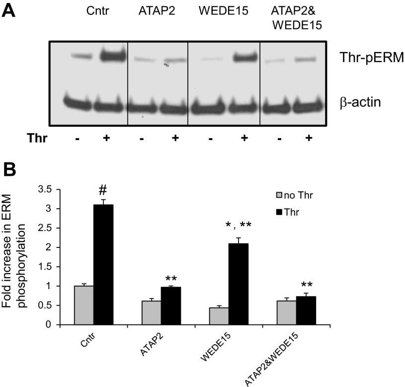
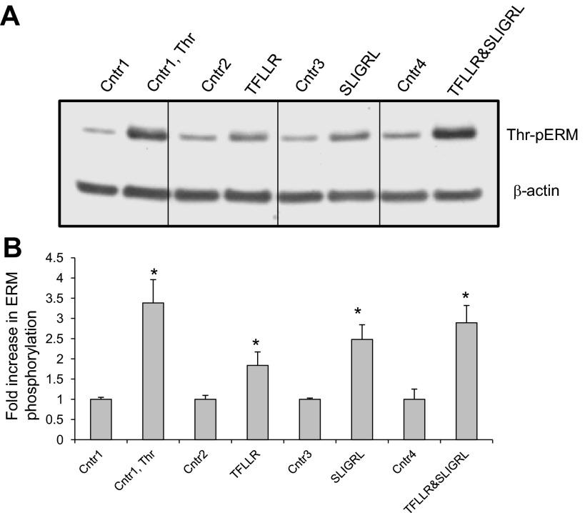
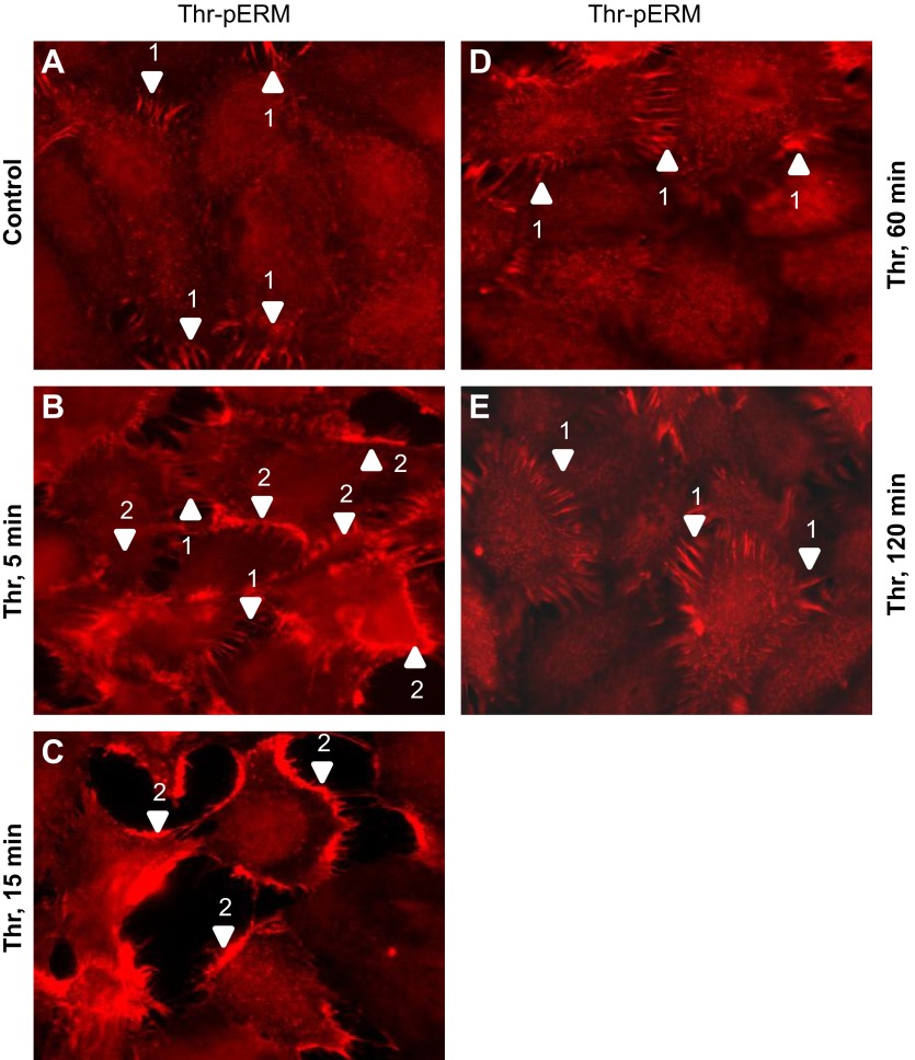
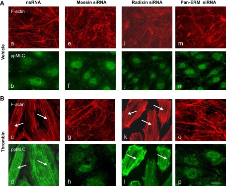
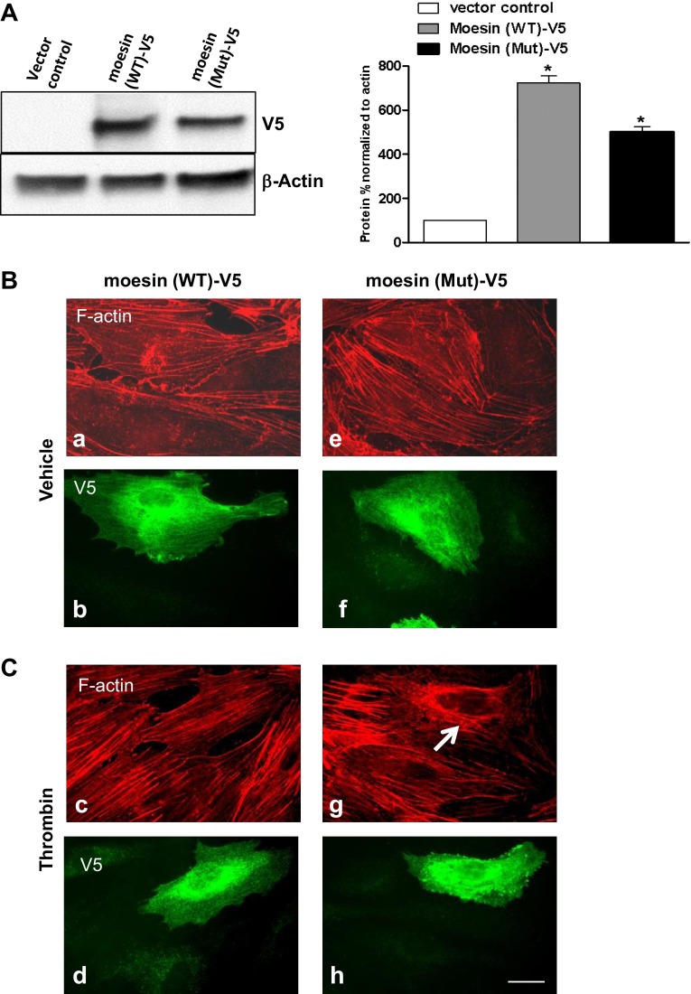
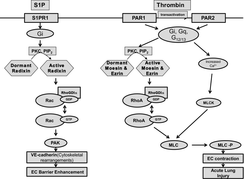
Endothelial cell (EC) barrier disruption induced by inflammatory agonists such as thrombin leads to potentially lethal physiological dysfunction such as alveolar flooding, hypoxemia, and pulmonary edema. Thrombin stimulates paracellular gap and F-actin stress fiber formation, triggers actomyosin contraction, and alters EC permeability through multiple mechanisms that include protein kinase C (PKC) activation. We previously have shown that the ezrin, radixin, and moesin (ERM) actin-binding proteins differentially participate in sphingosine-1 phosphate-induced EC barrier enhancement. Phosphorylation of a conserved threonine residue in the COOH-terminus of ERM proteins causes conformational changes in ERM to unmask binding sites and is considered a hallmark of ERM activation. In the present study we test the hypothesis that ERM proteins are phosphorylated on this critical threonine residue by thrombin-induced signaling events and explore the role of the ERM family in modulating thrombin-induced cytoskeletal rearrangement and EC barrier function. Thrombin promotes ERM phosphorylation at this threonine residue (ezrin Thr567, radixin Thr564, moesin Thr558) in a PKC-dependent fashion and induces translocation of phosphorylated ERM to the EC periphery. Thrombin-induced ERM threonine phosphorylation is likely synergistically mediated by protease-activated receptors PAR1 and PAR2. Using the siRNA approach, depletion of either moesin alone or of all three ERM proteins significantly attenuates thrombin-induced increase in EC barrier permeability (transendothelial electrical resistance), cytoskeletal rearrangements, paracellular gap formation, and accumulation of phospho-myosin light chain. In contrast, radixin depletion exerts opposing effects on these indexes. These data suggest that ERM proteins play important differential roles in the thrombin-induced modulation of EC permeability, with moesin promoting barrier dysfunction and radixin opposing it.

Keywords: ERM; PKC; barrier dysfunction; cytoskeleton; endothelial cells; phosphorylation; thrombin.


Figures

Fig. 1.
Fig. 1.
Relative quantity of moesin (MSN), ezrin (EZR), and radixin (RDX) (i.e., ERM) mRNA in human pulmonary artery endothelial cells (HPAEC). Total RNA was isolated from HPAEC and quantitative real-time reverse transcriptase-polymerase chain reaction (qRT-PCR) was performed by using target gene specific primers and probes and the relative amounts were expressed by standard curve method as described in materials and methods. Each value represents the mean of triplicates.
Fig. 2.
Fig. 2.
Time-dependent effects of thrombin on threonine phosphorylation of ERM. A: confluent HPAEC were treated either with control vehicle or thrombin (0.5 U/ml) for the indicated times, and phosphorylated ERM [phospho-ezrin (Thr567)/radixin (Thr564)/moesin (Thr558)] was detected via immunoblotting. B: bar graph represents relative densitometry. Data are presented as fold changes in phosphorylated ERM over vehicle-treated control and expressed as means ± SE from 3 independent experiments. *P < 0.05 vs. unstimulated control.
Fig. 3.
Fig. 3.
Thrombin-induced ERM phosphorylation requires activation of PKC. HPAEC were pretreated with either control vehicle or the following inhibitors: PKC inhibitors Ro-31-7549 (10 μM, A and C) for 30 min, bisindolylmaleimide (BIM, 1 μM, C) for 30 min, Go 6976 (1 μM, C) for 1 h, Ca2+ chelator BAPTA-AM (25 μM, A) for 1 h, p38 kinase inhibitor SB203580 (20 μM, A and B) for 30 min, Rho kinase inhibitors Y-27632 (10 μM, A and B) for 1 h and H-1152 (3 μM, B) for 1 h, phosphoinositide 3-kinase inhibitor LY294002 (10 μM, A) for 1 h. EC were then stimulated with EBM-2 medium alone or thrombin (0.5 U/ml) for the indicated time. Phosphorylation of ERM proteins and myosin light chain (MLC) were analyzed by immunoblotting of cell lysates with phospho-ERM (as in Fig. 2) or di-phospho-myosin light chain kinase (di-phospho-MLC) (Thr18/Ser19) specific Abs. GAPDH or β-actin Abs were used as a normalization control. Rearranged lanes from the same blot are outlined by vertical dotted line. Results of scanning densitometry of Western blots are shown as fold changes of ERM or MLC phosphorylation relative to vehicle treated EC stimulated by thrombin. Results are representative of 3–6 independent experiments. Values are means ± SE. *Significantly different from cells treated with vehicle (P < 0.05); **significantly different from cells stimulated with thrombin (P < 0.05).
Fig. 4.
Fig. 4.
Effects of thrombin on phosphorylation of PKC isoforms in HPAEC. Confluent HPAEC were treated either with control vehicle or thrombin (0.5 U/ml) for the indicated times, and phosphorylated PKCβ (A), PKCγ (B), PKCε (C), PKCζ (D), PKCθ (E), and PKCδ (F) were detected via immunoblotting. Bar graphs represent relative densitometry of fold changes in phosphorylated PKC isoforms after thrombin relative to vehicle-treated control and expressed as means ± SE from 3 independent experiments. *Significantly different from cells treated with EBM-2 (P < 0.05); #significantly different from cells treated with EBM-2 (P < 0.01).
Fig. 5.
Fig. 5.
Depletion of PKC isoforms by siRNA. PKCε (A) and PKCγ (B) depletion were induced by specific siRNA duplexes and assessed for silencing effects by immunoblotting with appropriate antibody (Ab), compared with treatment with nonspecific (ns) siRNA. Immunoblotting with β-actin Ab was used as a normalization control. Rearranged lanes from the same blot are outlined by vertical dotted line. Quantitative analysis of protein expression was performed by scanning densitometry and expressed in relative density units (RDU). Results are means ± SE for 3 independent experiments. #Significant difference (P < 0.01) compared with cells treated with ns siRNA.
Fig. 6.
Fig. 6.
Depletion of PKC isoforms inhibits thrombin-induced ERM and MLC phosphorylation. Confluent HPAEC were incubated with nonspecific and PKCβI-, PKCζ-, PKCθ-, PKCδ-, PKCγ-, and PKCε-specific siRNA (A) or with nonspecific, combinations of PKCδ- and PKCε-specific, combinations of PKCγ-, PKCδ-, and PKCε-specific, and combinations of PKCβI-, PKCθ-, and PKCζ-specific siRNAs (C) as described in materials and methods and then stimulated by thrombin (0.5 U/ml, 5 min) or vehicle. Total lysates were analyzed by immunoblotting for phospho-ERM or di-phospho-MLC (Thr18/Ser19). Immunoblotting with β-tubulin Ab was used as a normalization control. Rearranged lanes from the same blot are outlined by vertical dotted line. B, D, and E: bar graphs represent relative densitometry of fold changes in phosphorylated ERM and MLC after thrombin relative to vehicle-treated control. Results are means ± SE of 4 independent experiments. *Significantly different from cells treated with ns siRNA without thrombin (P < 0.01); #significantly different from cells treated with ns siRNA without thrombin (P < 0.05). **Significantly different from cells treated with ns siRNA and thrombin (P < 0.05).
Fig. 7.
Fig. 7.
Effects of ERM depletion on thrombin-induced ERM and MLC phosphorylation. Confluent HPAEC were incubated with nonspecific, moesin- (A), radixin- (B), or ezrin-specific (C) or combined siRNAs for ezrin, radixin, and moesin (D) as described in materials and methods then stimulated by thrombin (0.5 U/ml, 5 min) or vehicle. Total lysates were analyzed by immunoblotting with phospho-ERM or di-phospho-MLC (Thr18/Ser19) Abs. Immunoblotting with β-tubulin Ab was used as a normalization control. Bar graphs represent relative densitometry of fold changes in phosphorylated ERM and MLC after thrombin relative to vehicle-treated control. Results are means ± SE of 3 independent experiments. *P < 0.05, compared with corresponding pretreatment vehicle control.
Fig. 8.
Fig. 8.
Effects of ERM depletion on thrombin-induced endothelial barrier hyperpermeability. EC grown in chambers on gold microelectrodes were transfected with siRNA for moesin (A), radixin (B), ezrin (C), or combined siRNAs for ezrin, radixin, and moesin (D) or treated with ns siRNA, as described in materials and methods and used for transendothelial electrical resistance (TER) measurements. At time = 0, cells were stimulated with thrombin (0.5 U/ml) or vehicle control. Shown are pooled data of 5 independent experiments. Bar graph (E) depicts pooled TER data (n = 5) as maximal value of normalized TER elevation above baseline achieved within 30 min ± SE. *Significantly different from cells treated with ns siRNA reagent without thrombin (P < 0.05); **significantly different from control cells stimulated with thrombin (P < 0.05).
Fig. 9.
Fig. 9.
Effects of PAR1 blocking antibodies on thrombin-induced ERM phosphorylation. A: HPAEC were pretreated for 1 h with either control vehicle or the PAR1 blocking Abs ATAP2 (25 μg/ml) or WEDE15 (25 μg/ml) or with combination of ATAP2 and WEDE15, then stimulated by thrombin (0.5 U/ml, 5 min) or vehicle. Total lysates were analyzed by immunoblotting for phospho-ERM. Immunoblotting with β-actin Ab was used as a normalization control. B: bar graph represents relative densitometry of fold changes in phosphorylated ERM after thrombin relative to vehicle-treated control. Results are means ± SE of 4 independent experiments. *Significantly different from cells treated with ns siRNA without sphingosine-1 phosphate (S1P) (P < 0.05); #significantly different from cells treated with ns siRNA without thrombin (P < 0.01). **Significantly different from cells treated with ns siRNA and thrombin (P < 0.05).
Fig. 10.
Fig. 10.
Effects of PAR1- and PAR2-selective agonists on thrombin-induced ERM phosphorylation. A: EC were pretreated for 5 min with thrombin (0.5 U/ml), PAR1-selective agonist TFLLR-NH2 (50 μM), PAR2-selective agonist SLIGRL-NH2 (50 μM), or combination of TFLLR-NH2 and SLIGRL-NH2. Pretreatment with vehicle and reversed amino acid sequence peptides RLLFT-NH2 and LRGILS-NH2 used as controls. B: bar graph represents relative densitometry of fold changes in phosphorylated ERM after thrombin, TFLLR-NH2, or SLIGRL-NH2 relative to vehicle-treated control. Results are means ± SE of 3 independent experiments. *P < 0.05, compared with corresponding pretreatment controls.
Fig. 11.
Fig. 11.
Distribution of phospho-ERM in EC after thrombin. EC grown on glass coverslips and treated with 0.5 U/ml thrombin for indicated time (B–E) or nontreated control cells (A) were subjected to immunofluorescent staining with anti-phospho-ERM Ab. The phospho-ERM signal is very weak in quiescent monolayers and is evident only in spikelike structures in cell-cell border areas (A, arrow 1). Threonine-phosphorylated ERM proteins predominantly localized to the periphery of ECs following thrombin stimulation (5–15 min, B and C, arrow 2) and also are detectable in peripheral spikelike structures (B, arrow 1). After 1–2 h phosphorylated ERM localized in spikelike structures characteristic of quiescent cells and in cytoplasm (D and E). Images are representative of 3 independent experiments. Scale bar = 10 μm.
Fig. 12.
Fig. 12.
Effects of ERM depletion on thrombin-induced cytoskeletal remodeling. HPAEC grown on glass coverslips were incubated with siRNA to ezrin, radixin, moesin, or combination of siRNAs to all 3 proteins, or treated with nonspecific siRNA duplex as described in materials and methods followed by thrombin treatment (0.5 U/ml, 5 min). EC were subjected to double immunofluorescent staining with Texas red phalloidin to visualize F-actin (A and B, top) and anti-pp-MLC Ab (A and B, bottom). Incubation with siRNA to moesin (g and h) and combined siRNAs to ezrin, radixin, and moesin (o and p) almost completely abolishes thrombin-induced F-actin stress fiber and gap formation and MLC phosphorylation compared with control (nsRNA) incubation (c and d, arrows). In contrast, pretreatment with siRNA to radixin slightly enhances the thickness of stress fibers and MLC phosphorylation (k and l, arrows) compared with incubation with nsRNA. Bar = 10 μM. Images are representative of 3 independent experiments.
Fig. 13.
Fig. 13.
Effects of overexpression of the phosphorylation-deficient mutant of moesin (Thr558Ala) on thrombin-induced cytoskeletal remodeling. A: EC were transfected with empty vector (control), V5 (a peptide GKPIPNPLLGLDST, recognized by an antibody)-tagged wild-type (WT), or mutant (Mut) moesin, which were then detected via immunoblotting with V5 Ab. Results of scanning densitometry of Western blots are shown as % of moesin relative to control. Immunoblotting with β-actin Ab was used as a normalization control. B: after transfection with vectors expressing moesin (wild-type or mutant) tagged with V5, EC were grown on glass coverslips as described in materials and methods followed by thrombin treatment (0.5 U/ml, 5 min). EC were subjected to double immunofluorescent staining with Texas red phalloidin to visualize F-actin (B and C, top) and V5 Ab (B and C, bottom). Overexpression with mutant moesin abolishes thrombin-induced F-actin stress fibers and induces cortical actin formation (C, image g) compared with EC overexpressed with wild-type moesin (C, image c). Arrow indicates cell transfected with mutant moesin EC (C, image g). Images are representative of 3 independent experiments. Scale bar = 10 μm.
Fig. 14.
Fig. 14.
Proposed model of ERM-dependent signaling in thrombin- and S1P-challenged lung endothelium (see explanation in Conclusion).

References

    1. Adyshev DM, Moldobaeva NK, Elangovan VR, Garcia JG, Dudek SM. Differential involvement of ezrin/radixin/moesin proteins in sphingosine 1-phosphate-induced human pulmonary endothelial cell barrier enhancement. Cell Signal 23: 2086–2096, 2011 - PMC - PubMed
    1. Arpin M, Chirivino D, Naba A, Zwaenepoel I. Emerging role for ERM proteins in cell adhesion and migration. Cell Adh Migr 5: 199–206, 2011 - PMC - PubMed
    1. Belkina NV, Liu Y, Hao JJ, Karasuyama H, Shaw S. LOK is a major ERM kinase in resting lymphocytes and regulates cytoskeletal rearrangement through ERM phosphorylation. Proc Natl Acad Sci USA 106: 4707–4712, 2009 - PMC - PubMed
    1. Berryman M, Franck Z, Bretscher A. Ezrin is concentrated in the apical microvilli of a wide variety of epithelial cells whereas moesin is found primarily in endothelial cells. J Cell Sci 105: 1025–1043, 1993 - PubMed
    1. Birukova AA, Birukov KG, Smurova K, Adyshev D, Kaibuchi K, Alieva I, Garcia JG, Verin AD. Novel role of microtubules in thrombin-induced endothelial barrier dysfunction. FASEB J 18: 1879–1890, 2004 - PubMed

Publication types

MeSH terms

LinkOut - more resources