. 2013 Sep;27(9):3730-40.
doi: 10.1096/fj.13-232892. Epub 2013 May 31.

Perivascular macrophage-like melanocyte responsiveness to acoustic trauma--a salient feature of strial barrier associated hearing loss

Affiliations

Perivascular macrophage-like melanocyte responsiveness to acoustic trauma--a salient feature of strial barrier associated hearing loss

Fei Zhang et al. FASEB J. 2013 Sep.

Abstract

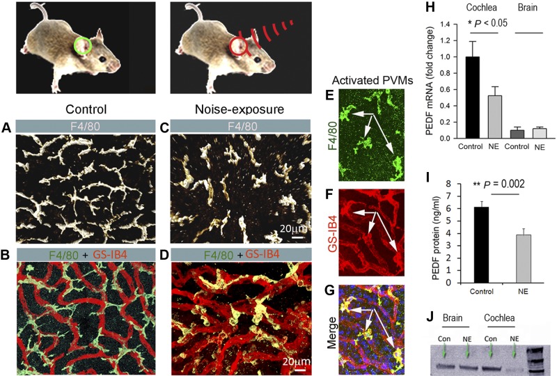
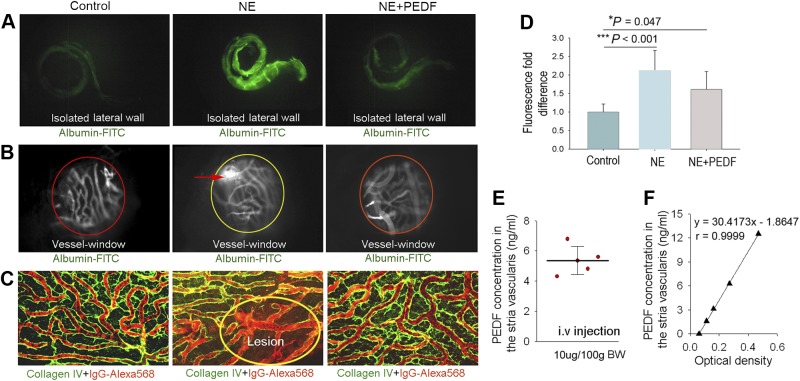
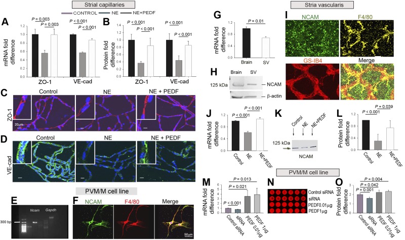
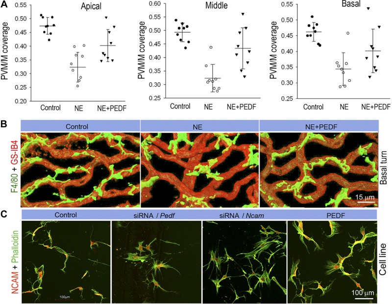
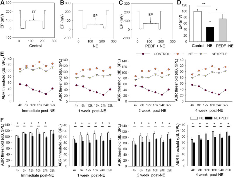
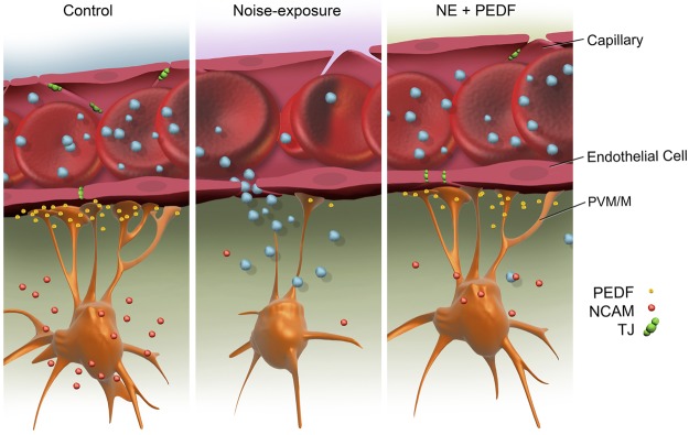
Tissue perivascular resident macrophages (PVM/Ms), a hybrid cell type with characteristics of both macrophages and melanocytes, are critical for establishing and maintaining the endocochlear potential (EP) required for hearing. The PVM/Ms modulate expression of tight- and adherens-junction proteins in the endothelial barrier of the stria vascularis (intrastrial fluid-blood barrier) through secretion of a signaling molecule, pigment epithelium growth factor (PEDF). Here, we identify a significant link between abnormalities in PVM/Ms and endothelial barrier breakdown from acoustic trauma to the mouse ear. We find that acoustic trauma causes activation of PVM/Ms and physical detachment from capillary walls. Concurrent with the detachment, we find loosened tight junctions between endothelial cells and decreased production of tight- and adherens-junction protein, resulting in leakage of serum proteins from the damaged barrier. A key factor in the intrastrial fluid-blood barrier hyperpermeability exhibited in the mice is down-regulation of PVM/M modulated PEDF production. We demonstrate that delivery of PEDF to the damaged ear ameliorates hearing loss by restoring intrastrial fluid-blood barrier integrity. PEDF up-regulates expression of tight junction-associated proteins (ZO-1 and VE-cadherin) and PVM/M stabilizing neural cell adhesion molecule (NCAM-120). These studies point to the critical role PVM/Ms play in regulating intrastrial fluid-blood barrier integrity in healthy and noise-damaged ears.

Keywords: acoustic trauma; endothelial cell; instrastrial fluid-blood barrier; mouse cochlea; paracellular permeability.


Figures

Figure 1.
Figure 1.
NE affects PVM/M morphology and secretion of PEDF. A) Confocal image shows PVM/M morphology. B) PVM/M distribution on strial capillaries labeled with GS-IB4 in a control animal. C) PVM/Ms in noise-exposed animals show reduced branching and withdrawal of ramifications. D) PVM/Ms display less physical contact with capillaries in noise-exposed animals. E–G) Triple-labeled whole-mounted stria vascularis shows PVM/Ms labeled with antibody for F4/80 also positive for GS-IB4. H) qRT-PCR analysis shows decreased mRNA expression for Pedf in noise-exposed mice relative to controls (n=3). *P < 0.05. I) ELISA analysis shows significantly decreased production of PEDF in noise-exposed groups relative to controls (n=6). **P < 0.01; 1-way ANOVA. J) Western blot analysis shows NE to dramatically decrease PEDF protein in the stria vascularis, but not in brain tissue.
Figure 2.
Figure 2.
Down-regulation of PEDF causes disruption of the intrastrial fluid-blood barrier. A) Fluorescent tracers (albumin-FITC) accumulate in the cochlear lateral wall of noise-exposed animals (middle panel) but not in control animals (left panel). The accumulation is attenuated in PEDF treated noise-exposed animals (rignt panel). B) Albumin-FITC tracer is shown to extravasate from capillaries in noise-exposed living animals (open “vessel-window” method, middle panel) but not in control animals (left panel). Less extravasation is seen in PEDF-treated noise-exposed animals (right panel). C) High-molecular-weight IgG-Alexa Fluor 568 tracer, visualized by collagen IV immunostaining (green), extravasates from capillaries in noise-exposed mice (middle panel) but not in control (left panel) and PEDF-treated animals (right panel). Fluorescence IgG-Alexa fluor 568 tracer (red) leaks from lesioned capillaries (oval circle region). D) Treatment with PEDF significantly reduces capillary leakage (n=9). Data are expressed as means ± sd. *P < 0.05, ***P < 0.01; 1-way ANOVA. E) Localized PEDF concentration in the stria vascularis was assessed for a range of intravenously administered PEDF doses. F) Representative ELISA standard calibration curve for PEDF.
Figure 3.
Figure 3.
PEDF ameliorates noise-induced suppression of TJ proteins. A) qRT-PCR shows PEDF to attenuate noise-induced down-regulation of ZO-1 (n=3) and VE-cadherin mRNA (n=3). B) PEDF attenuates down-regulation of ZO-1 (n=9) and VE-cadherin protein (n=9). C, D) PEDF-treated noise-exposed groups show reduced suppression of ZO-1 (C, red) and VE-cadherin (D, green). E) Agarose gel of Ncam mRNA in the PVM/M cell line. F) PVM/M cell line labeled for NCAM (green), F4/80 (red), and nuclei (Hoechst, blue). G) qRT-PCR Ncam mRNA in strial tissue and brain tissue control. H) Western blot of NCAM protein in strial tissue and brain tissue control. I) NCAM (green) labeled whole-mounted stria vascularis, capillaries labeled by GS-IB4 (red) and PVM/Ms for F4/80 (yellow). J) qRT-PCR Ncam mRNA in the stria vascularis, transcript mRNA for Ncam expression in the 3 groups (n=3). K) Western blot of NCAM in the 3 groups. L) PEDF restores NCAM in noise-exposed groups (n=4). M–O) Concurrent expression of PEDF and NCAM transcript mRNA (M, N) and protein (O) (n=6).
Figure 4.
Figure 4.
PEDF has a restorative effect on blood tissue barrier architecture after noise damage. A) Noise-exposed groups display significantly reduced PVM/M density on blood vessels in apical, middle, and basal regions in noise exposed groups. PEDF treatment promotes PVM/M coverage in apical (n=9; P < 0.001 for NE vs. control, P = 0.03 for NE+PEDF vs. NE; 1-way ANOVA test), middle (n=9; P < 0.01 for NE vs. control, P < 0.001 for NE+PEDF vs. NE; 1-way ANOVA test), and basal (n=9; P < 0.001 for NE vs. control, P = 0.008 for NE+PEDF vs. NE; 1-way ANOVA test) regions. Data are expressed as means ± sd. B) Three-dimensional rendering of confocal z stacks highlights the degree of ramification of PVM/M processes in noise-exposed, and PEDF-treated noise-exposed groups. PVM/Ms are labeled with antibody for F4/80 and GS-IB4. C) Control in vitro PVM/Ms are double labeled with phalloidin for F-actin and an antibody for NCAM-120 to show the branched morphology. PVM/Ms in vitro show withdrawal of ramified branches when Pedf and Ncam expressions are down-regulated with siRNA. PEDF treatment promotes ramified branching in PVM/M.
Figure 5.
Figure 5.
PEDF restores endocochlear potential and hearing function. A–C) Representative EP values in control (A), noise-exposed (B), and PEDF-treated noise-exposed animals. D) Mean value of EP in the 3 groups. PEDF significantly attenuates the drop in EP (n=6). *P < 0.05, ***P < 0.001; 1-way ANOVA test. E) Hearing function is assessed by ABR at 1, 2, and 4 after NE. F) Mean ABR threshold in PEDF-treated mice is significantly lower than in untreated noise-exposed mice (n=6). *P < 0.05; Student's t test.
Figure 6.
Figure 6.
PEDF remodeling of the intrastrial fluid–blood barrier. Under normal conditions, tight-adherens junctions between capillary endothelial cells link adjacent cells and limit paracellular transport. PEDF secreted by PVM/Ms regulates vascular permeability by affecting expression of tight- and adherens-junction proteins. The branched (dendrite) morphology of PVM/Ms maximizes coverage of the capillary wall. Acoustic trauma causes down-regulation of PEDF, detachment of PVM/M end-feet from endothelial cells, and decreased expression of TJ and adherens-junction proteins, disrupting the intrastrial fluid-blood barrier. Leakage of serum proteins from loosened TJs results in cochlear edema. Delivery of PEDF to the damaged ear ameliorates hearing loss by restoring intrastrial fluid-blood barrier integrity. PEDF up-regulates the expression of TJ and adherens-junction proteins, including ZO-1 and VE-cadherin, and adhesive proteins such as NCAM, and restores intrastrial fluid-blood barrier integrity.

Similar articles

Cited by

References

    1. Juhn S. K., Hunter B. A., Odland R. M. (2001) Blood-labyrinth barrier and fluid dynamics of the inner ear. Int. Tinnitus J. 7, 72–83 - PubMed
    1. Juhn S. K., Rybak L. P. (1981) Labyrinthine barriers and cochlear homeostasis. Acta Otolaryngol. 91, 529–534 - PubMed
    1. Lin D. W., Trune D. R. (1997) Breakdown of stria vascularis blood-labyrinth barrier in C3H/lpr autoimmune disease mice. Otolaryngol. Head Neck Surg. 117, 530–534 - PubMed
    1. Doi K., Mori N., Matsunaga T. (1992) Adenylate cyclase modulation of ion permeability in the guinea pig cochlea: a possible mechanism for the formation of endolymphatic hydrops. Acta Otolaryngol. 112, 667–673 - PubMed
    1. McMenomey S. O., Russell N. J., Morton J. I., Trune D. R. (1992) Stria vascularis ultrastructural pathology in the C3H/lpr autoimmune strain mouse: a potential mechanism for immune-related hearing loss. Otolaryngol. Head Neck Surg. 106, 288–295 - PubMed

Publication types

MeSH terms