. 2013 Jul 12;288(28):20558-67.
doi: 10.1074/jbc.M113.482992. Epub 2013 May 31.

Identification and functional analysis of the primary pantothenate transporter, PfPAT, of the human malaria parasite Plasmodium falciparum

Affiliations

Identification and functional analysis of the primary pantothenate transporter, PfPAT, of the human malaria parasite Plasmodium falciparum

Yoann Augagneur et al. J Biol Chem. .

Abstract

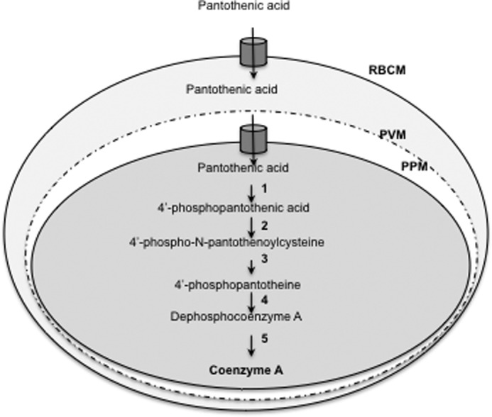
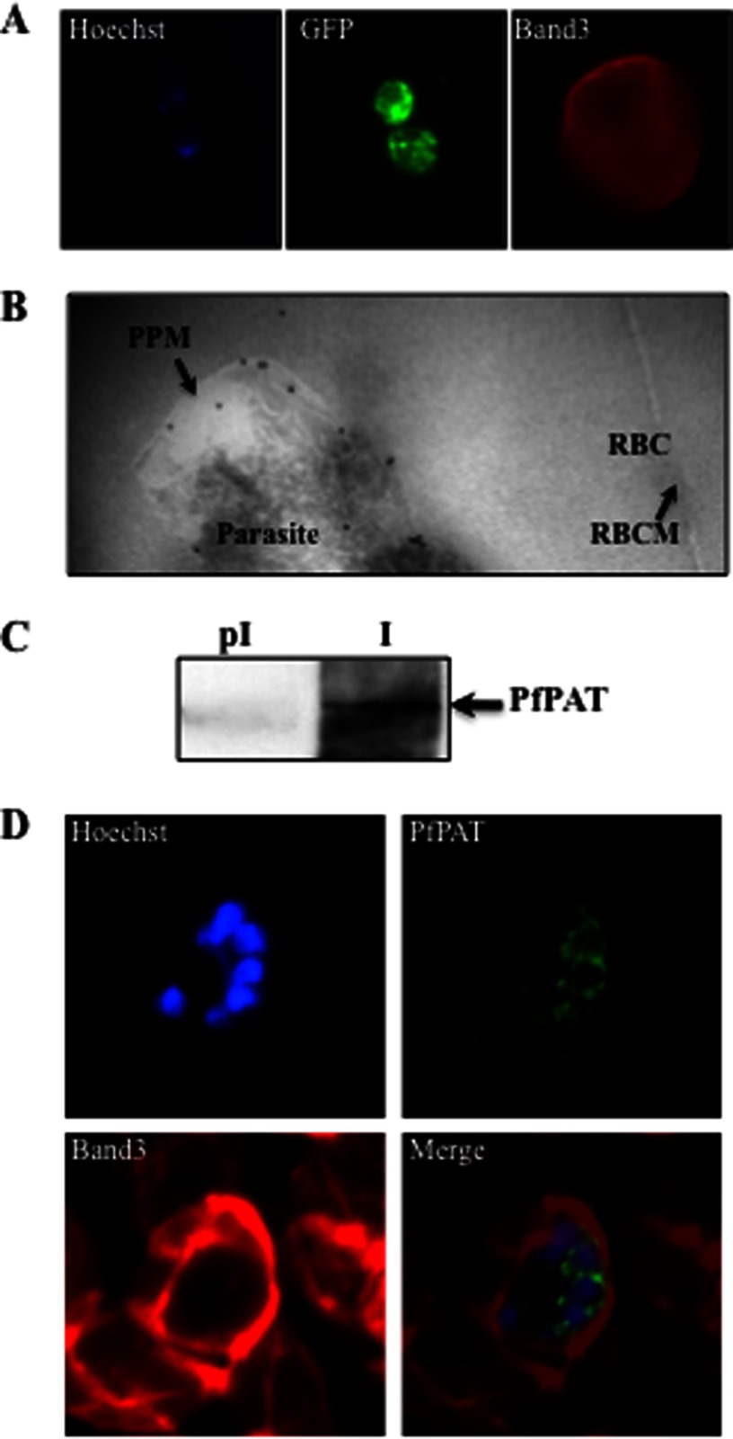
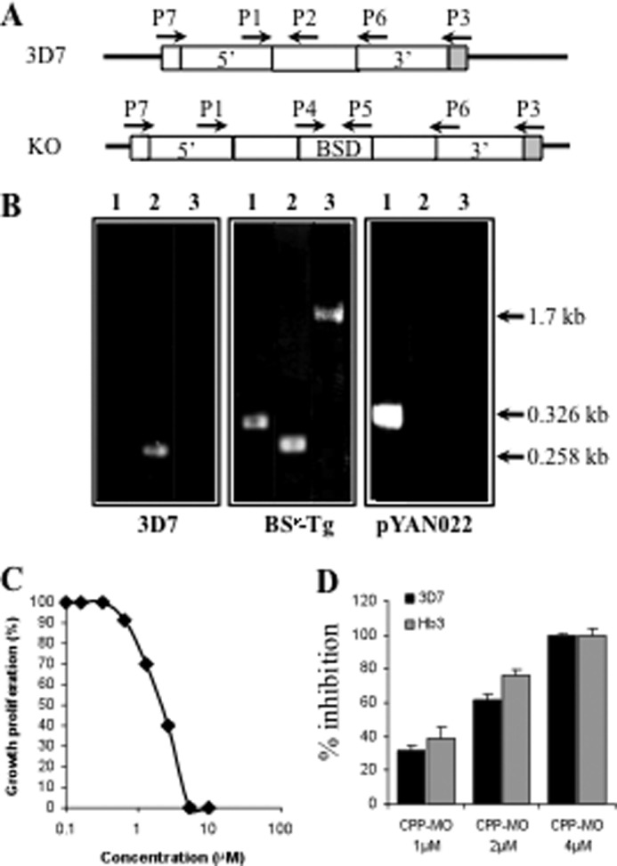
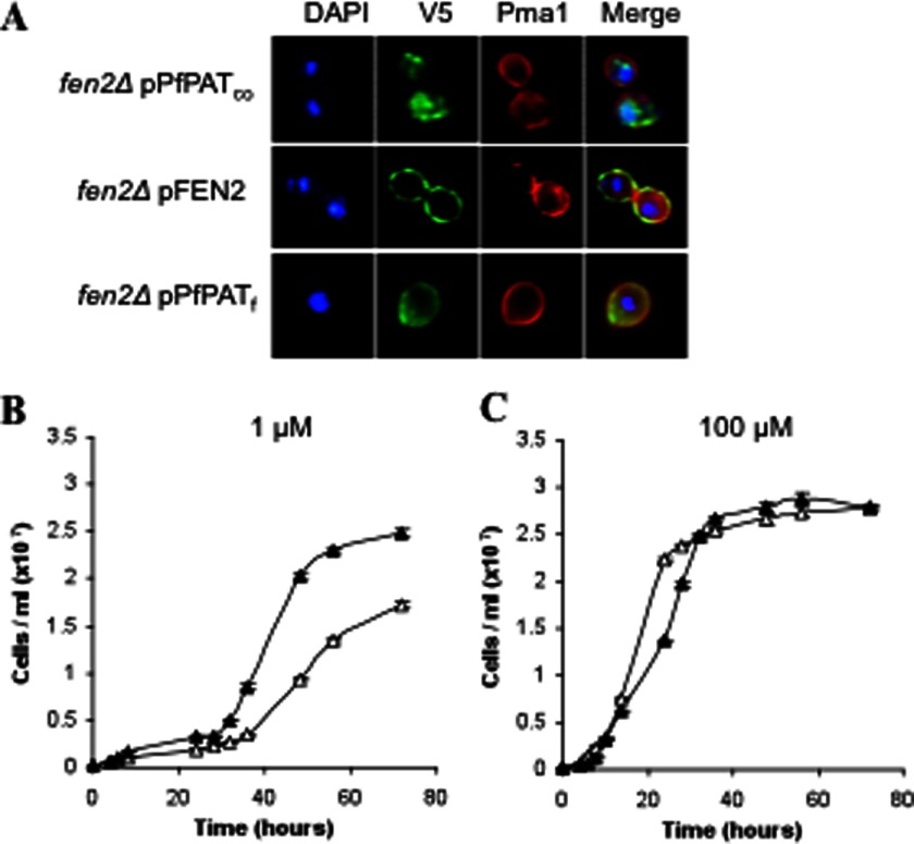
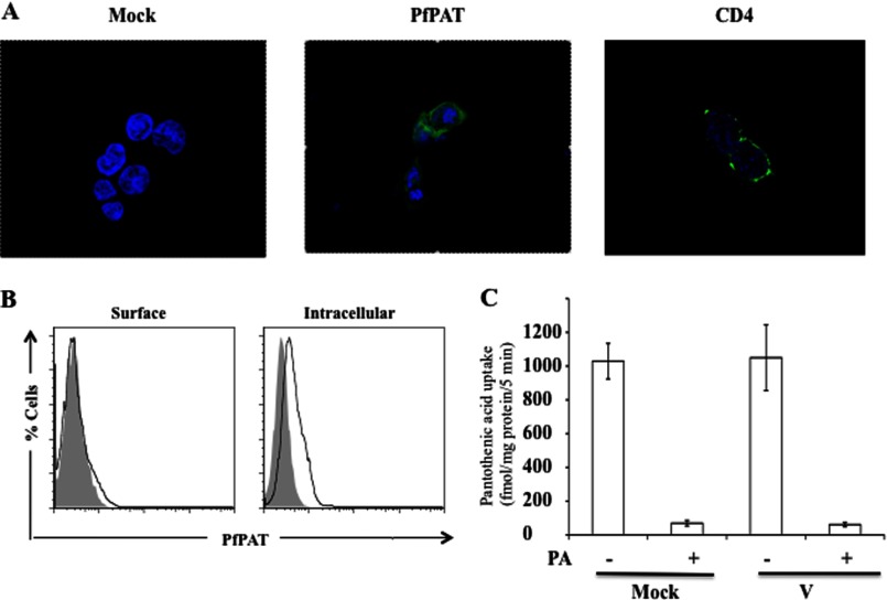
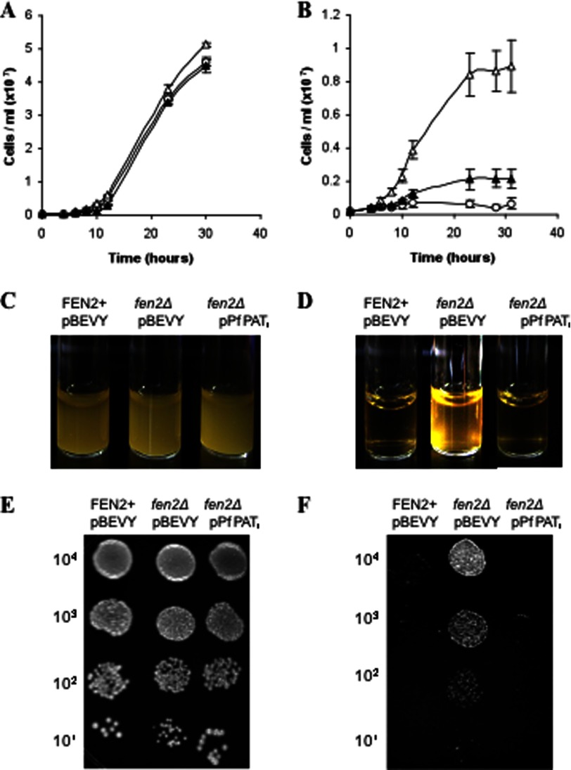
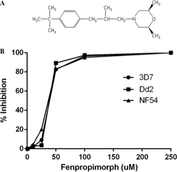
The human malaria parasite Plasmodium falciparum is absolutely dependent on the acquisition of host pantothenate for its development within human erythrocytes. Although the biochemical properties of this transport have been characterized, the molecular identity of the parasite-encoded pantothenate transporter remains unknown. Here we report the identification and functional characterization of the first protozoan pantothenate transporter, PfPAT, from P. falciparum. We show using cell biological, biochemical, and genetic analyses that this transporter is localized to the parasite plasma membrane and plays an essential role in parasite intraerythrocytic development. We have targeted PfPAT to the yeast plasma membrane and showed that the transporter complements the growth defect of the yeast fen2Δ pantothenate transporter-deficient mutant and mediates the entry of the fungicide drug, fenpropimorph. Our studies in P. falciparum revealed that fenpropimorph inhibits the intraerythrocytic development of both chloroquine- and pyrimethamine-resistant P. falciparum strains with potency equal or better than that of currently available pantothenate analogs. The essential function of PfPAT and its ability to deliver both pantothenate and fenpropimorph makes it an attractive target for the development and delivery of new classes of antimalarial drugs.

Keywords: Drug Delivery; Malaria; Plasmodium; Transport; Yeast.


Figures

FIGURE 1.
FIGURE 1.
Schematic representation of pantothenate transport and CoA biosynthesis in P. falciparum-infected erythrocytes. Pantothenate is transported across the plasma membrane by the parasite pantothenate transporter and converted into CoA by the enzymes pantothenate kinase (1), phosphopantothenoylcysteine synthetase (2), phosphopantothenoylcysteine decarboxylase (3), phosphopantotheine adenyltransferase (4), and dephospho-CoA kinase (5). PPM, parasite plasma membrane; PVM, parasitophorous vacuolar membrane; RBCM, red blood cells membrane.
FIGURE 2.
FIGURE 2.
Essential role of exogenous pantothenate in P. falciparum intraerythrocytic development. Growth of P. falciparum parasites in the absence (pantothenic acid-free medium) (A) or presence of 10 μm (B), 100 μm (C), and 200 μm (D) pantothenate is shown. Parasite cultures were initiated at 1% parasitemia and 2% hematocrit and maintained for three developmental cycles. Cultures were diluted at days 3 and 6 to 1% when parasitemia reached 8–10%.
FIGURE 3.
FIGURE 3.
PfPAT localization in P. falciparum-infected erythrocytes. A, localization of PfPAT-GFP by immunofluorescence analysis using anti-GFP (green). The red blood cell membrane marker Band3 (red) was detected using an anti-Band3 monoclonal antibody. The parasite nucleus was visualized using the Hoescht 33258 dye (blue). B, transmission electron micrograph of ultrathin cryosections of the intraerythrocytic early trophozoite stage of P. falciparum PfPAT-GFP transgenic parasites using anti-GFP antibody (18-nm gold particles; indicated with arrows). PPM, parasite plasma membrane; RBC, red blood cell; RBCM, red blood cell membrane. C, detection of native PfPAT using affinity-purified PfPAT antibodies (I). Preimmune serum (pI) is used as a control. D, localization of PfPAT in P. falciparum 3D7 parasites at the schizont stage using anti-PfPAT antibodies (green). The red blood cell membrane marker Band3 (red) was detected using an anti-Band3 monoclonal antibody. The parasite nucleus was visualized using the Hoescht 33258 dye (blue).
FIGURE 4.
FIGURE 4.
Genetic evidence for an essential role of PfPAT in P. falciparum. A, schematic representation of the PfPAT locus in 3D7 and expected knock-out parasites. B, PCR for screening knock-out parasite using primers pairs P4 and P5 (lanes 1), P1 and P2 (lanes 2), and P3 and P4 (lanes 3). Transgenic parasites harboring the pYAN022 vector were selected on blasticidin-containing medium and further subjected to several cycles of growth on media lacking or supplemented with blasticidin and/or ganciclovir. 35 clones were isolated by limited dilution and tested by PCR using the primer pairs P3 and P4. C and D, inhibition of parasite proliferation using a cell-penetrating peptide morpholino oligomer (CPP-MO), which mediates selective cleavage of the PfPAT mRNA. C, growth of 3D7 parasites in the absence of or with increasing concentrations of PfPAT PMO. D, effect of 1, 2, and 4 μm concentrations of PfPAT PMO on the growth of 3D7 and HB3 strains.
FIGURE 5.
FIGURE 5.
Expression and functional characterization of PfPAT in yeast. A, targeting PfPAT to the yeast plasma membrane for functional analysis. fen2Δ cells were transformed with plasmids harboring the codon optimized version of PfPAT (PfPATco), the yeast pantothenate transporter Fen2 (pFEN2) or a chimeric form (PfPATf). All constructs result in a V5 epitope in the C-terminal region of the fusion proteins. Plasma membrane localization was confirmed using anti-Pma1 antibodies. Nuclear staining was achieved using DAPI. B and C, complementation of fen2Δ pantothenate uptake defect by PfPAT. fen2Δ cells harboring the empty pBEVY-U vector (open triangles) or the pPFPATf (closed triangles) expression vector were precultured in SV medium supplemented with 100 μm of pantothenate, washed twice, and inoculated to an initial cell density of 2 × 10⁵ cells/ml in medium supplemented with 1 μm (B) or 100 μm (C) of pantothenate. All cultures were initiated at pH 5.7.
FIGURE 6.
FIGURE 6.
A, confocal microscopy of HEK-293T cells transfected with PfPAT-FLAG (middle) and stained with anti-FLAG FITC (green) or transfected with CD4-GFP as a control (right). Cell nucleus was visualized by staining with Hoescht 33258 dye (blue). B, flow cytometric analysis of HEK-293T cells mock transfected (gray) or transfected with PfPAT-FLAG (black) and surface stained or permeabilized for intracellular staining with anti-FLAG FITC. C, uptake of [3H]pantothenic acid (PA) by ARPE19 cells transiently transfected with pCMV-3Tag vector alone or with PfPAT cloned in the vector examined 48 h after transfection. Mock denotes the endogenous pantothenic acid transport by ARPE19 cells. The presence of carrier was confirmed by competing the [3H]pantothenic acid uptake by excess of unlabeled pantothenic acid. The data shown are means ± S.E. of five independent sets of experiments.
FIGURE 7.
FIGURE 7.
PfPAT mediates entry of fenpropimorph into yeast cells. BY4741-derived end3Δ strain lacking the END3 gene harboring the pBEVY-U vector (open circles), end3Δfen2Δ pBEVY-U (open triangles), and end3Δfen2Δ-pPFPATf (closed triangles) were precultured in SV medium supplemented with 100 μm of pantothenate, washed twice, and inoculated to an initial cell density of 2 105 cells/ml in YPD medium in the absence (A and C) or presence (B and D) of 1 μg/ml fenpropimorph. E and F, for agar assay, cells were cultured in SV medium supplemented with 100 μm of pantothenate and washed twice and 10-fold serial dilutions of cells were plated onto YPD medium (E) or YPD medium supplemented with 1 μg/ml fenpropimorph (F) and incubated at 30 °C.
FIGURE 8.
FIGURE 8.
P. falciparum parasites are sensitive to fenpropimorph. A, chemical structure of fenpropimorph. B, growth of P. falciparum 3D7, NF54, and Dd2 strains in the absence or presence of increasing concentrations of fenpropimorph using the SYBR green proliferation assay (43).

References

    1. World Malaria Report (2011) World Health Organization, Geneva
    1. Singh B., Kim Sung L., Matusop A., Radhakrishnan A., Shamsul S. S., Cox-Singh J., Thomas A., Conway D. J. (2004) A large focus of naturally acquired Plasmodium knowlesi infections in human beings. Lancet 363, 1017–1024 - PubMed
    1. Murray C. J., Rosenfeld L. C., Lim S. S., Andrews K. G., Foreman K. J., Haring D., Fullman N., Naghavi M., Lozano R., Lopez A. D. (2012) Global malaria mortality between 1980 and 2010. A systematic analysis. Lancet 379, 413–431 - PubMed
    1. Saliba K. J., Kirk K. (2001) Nutrient acquisition by intracellular apicomplexan parasites. Staying in for dinner. Int. J. Parasitol. 31, 1321–1330 - PubMed
    1. Downie M. J., El Bissati K., Bobenchik A. M., Nic Lochlainn L., Amerik A., Zufferey R., Kirk K., Ben Mamoun C. (2010) PfNT2, a permease of the equilibrative nucleoside transporter family in the endoplasmic reticulum of Plasmodium falciparum. J. Biol. Chem. 285, 20827–20833 - PMC - PubMed

Publication types

MeSH terms