. 2013 Jul 19;288(29):20927-20941.
doi: 10.1074/jbc.M112.440115. Epub 2013 May 31.

Inducible interleukin 32 (IL-32) exerts extensive antiviral function via selective stimulation of interferon λ1 (IFN-λ1)

Affiliations

Inducible interleukin 32 (IL-32) exerts extensive antiviral function via selective stimulation of interferon λ1 (IFN-λ1)

Yongkui Li et al. J Biol Chem. .

Abstract

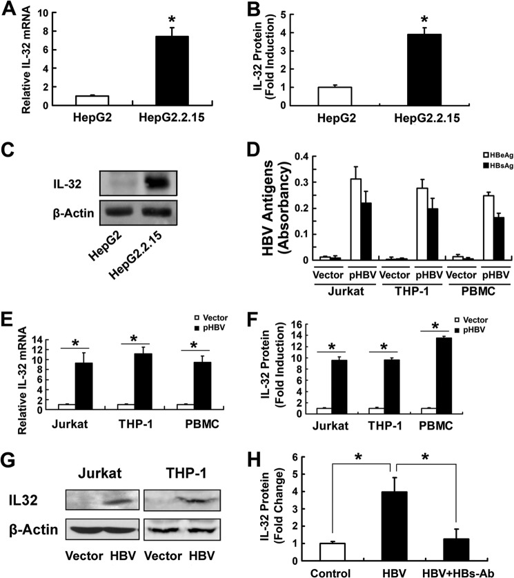
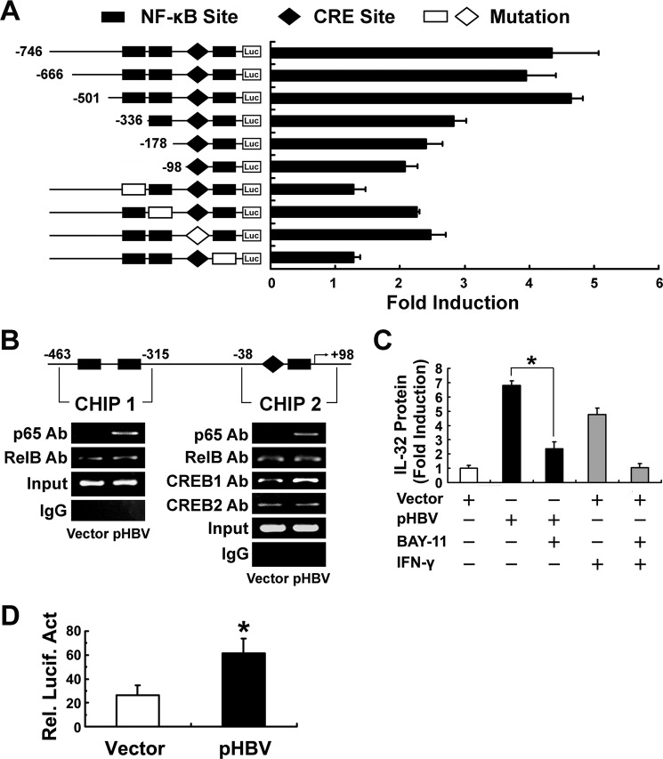
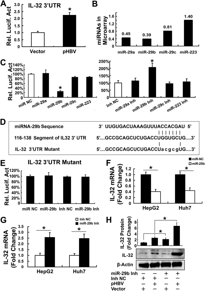
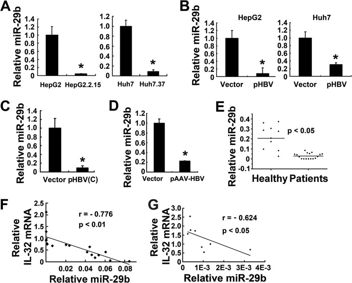
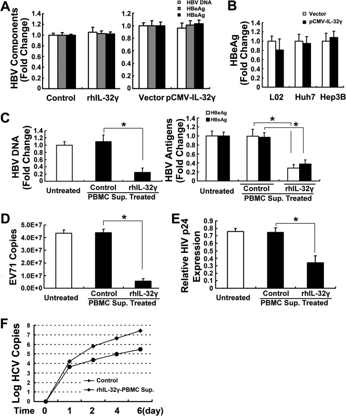
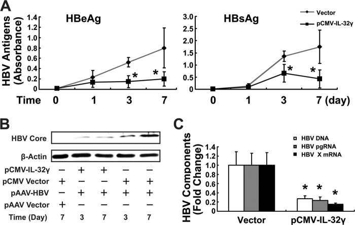
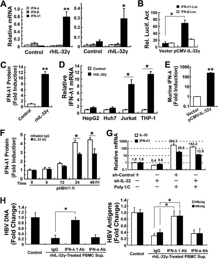
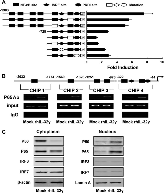
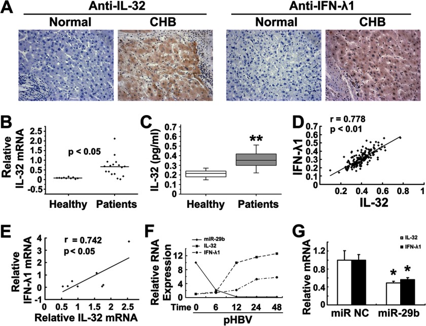
Interleukin (IL)-32 has been recognized as a proinflammatory cytokine that participates in responses to viral infection. However, little is known about how IL-32 is induced in response to viral infection and the mechanisms of IL-32-mediated antiviral activities. We discovered that IL-32 is elevated by hepatitis B virus (HBV) infection both in vitro and in vivo and that HBV induced IL-32 expression at the level of both transcription and post-transcription. Furthermore, microRNA-29b was found to be a key factor in HBV-regulated IL-32 expression by directly targeting the mRNA 3'-untranslated region of IL-32. Antiviral analysis showed that IL-32 was not sufficient to alter HBV replication in HepG2.2.15 cells. To mimic the viremic phase of viral infection, freshly isolated peripheral blood mononuclear cells were treated with IL-32γ, the secretory isoform, and the supernatants were used for antiviral assays. Surprisingly, these supernatants exhibited extensive antiviral activity against multiplex viruses besides HBV. Thus, we speculated that the IL-32γ-treated peripheral blood mononuclear cells produced and secreted an unknown antiviral factor. Using antibody neutralization assays, we identified the factor as interferon (IFN)-λ1 and not IFN-α. Further studies indicated that IL-32γ effectively inhibited HBV replication in a hydrodynamic injection mouse model. Clinical data showed that elevated levels of IFN-λ1 both in serum and liver tissue of HBV patients were positively correlated to the increased levels of IL-32. Our results demonstrate that elevated IL-32 levels during viral infection mediate antiviral effects by stimulating the expression of IFN-λ1.

Keywords: Cytokines/Interferon; Gene Regulation; Host-Pathogen Interactions; Infectious Diseases; Interleukin; Viral Immunology.


Figures

FIGURE 1.
FIGURE 1.
Induction of IL-32 by HBV in various cell types. A, IL-32 mRNA levels in HepG2 and HepG2.2.15 cells were analyzed by real time PCR. B and C, IL-32 protein in the supernatants of HepG2 and HepG2.2.15 cells was measured by ELISA (B) and Western blot (C). D–G, Jurkat cells, THP-1 cells, and PBMCs were electroporated with the HBV-expressing plasmid pHBV or an empty vector. Forty-eight hours after transfection, HBeAg and HBsAg in the supernatants of Jurkat cells, THP-1 cells, and PBMCs were measured by ELISA. Data are expressed as OD values (D). IL-32 mRNA levels were analyzed by real time PCR (E). The secreted (F) and intracellular (G) IL-32 protein levels were measured by ELISA and Western blot, respectively. H, freshly isolated PBMCs were stimulated by culture supernatants from HepG2.2.15 cells that contain HBV (8.5 × 10⁴ copies/ml) for 24 h followed by washing with PBS twice and incubating with fresh RPMI 1640 medium for another 24 h. IL-32 expression was then detected by ELISA. The supernatants from HepG2.2.15 cells were pretreated with or without anti-HBs, an HBV-neutralizing antibody, for 4 h at 37 °C. The supernatant from HepG2 cells was used as control. All the IL-32 protein levels are normalized to control and are shown as -fold induction.*, p < 0.05. Error bars represent S.D.
FIGURE 2.
FIGURE 2.
IL-32 transcription was induced mainly through NF-κB activation. A, pHBV or a vector control was individually co-transfected into HepG2 cells with luciferase (Luc) reporter plasmids, including the wild-type IL-32 promoter (−746/+25), truncated mutants, and site-specific mutants. The induction of luciferase activity was calculated compared with the vector control (-fold change). CRE, cAMP response element. B, Jurkat cells were electroporated with pHBV or an empty vector. Forty-eight hours after transfection, ChIP assays were performed using 5 μg of anti-NF-κB p65, anti-NF-κB RelB, anti-CREB1, or anti-CREB2 antibodies. Normal rabbit IgG was used as a control. Immunoprecipitated DNA or control DNA was collected and amplified using specific primers. C, PBMCs were electroporated with pHBV or an empty vector. Four hours after transfection, the medium was removed and replaced by fresh RPMI 1640 medium, and simultaneously some samples as shown were treated with IFN-γ (100 units/ml) or Bay-11-7802 (10 μm). After 24 h, IL-32 levels in the supernatant were measured by ELISA and normalized to the vector control. D, Jurkat cells were electroporated with an NF-κB-Luc reporter plasmid and pHBV or empty vector. Luciferase activities were analyzed 48 h after electroporation. Rel. Lucif. Act, relative luciferase activity. *, p < 0.05. Error bars represent S.D.
FIGURE 3.
FIGURE 3.
Involvement of miRNA-29b in the regulation of IL-32 expression. A, luciferase reporter plasmid containing the IL-32 3′-UTR was co-transfected into HepG2 cells with the HBV-expressing plasmid pHBV or a vector control. Luciferase activities were analyzed at 48 h. B, changes of the expression of four miRNAs caused by HBV in microarray assays in which miRNA levels in both HepG2.2.15 and HepG2 cells were detected. Data shown are -fold changes of miRNA levels in HepG2.2.15 cells relative to those in HepG2. C, IL-32 3′-UTR luciferase reporter plasmids were co-transfected into HepG2 cells along with the mimics of miR-29a, miR-29b, miR-29c, and miR-223 or nonsense control miRNA (50 nm), respectively. After serum starvation for 24 h, the luciferase activities were analyzed (left panel). The inhibitors of these four miRNAs and nonsense control inhibitor (100 nm) were used to perform the same experiments (right panel). D, the miR-29b potential target region on IL-32 3′-UTR was identified by a targeting prediction program, and the potential target site mutation was generated from the IL-32 3′-UTR luciferase reporter plasmids. E, the mutant IL-32 3′-UTR reporter plasmid was co-transfected into HepG2 cells with miR-29b, the miR-29b inhibitor, or nonsense controls, and the luciferase activities were analyzed. F and G, miR-29b mimics (F), miR-29b inhibitor (G), or the control RNAs were transfected into HepG2 and Huh7 cells. Relative IL-32 mRNA levels were analyzed at 48 h and normalized to the control. H, HepG2 cells were transfected with miR-29b inhibitor and pHBV. Nonsense miRNA inhibitor and empty vector were used as controls. Intracellular and secretory IL-32 proteins were analyzed by Western blot and ELISA, respectively. Rel. Lucif. Act, relative luciferase activity; Inh, inhibitor; NC, nonsense control. *, p < 0.05. Error bars represent S.D.
FIGURE 4.
FIGURE 4.
Correlation of IL-32 expression and suppressed miR-29b levels during HBV infection. A, relative miR-29b levels in the HBV-positive cell lines HepG2.2.15 and Huh7.37 were compared with control HepG2 and Huh7 cells. B, the HBV-expressing plasmid pHBV or control vector was co-transfected into HepG2 and Huh7 cells. Relative miR-29b levels were detected at 48 h. C, a pHBV (Genotype C) plasmid and the control vector were transfected into HepG2 cells. The relative miR-29b levels were detected by real time PCR. D, C57BL/6 mice (six males for each group) were treated by hydrodynamic injection of 10 μg of pAAV-HBV or vector. On the 7th day, miR-29b levels in the liver tissues were analyzed. E, PBMCs were isolated from blood samples from healthy individuals (n = 10) and HBV-infected patients (n = 17) listed in supplemental Table 2. The relative miR-29b levels in these samples were analyzed by real time PCR. F, the relative IL-32 mRNA and miR-29b levels in the PBMC samples from HBV patients were subjected to Pearson correlation analysis (n = 17). G, relative IL-32 mRNA and miR-29b levels were detected in eight HBV-infected liver tissues (supplemental Table 3), and the data were subjected to Pearson correlation analysis (n = 8). *, p < 0.05. Error bars represent S.D.
FIGURE 5.
FIGURE 5.
Antiviral activity of IL-32γ in vitro. A, HepG2.2.15 cells were treated with rhIL-32γ (left panel) or transfected with pCMV-IL-32γ/vector plasmid (right panel). Forty-eight hours later, the intracellular HBV DNA was measured by real time PCR, and HBsAg/HBeAg levels in the cell culture supernatants were measured by ELISA. B, pCMV-IL-32γ or vector was transfected into L02, Huh7, and Hep3B cells, and HBeAg was analyzed 48 h after transfection. C, freshly isolated PBMCs from healthy volunteers were cultured in RPMI 1640 medium containing 5 ng/ml rhIL-32γ for 24 h. The supernatants were then collected and used for antiviral assays. RPMI 1640 medium incubated with freshly isolated PBMCs for 24 h was collected and used as control. The antiviral assays were performed in pHBV-transfected Hep3B cells by incubation with the collected supernatants for 48 h. The intracellular HBV DNA (left panel) and HBe/sAg (right panel) were analyzed by real time PCR and ELISA, respectively. The pHBV-transfected Hep3B cells cultured with fresh RPMI 1640 medium were set as untreated samples. D, EV71-infected RD cells were cultured in 6-well plates, washed three times with PBS after medium was removed, and incubated with the supernatants in C or fresh RPMI 1640 medium for 12 h. The supernatants were then collected and used for EV71 copy testing by real time PCR. E, HEK293 cells were transfected with the recombinant HIV plasmid PNL4-3, and the medium was removed 4 h after transfection. The transfected cells were incubated with the supernatants in C or fresh RPMI 1640 medium for 48 h, and HIV p24 expression was detected by ELISA. F, Huh7.5.1 cells were infected with HCV (multiplicity of infection of 1) and cultured for 48 h. The DMEM was then removed, and the cells were incubated with the supernatants in C or fresh RPMI 1640 medium for 0, 1, 2, 4, and 6 days. The medium supernatants for each sample were collected and subjected to HCV copy testing by real time PCR. Sup., supernatant. *, p < 0.05. Error bars represent S.D.
FIGURE 6.
FIGURE 6.
IL-32γ inhibits HBV in hydrodynamic injection mouse model. C57BL/6 mice (six males for each group) were treated by hydrodynamic injection of pAAV-HBV1.3 and pCMV-IL-32γ with empty vectors as a control. A, after 1, 3, and 7 days, blood was taken from fossa orbitalis and used for HBe/sAg measurement. B, on the 3rd and 7th days, the mice were sacrificed, and liver tissues were used for Western blot analysis of HBV core protein. C, HBV DNA, pregenomic RNA, and HBx mRNA in the 7th-day livers were analyzed by real time PCR. *, p < 0.05. Error bars represent S.D.
FIGURE 7.
FIGURE 7.
IFN-λ1 induction by IL-32γ. A, freshly isolated PBMCs from healthy volunteers (left panel) and the six chronic HBV-infected patients (supplemental Table 4) (right panel) were treated with 5 ng/ml rhIL-32γ or control for 24 h. The relative mRNA levels of IFN-α, IFN-β, and IFN-λ1 were analyzed by real time PCR. B, luciferase reporter plasmids for IFN-λ1 or IFN-β promoters were separately electroporated into Jurkat cells along with pCMV-IL-32γ or vector. Twenty-four hours after transfection, the cells were harvested for luciferase activity analysis. C, the secreted IFN-λ1 in A was measured by ELISA and normalized to the control. D, four cell lines (HepG2, Huh7, Jurkat, and THP-1) were treated with rhIL-32γ (5 ng/ml) or vehicle control for 24 h, respectively. Relative mRNA levels were detected by real time PCR. E, C57BL/6 mice (six males for each group) were treated by hydrodynamic injection of pCMV-IL-32γ or vector. On the 7th day, relative mRNA levels of murine IFN-λ was detected by real time PCR. F, freshly isolated PBMCs were electroporated with pHBV and cultured in different wells. Simultaneously, IL-32 neutralization antibody or control rabbit IgG was added into the wells at a final concentration of 5 μg/ml. The medium supernatants were harvested at 6, 12, 24, and 48 h after electroporation and used for IFN-λ1 measurement. Supernatants incubated with untreated PBMCs for 6 h were used as the samples at 0 h, and the data were normalized to the IgG control at 0 h. G, PBMCs were electroporated with specific shRNA for IL-32 or irrelevant shRNA control and treated with or without 50 mg/ml poly(I-C), and relative mRNA levels of IL-32 and IFN-λ1 were analyzed by real time PCR 24 h after treatment. H, the supernatants described in Fig. 5C were neutralized with IFN-λ1 Abs (5 μg/ml), IFN-α Abs (5 μg/ml), or control IgG, respectively. Hep3B cells were transfected with pHBV for 24 h and then incubated with the neutralized supernatants for 48 h, and the intracellular HBV DNA was measured by real time PCR (left panel). The secretion of HBs/eAg was detected by ELISA (right panel). Rel. Lucif. Act, relative luciferase activity; Sup., supernatant; sh, shRNA. *, p < 0.05; **, p < 0.01. Error bars represent S.D.
FIGURE 8.
FIGURE 8.
Pathway of IL-32γ-mediated induction of IFN-λ1 expression. A, luciferase reporter plasmids containing the IFN-λ1 wild-type promoter or truncation and site-specific mutants were electroporated into Jurkat cells with the pCMV-IL-32γ construct or a control vector. Forty-eight hours after transfection, luciferase activities were analyzed. The induction of IFN-λ1 (-fold change) was calculated compared with the vector control. Error bars represent S.D. B, PBMCs were treated with rhIL-32γ (5 ng/ml) or a vehicle for 24 h. ChIP assays were performed using 5 μg of anti-NF-κB p65 antibodies. Normal rabbit IgG was used as a control. Immunoprecipitated DNA or control DNA was collected and amplified using four pairs of primer. C, Jurkat cells were treated with rhIL-32γ (5 ng/ml) or vehicle for 24 h. The nuclear and cytoplasmic fractions were then extracted and subjected to analysis by Western blot with the indicated antibodies for transcription factors and an internal control. ISRE, IFN-stimulated response element; PRDI, positive regulatory domain I.
FIGURE 9.
FIGURE 9.
Expression of IL-32 and IFN-λ1 during chronic HBV infection. A, IL-32 (left panel) and IFN-λ1 (right panel) expression was detected by immunohistochemical assays with normal liver biopsies (n = 16) and chronic hepatitis B-infected liver biopsies (n = 16). One representative photograph is shown in each group. B, IL-32 mRNA levels in PBMCs from healthy individuals (n = 10) and chronic HBV-infected patients (n = 17; shown in supplemental Table 2) were analyzed by real time PCR. C, serum IL-32 levels in healthy individuals (n = 40) and HBV-infected patients (n = 174; shown in supplemental Table 5) were compared. D, IFN-λ1 protein levels in the sera of HBV patients were measured by ELISA. The IL-32 and IFN-λ1 levels were subjected to a Pearson correlation analysis. The protein levels in C and D are expressed as observed OD value. E, relative IL-32 and IFN-λ1 mRNA levels in eight HBV-infected liver tissues (supplemental Table 3) were measured by real time PCR and subjected to a Pearson correlation analysis. F, freshly isolated PBMCs were electroporated with pHBV or an empty vector, and the cells were harvested at the indicated time points (0, 6, 12, 24, and 48 h). The relative levels of miR-29b and the mRNA levels of IL-32 and IFN-λ1 were analyzed by real time PCR. The data shown were normalized to the vector control. G, miR-29b (or nonsense miRNA) and pHBV were electroporated into freshly isolated PBMCs. The relative mRNA levels of IL-32 and IFN-λ1 were analyzed by real time PCR. CHB, chronic hepatitis B; NC, nonsense control. *, p < 0.05; **, p < 0.01. Error bars represent S.D.

Similar articles

Cited by

References

    1. Joosten L. A., Netea M. G., Kim S. H., Yoon D. Y., Oppers-Walgreen B., Radstake T. R., Barrera P., van de Loo F. A., Dinarello C. A., van den Berg W. B. (2006) IL-32, a proinflammatory cytokine in rheumatoid arthritis. Proc. Natl. Acad. Sci. U.S.A. 103, 3298–3303 - PMC - PubMed
    1. Andoh A. (2008) Mucosal cytokine network in inflammatory bowel disease. World J. Gastroenterol. 14, 5154–5161 - PMC - PubMed
    1. Netea M. G., Azam T., Lewis E. C., Joosten L. A., Wang M., Langenberg D., Meng X., Chan E. D., Yoon D.-Y., Ottenhoff T., Kim S.-H., Dinarello C. A. (2006) Mycobacterium tuberculosis induces interleukin-32 production through a caspase-1/IL-18/interferon-γ-dependent mechanism. PLoS Med. 3, e277. - PMC - PubMed
    1. Nold M. F., Nold-Petry C. A., Pott G. B., Zepp J. A., Saavedra M. T., Kim S. H., Dinarello C. A. (2008) Endogenous IL-32 controls cytokine and HIV-1 production. J. Immunol. 181, 557–565 - PubMed
    1. Netea M. G. (2005) IL-32 synergizes with nucleotide oligomerization domain (NOD) 1 and NOD2 ligands for IL-1 and IL-6 production through a caspase 1-dependent mechanism. Proc. Natl. Acad. Sci. U.S.A. 102, 16309–16314 - PMC - PubMed

Publication types

MeSH terms