. 2012 Jun;29(2):81-9.
doi: 10.1055/s-0032-1312568.

Portal vein embolization: rationale, technique, and current application

Affiliations

Portal vein embolization: rationale, technique, and current application

Benjamin J May et al. Semin Intervent Radiol. 2012 Jun.

Abstract

Portal vein embolization (PVE) is a technique used before hepatic resection to increase the size of liver segments that will remain after surgery. This therapy redirects portal blood to segments of the future liver remnant (FLR), resulting in hypertrophy. PVE is indicated when the FLR is either too small to support essential function or marginal in size and associated with a complicated postoperative course. When appropriately applied, PVE has been shown to reduce postoperative morbidity and increase the number of patients eligible for curative intent resection. PVE is also being combined with other therapies in novel ways to improve surgical outcomes. This article reviews the rationale, technical considerations, and current use of preoperative PVE.

Keywords: Portal vein; embolization; liver malignancy; liver resection.


Figures
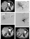
Figure 1
Figure 1
Transhepatic ipsilateral right portal vein embolization (PVE) using trisacryl particles and coils performed in a 49-year-old patient with colon cancer metastatic to the liver. (A) Computed tomography (CT) scan obtained prior to PVE shows marginal future liver remnant (FLR) (FLR-to-TELV [total estimated liver volume] ratio: 25%). (B) Anteroposterior flush portogram demonstrates a 6F vascular sheath in a right portal vein branch (arrowheads) and a 5F flush catheter within the main portal vein (arrow). (C) Selective right portogram with use of reverse-curve catheter placed prior to right PVE. (D) Final portogram shows occlusion of the portal vein branches to segments V through VIII with continued patency of the veins supplying the left lateral lobe. (E) CT scan obtained 1 month after right PVE shows substantial FLR hypertrophy (FLR-to-TELV ratio: 50%). The degree of hypertrophy is 25%. (F) CT scan after right hepatectomy demonstrates hypertrophy of remnant liver. (Used with permission from Avritscher R, de Baere T, Murthy R, Deschamps F, Madoff DC. Percutaneous transhepatic portal vein embolization: rationale, technique, and outcomes. Semin Intervent Radiol 2008;25(2):132–145.)
Figure 2
Figure 2
Transhepatic ipsilateral right portal vein embolization (PVE) extended to segment IV using trisacryl particles and coils in a 59-year-old woman with a history of gallbladder carcinoma. (A) Computed tomography (CT) scan obtained prior to PVE shows marginal future liver remnant (FLR) (FLR-to-TELV [total estimated liver volume] ratio: 17%). (B) Anteroposterior flush portogram demonstrates a 6F vascular sheath in a right portal vein branch (arrowheads) and a 5F flush catheter within the main portal vein (arrow). (C) Selective left portogram prior to segment IV embolization shows the veins that supply segments II, III, and IV. (D) Selective portography via a 3F microcatheter (arrowheads) during embolization of segment IVa. (E) Selective left portogram via a 5F catheter (arrow) after segment IV embolization (arrowheads show coils within proximal branches of segment IV). (F) Selective venogram via a 5F reverse-curve catheter with tip in the right portal vein (arrow). Note multiple previously placed coils within segment IV branches (arrowheads). (G) Final portal venogram shows occlusion of the portal vein branches to segments IV through VIII with continued patency of the veins supplying the left lateral lobe. (H) CT scan obtained 1 month after PVE shows substantial atrophy of right liver and segment IV, and FLR hypertrophy (FLR-to-TELV ratio: 26%). The degree of hypertrophy is 9%. The patient underwent successful extended hepatectomy. (Used with permission from Avritscher R, de Baere T, Murthy R, Deschamps F, Madoff DC. Percutaneous transhepatic portal vein embolization: rationale, technique, and outcomes. Semin Intervent Radiol 2008;25(2):132–145.)
Figure 3
Figure 3
Technique of transhepatic contralateral approach to right portal vein embolization using n-butyl cyanoacrylate mixed with ethiodized oil. (A) Ultrasound view of needle puncture of a segment III portal branch (arrowheads). (B) Anteroposterior flush portogram demonstrates a 5F flush catheter within the main portal vein (arrow). Note portal vein access through the left portal vein (arrowheads). (C) Single image obtained during fluoroscopy shows cast of embolic material (cyanoacrylate and lipiodol) within right portal vein branches (arrowheads). (D) Final portogram shows occlusion of the portal vein branches to segments V through VIII with continued patency of the veins supplying the left lateral lobe. The cast of embolic material is visualized in the treated right portal branches (arrows). (Used with permission from Avritscher R, de Baere T, Murthy R, Deschamps F, Madoff DC. Percutaneous transhepatic portal vein embolization: rationale, technique, and outcomes. Semin Intervent Radiol 2008;25(2):132–145.)

Similar articles

Cited by

References

    1. Jemal A, Siegel R, Xu J, Ward E. Cancer statistics, 2010. CA Cancer J Clin. 2010;60(5):277–300. - PubMed
    1. Bosch F X, Ribes J, Díaz M, Cléries R. Primary liver cancer: worldwide incidence and trends. Gastroenterology. 2004;127(5, Suppl 1):S5–S16. - PubMed
    1. Ng K K, Vauthey J N, Pawlik T M. et al.Is hepatic resection for large or multinodular hepatocellular carcinoma justified? Results from a multi-institutional database. Ann Surg Oncol. 2005;12(5):364–373. - PubMed
    1. Shoup M, Gonen M, D'Angelica M, Jamagin W R, DeMatteo R P, Schwartz L H. et al.Volumetric analysis predicts hepatic dysfunction in patients undergoing major liver resection. J Gastrointest Surg. 2003;7(3):325–330. - PubMed
    1. Ribero D, Abdalla E K, Madoff D C, Donadon M, Loyer E M, Vauthey J N. Portal vein embolization before major hepatectomy and its effects on regeneration, resectability and outcome. Br J Surg. 2007;94(11):1386–1394. - PubMed