. 2012 Jun;29(2):147-9.
doi: 10.1055/s-0032-1312577.

Splenic artery embolization in blunt trauma

Affiliations

Splenic artery embolization in blunt trauma

Brian F Imbrogno et al. Semin Intervent Radiol. 2012 Jun.
No abstract available


Figures

Figure 1
Figure 1
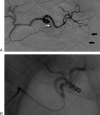
Distal splenic embolization in a 30-year-old man with lower pole grade V splenic injury. (A) Splenic arteriogram, anteroposterior (AP) projection, midarterial phase, demonstrating attenuation of intrasplenic branches supplying the inferior pole of the spleen, as well as mass effect by the intrasplenic hematoma evidence by splaying of the inferior pole branches (black arrows). Note the dorsal pancreatic artery (white arrow) and arterial pancreatica magna (open arrow), both of which provide important collateral pathways in the setting of proximal splenic artery embolizations. (B) Splenic arteriogram, AP projection, midarterial phase, after selective coil embolization of the lower pole splenic arterial branch. This is considered a distal splenic artery embolization because the embolization occurred beyond the splenic hilum and distal to any major potential collateral pathways.
Figure 2
Figure 2
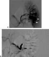
Proximal splenic embolization in a 42-year-old man after blunt traumatic injury to his spleen. A contrast-enhanced computed tomography scan (not shown) demonstrated a hypervascular enhancing structure just distal to the splenic hilum. (A) Splenic arteriogram, anteroposterior (AP) projection, midarterial phase, demonstrating hypervascular structure within the splenic parenchyma (white arrows) and an early draining splenic vein (open arrow). (B) Splenic arteriogram, AP projection, midarterial phase, following proximal coil embolization of the main splenic artery. Note the coils are placed just distal to the dorsal pancreatic artery (arrow).

References

    1. Smith H E Biffl W L Majercik S D Jednacz J Lambiase R Cioffi W G Splenic artery embolization: Have we gone too far? J Trauma 2006613541–544.; discussion 545–546 - PubMed
    1. Tinkoff G, Esposito T J, Reed J. et al.American Association for the Surgery of Trauma Organ Injury Scale I: spleen, liver, and kidney, validation based on the National Trauma Data Bank. J Am Coll Surg. 2008;207(5):646–655. - PubMed
    1. Shanmuganathan K, Mirvis S E, Boyd-Kranis R, Takada T, Scalea T M. Nonsurgical management of blunt splenic injury: use of CT criteria to select patients for splenic arteriography and potential endovascular therapy. Radiology. 2000;217(1):75–82. - PubMed
    1. Bessoud B, Denys A, Calmes J M. et al.Nonoperative management of traumatic splenic injuries: is there a role for proximal splenic artery embolization? AJR Am J Roentgenol. 2006;186(3):779–785. - PubMed
    1. Schnüriger B, Inaba K, Konstantinidis A, Lustenberger T, Chan L S, Demetriades D. Outcomes of proximal versus distal splenic artery embolization after trauma: a systematic review and meta-analysis. J Trauma. 2011;70:242–260. - PubMed