

Intramural hematoma of the esophagus
- PMID: 23730267
- PMCID: PMC3668800
- DOI: 10.1159/000341808
Intramural hematoma of the esophagus
Abstract
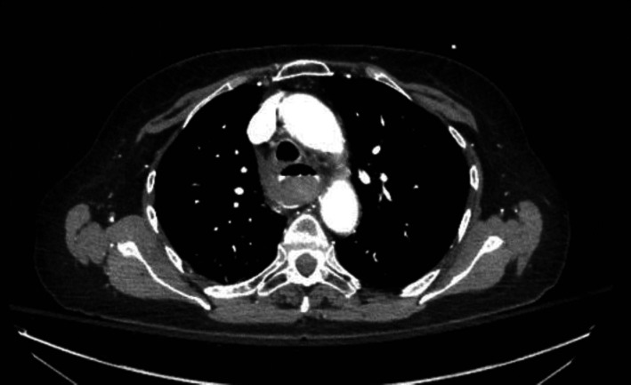
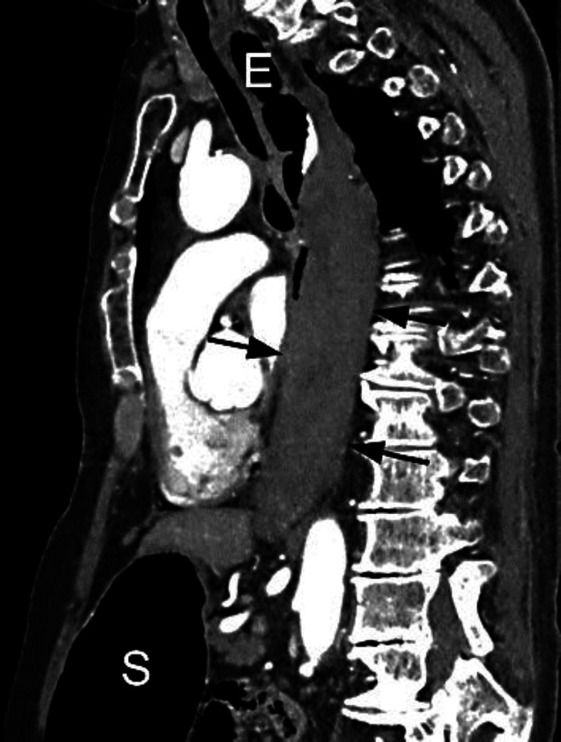
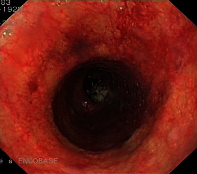
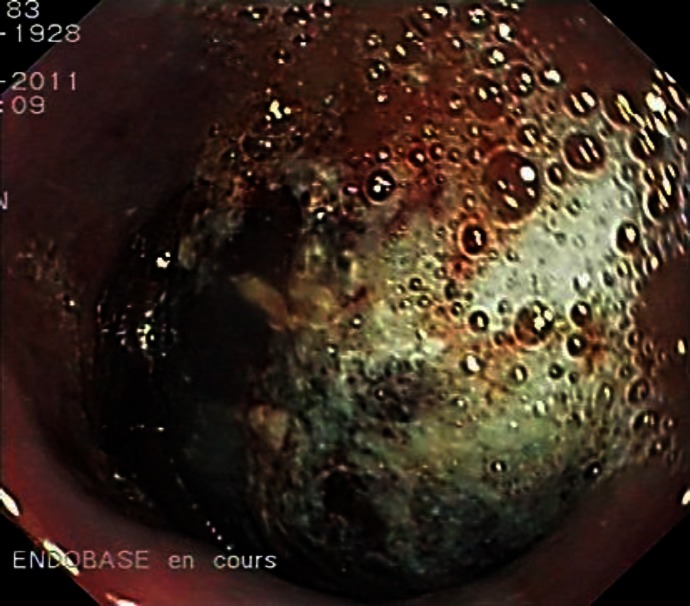
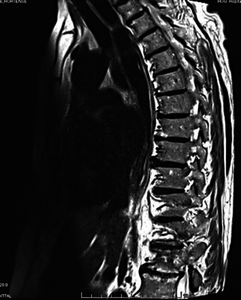
We report the case of a patient with an intramural hematoma of the esophagus. This rare condition is more common in elderly women and can be misdiagnosed as cardiovascular or other digestive emergent disease. The classical clinical triad includes chest pain, sudden dysphagia or odynophagia and minor hematemesis. Known precipitating factors are Valsalva maneuver, blunt, direct or iatrogenic injuries, but spontaneous cases have also been described. Chest imaging including computed tomography or magnetic resonance imaging as well as upper gastrointestinal endoscopy are useful tools for diagnosis. The treatment is conservative and the prognosis usually excellent with complete resolution within a few weeks.
Keywords: Esophagus; Hematoma; Intramural.
Figures
References
-
- Cullen SN, McIntyre AS. Dissecting intramural haematoma of the esophagus. Eur J Gastroenterol Hepatol. 2000;12:1151–1162. - PubMed
-
- Meulman N, Evans J, Watson A. Spontaneous intramural haematoma of the oesophagus; a report of three cases and review of the literature. Aust N Z J Surg. 1994;64:190–193. - PubMed
-
- Ackert JJ, Sherman A, Lustbader IJ, McCauley DI. Spontaneous intramural hematoma of the esophagus. Am J Gastroenterol. 1989;84:1325–1328. - PubMed
-
- Katabathina V, Restrepo CS, Martinez-Jimenez S, Riascos RF. Nonvascular, nontraumatic mediastinal emergencies in adults: a comprehensive review of imaging findings. Radiographics. 2011;31:1141–1160. - PubMed
-
- Van Laethem JL, Devière J, Cremer M. Serial endoscopic findings of spontaneous intramural hematoma of the esophagus. Endoscopy. 1997;29:44–46. - PubMed
Publication types
LinkOut - more resources
Full Text Sources
