

doi: 10.1055/s-0032-1313363.
Superior orbital fissure syndrome: a case report
Affiliations
- PMID: 23730429
- PMCID: PMC3444026
- DOI: 10.1055/s-0032-1313363
Item in Clipboard
Superior orbital fissure syndrome: a case report
Craniomaxillofac Trauma Reconstr.
2012 Jun.
Abstract
Superior orbital fissure syndrome is an infrequently encountered entity with a unique presentation and significant morbidity. This article reviews the background of the syndrome, treatments in the literature, and discusses a recent case with treatment strategy.
Keywords: midface; superior orbital fissure syndrome; trauma; zygomaticomaxillary complex fracture.
Figures
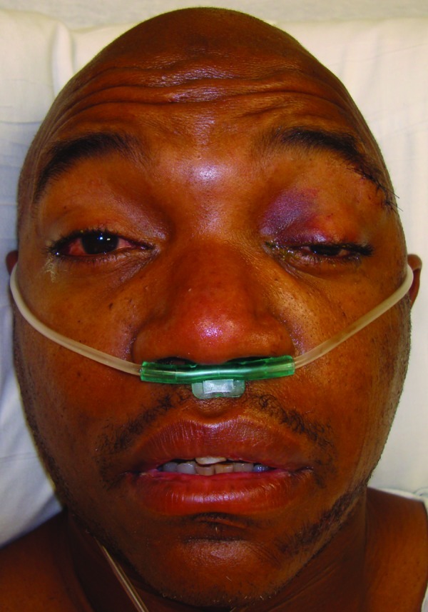
Initial presentation of the patient s/p fall. Note the typical presentation of the superior orbital fissure syndrome.
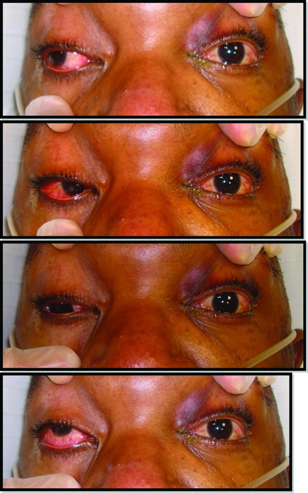
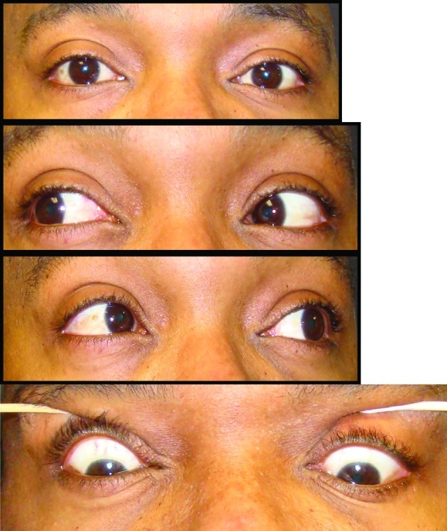
Clinical examination photos documenting ophthalmoplegia of the left eye.
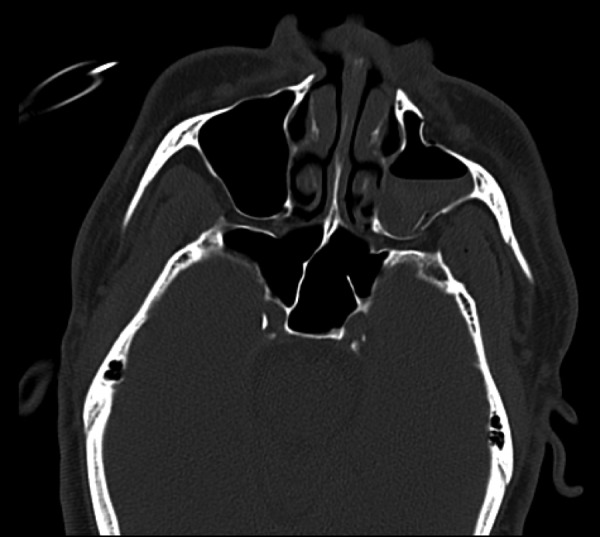
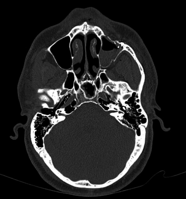
Maxillofacial computed tomography scan:, axial cut, bony window, at the level of the zygomatic arches showing left displaced zygomaticomaxillary complex fracture.
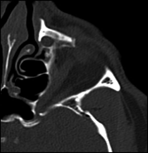
Enhanced view of the left superior orbital fissure from the previous image. Note the constriction and impingement of the superior orbital fissure.
Postoperative maxillofacial computed tomography showing reduction of the left zygomaticomaxillary complex fractures.
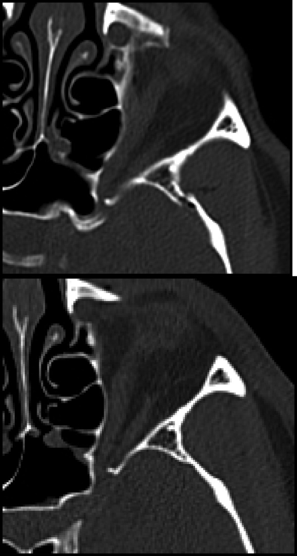
Comparison of the pre- and postoperative maxillofacial computed tomographic scans. Both images are axial cuts, bony windows, at the approximate level of the superior orbital fissure. Note the second image demonstrating widening of the superior orbital fissure, verified by radiological report, with decreased bony compression and excellent reduction of the fracture segment.
Postoperative clinical photos. Note the resolved periorbital edema and ecchymosis, complete resolution of anisocoria, and normal ocular movement.
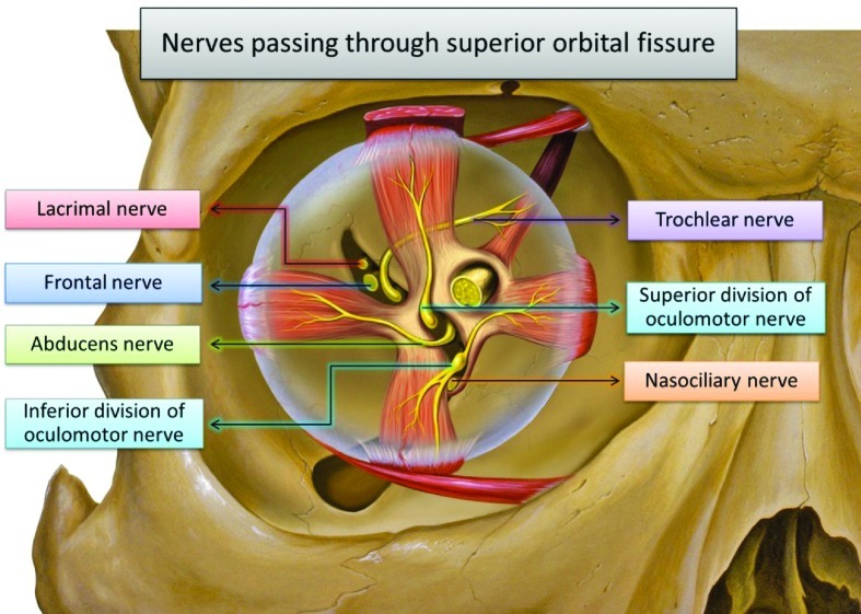
Anatomy of the superior orbital fissure and its contents.
References
-
- Kurzer A, Patel M P. Superior orbital fissure syndrome associated with fractures of the zygoma and orbit. Plast Reconstr Surg. 1979;64(5):715–719. - PubMed
-
- Acartürk S, Seküçoğlu T, Kesiktäs E. Mega dose corticosteroid treatment for traumatic superior orbital fissure and orbital apex syndromes. Ann Plast Surg. 2004;53(1):60–64. - PubMed
-
- Rohrich R J, Hackney F L, Parikh R S. Superior orbital fissure syndrome: current management concepts. J Craniomaxillofac Trauma. 1995;1(2):44–48. - PubMed
-
- Fujiwara T, Matsuda K, Kubo T, Tomita K, Yano K, Hosokawa K. Superior orbital fissure syndrome after repair of maxillary and naso-orbito-ethmoid fractures: a case study. J Plast Reconstr Aesthet Surg. 2009;62(12):e565–e569. - PubMed
-
- Hedstrom J, Parsons J, Maloney P L, Doku H C. Superior orbital fissure syndrome: report of case. J Oral Surg. 1974;32(3):198–201. - PubMed
LinkOut - more resources
Full Text Sources