. 2013 Aug;81(8):2920-30.
doi: 10.1128/IAI.00258-13. Epub 2013 Jun 3.

Gallbladder epithelium as a niche for chronic Salmonella carriage

Affiliations

Gallbladder epithelium as a niche for chronic Salmonella carriage

Geoffrey Gonzalez-Escobedo et al. Infect Immun. 2013 Aug.

Abstract

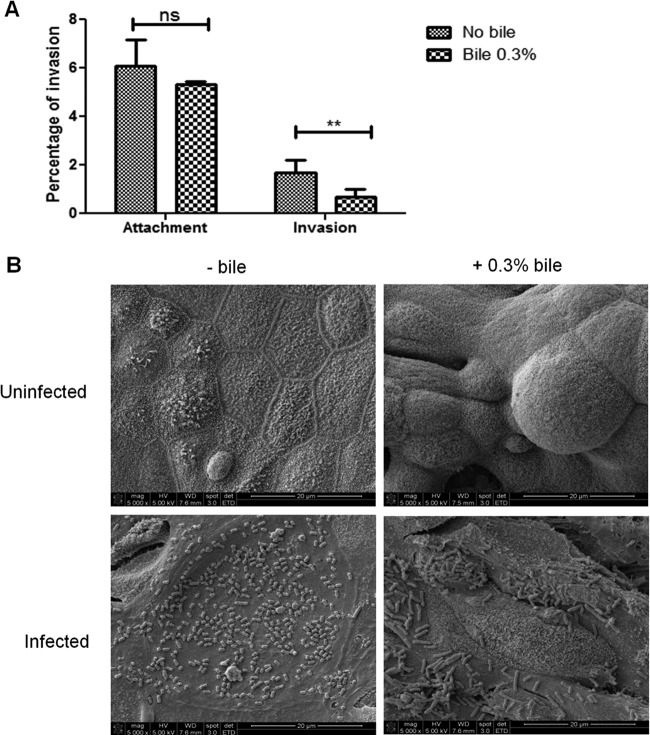
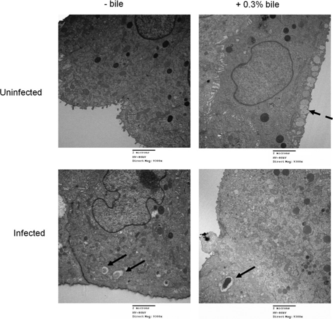
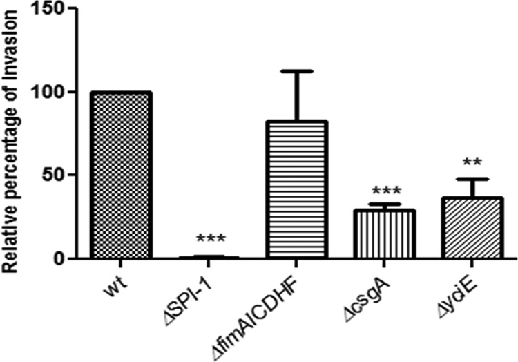
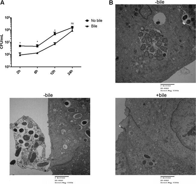
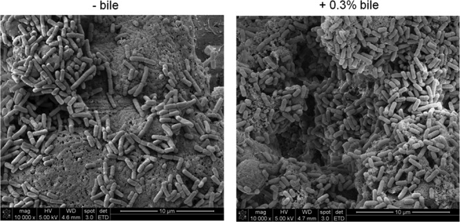
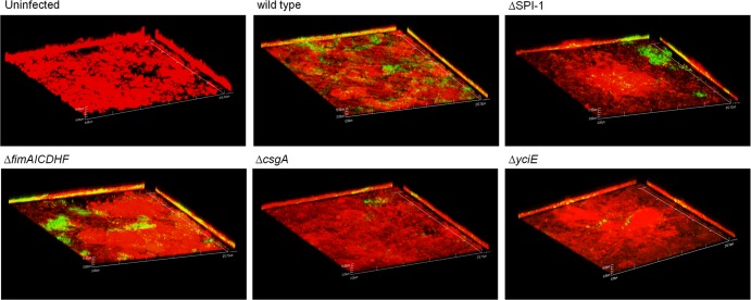
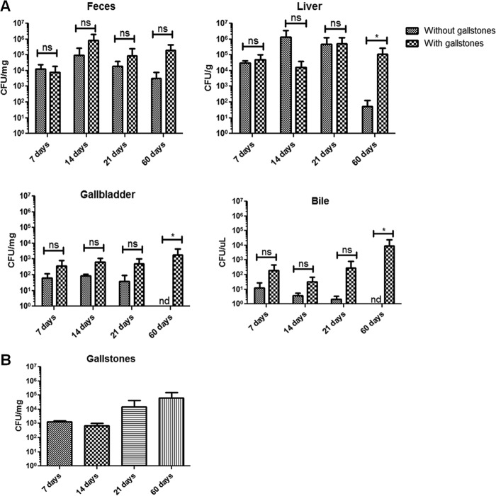
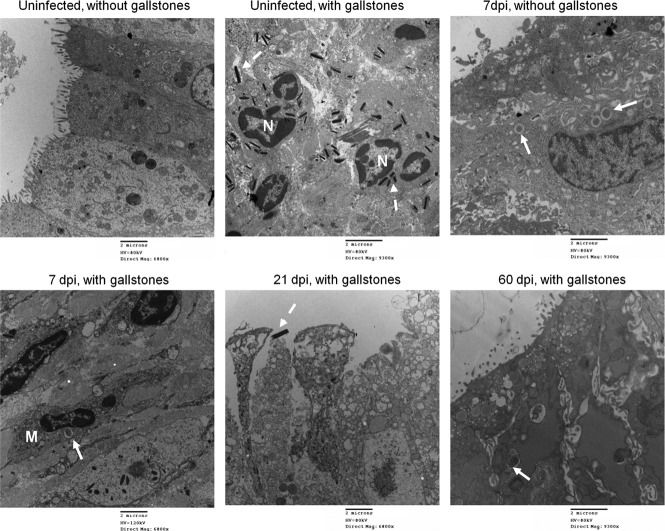
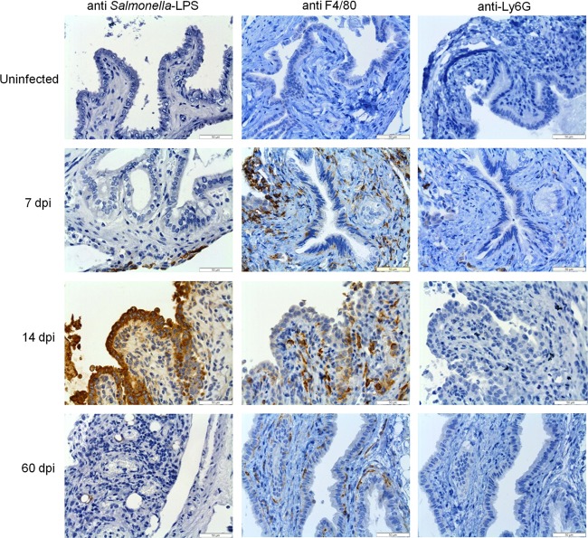
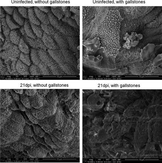
Although typhoid fever has been intensively studied, chronic typhoid carriage still represents a problem for the transmission and persistence of the disease in areas of endemicity. This chronic state is highly associated with the presence of gallstones in the gallbladder of infected carriers upon which Salmonella can form robust biofilms. However, we hypothesize that in addition to gallstones, the gallbladder epithelium aids in the establishment/maintenance of chronic carriage. In this work, we present evidence of the role of the gallbladder epithelium in chronic carriage by a mechanism involving invasion, intracellular persistence, and biofilm formation. Salmonella was able to adhere to and invade polarized gallbladder epithelial cells apically in the absence and presence of bile in a Salmonella pathogenicity island 1 (SPI-1)-dependent manner. Intracellular replication of Salmonella was also evident at 12 and 24 h postinvasion. A flowthrough system revealed that Salmonella is able to adhere to and form extensive bacterial foci on gallbladder epithelial cells as early as 12 h postinoculation. In vivo experiments using a chronic mouse model of typhoid carriage showed invasion and damage of the gallbladder epithelium and lamina propria up to 2 months after Salmonella infection, with an abundant presence of macrophages, a relative absence of neutrophils, and extrusion of infected epithelial cells. Additionally, microcolonies of Salmonella cells were evident on the surface of the mouse gallbladder epithelia up to 21 days postinfection. These data reveal a second potential mechanism, intracellular persistence and/or bacterial aggregation in/on the gallbladder epithelium with luminal cell extrusion, for Salmonella maintenance in the gallbladder.


Figures

Fig 1
Fig 1
S. Typhimurium attaches and invades into/on polarized DGEC in vitro. (A) Apical attachment and invasion (mean plus standard deviation) of S. Typhimurium bacteria on/into DGEC previously exposed or not exposed to 0.3% bile. Invasion after 2.5 h is shown as a percentage of the inoculum. Exposure of the epithelial cells to bile significantly decreased bacterial invasion but not attachment. Means between conditions with (+) and without (−) bile treatment were compared by a Student's t test (**, P < 0.01; ns, not significant). Experiments were performed in triplicate and repeated four times. (B) S. Typhimurium attaches to the surface of polarized DGEC, resembling microcolonies. Representative SEM images of uninfected and infected DGEC in the absence and presence of 0.3% bile are shown. Note the differentiation when cells were exposed to bile. Cells were fixed and processed at 2 h postinfection.
Fig 2
Fig 2
Exposure of DGEC to bile decreases invasion by S. Typhimurium. Representative TEM images showing uninfected or S. Typhimurium-infected DGEC in the absence or presence of bile are shown. Solid arrows, Salmonella-containing vacuoles; dashed arrows, mucus granules that were only present in DGEC exposed to bile. Cells were fixed and processed at 2 h postinfection.
Fig 3
Fig 3
SPI-1, curli fimbriae, and yciE but not type 1 fimbria mutants decrease invasion into DGEC in vitro. Invasion relative to the wild-type (wt) level (mean plus standard deviation) of S. Typhimurium mutants into DGEC exposed to 0.3% bile at 2.5 h postinfection. Means of wild-type and mutant values were compared by a Student t test (**, P < 0.01; ***, P < 0.001). Assays were performed in triplicate on two separate occasions.
Fig 4
Fig 4
S. Typhimurium can replicate intracellularly in DGEC. (A) Intracellular replication of S. Typhimurium in DGEC cultured with or without bile measured as CFU/ml. There is statistical significance between no bile and bile treatments at all time points except 24 h postinfection (*, P < 0.05, by a Student's t test). (B) Representative TEM images showing infected DGEC at 9 h postinfection with S. Typhimurium in the absence or presence of bile. In the absence of bile, bacteria were seen replicating inside SCV in higher numbers than in cells exposed to bile. Cell/cell debris being released to the extracellular environment was also observed.
Fig 5
Fig 5
S. Typhimurium forms extensive cellular aggregations/biofilms on the surface of DGEC. Representative SEM images of S. Typhimurium biofilms on DGEC at 12 h postinoculation in a flow chamber in the absence or presence of 0.3% bile are shown. The presence of bile modestly increased biofilm formation.
Fig 6
Fig 6
Curli and yciE affect biofilm formation on DGEC. Representative confocal images are shown of DGEC uninfected or infected with wild-type S. Typhimurium and mutants at 9 h postinfection in the presence of bile. All bacterial strains harbor the plasmid pFPV25.1 constitutively expressing GFP. DGEC are stained with Cell Tracker Red CMPTX. All images have a magnification of ×60.
Fig 7
Fig 7
The presence of gallstones enhances Salmonella colonization at 60 days postinfection. (A) S. Typhimurium enumeration in different mouse sites at 7, 14, 21, and 60 days postinfection. Although all sites showed more bacteria in mice harboring gallstones, the difference was only significant by a Student's t test at 60 dpi in the liver, gallbladder tissue, and bile (*, P < 0.05). (B) S. Typhimurium enumeration on gallstones at different days postinfection. Bacteria were recovered in higher numbers from gallstones at later time points (21 and 60 dpi), but this difference was not significant (analysis of variance test) from amounts at earlier time points (7 and 14 dpi). Data were gathered from two different experiments. ns, not significant. nd, not detected.
Fig 8
Fig 8
In vivo assays recapitulate invasion, intracellular survival, and extrusion. Representative TEM images of the mouse gallbladder epithelium in the absence and the presence of gallstones are shown. Damage of the gallbladder epithelium is evident at all days postinfection including in uninfected mice with gallstones. SCV are indicated with solid arrows and were observed containing only one or two bacteria. Dashed arrows indicate cholesterol crystals. Macrophages (M) were sometimes observed harboring SCV. Neutrophils (N) were actively seen engulfing cholesterol crystals. The release of epithelial cells was commonly seen, especially at 21 dpi. Regeneration of tight junctions was evident especially by 60 dpi. However, Salmonella was still seen intracellularly in mice harboring gallstones.
Fig 9
Fig 9
Inflammation of the gallbladder is mostly macrophage related during infection in the absence of gallstones. Images show immunohistochemical analysis of representative sections of the gallbladder epithelium in the absence of gallstones from uninfected or infected mice at 7, 14, and 60 dpi by using anti-Salmonella LPS, anti-F4/80 (macrophage marker), and anti-Ly6G (neutrophil marker) antibodies. Brown areas are positive. The recruitment of macrophages was the hallmark throughout the course of infection.
Fig 10
Fig 10
S. Typhimurium can form microcolonies on the surface of the mouse gallbladder epithelium. Representative SEM images of uninfected/infected mouse gallbladder epithelium in the absence or presence of gallstones are shown. Salmonella microcolonies were observed on the epithelium only of mice harboring gallstones and only at 21 dpi.

References

    1. Crump JA, Luby SP, Mintz ED. 2004. The global burden of typhoid fever. Bull. World Health Organ. 82:346–353 - PMC - PubMed
    1. Vazquez-Torres A, Jones-Carson J, Baumler AJ, Falkow S, Valdivia R, Brown W, Le M, Berggren R, Parks WT, Fang FC. 1999. Extraintestinal dissemination of Salmonella by CD18-expressing phagocytes. Nature 401:804–808 - PubMed
    1. Levine MM, Black RE, Lanata C. 1982. Precise estimation of the numbers of chronic carriers of Salmonella Typhi in Santiago, Chile, an endemic area. J. Infect. Dis. 146:724–726 - PubMed
    1. Merselis JG, Jr, Kaye D, Connolly CS, Hook EW. 1964. Quantitative bacteriology of the typhoid carrier state. Am. J. Trop. Med. Hyg. 13:425–429 - PubMed
    1. Vogelsang TM, Boe J. 1948. Temporary and chronic carriers of Salmonella Typhi and Salmonella Paratyphi B. J. Hyg. (Lond.) 46:252–261 - PMC - PubMed

Publication types

LinkOut - more resources