Review
. 2013 Jun 1;5(6):a011072.
doi: 10.1101/cshperspect.a011072.

Mechanisms of mitochondrial fission and fusion

Affiliations
Review

Mechanisms of mitochondrial fission and fusion

Alexander M van der Bliek et al. Cold Spring Harb Perspect Biol. .

Abstract

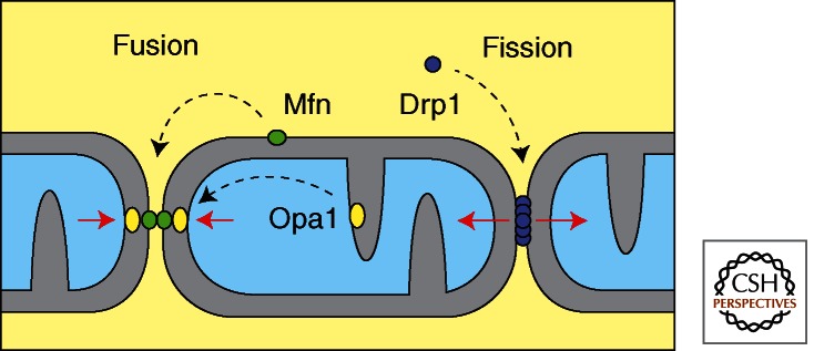
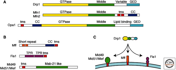
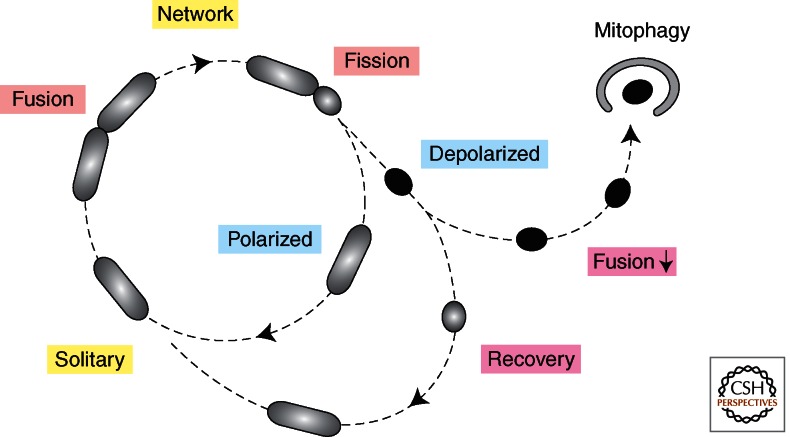
Mitochondria continually change shape through the combined actions of fission, fusion, and movement along cytoskeletal tracks. The lengths of mitochondria and the degree to which they form closed networks are determined by the balance between fission and fusion rates. These rates are influenced by metabolic and pathogenic conditions inside mitochondria and by their cellular environment. Fission and fusion are important for growth, for mitochondrial redistribution, and for maintenance of a healthy mitochondrial network. In addition, mitochondrial fission and fusion play prominent roles in disease-related processes such as apoptosis and mitophagy. Three members of the Dynamin family are key components of the fission and fusion machineries. Their functions are controlled by different sets of adaptor proteins on the surface of mitochondria and by a range of regulatory processes. Here, we review what is known about these proteins and the processes that regulate their actions.


Figures

Figure 1.
Figure 1.
Functions of the mitochondrial Dynamin family members. Mitofusins mediate mitochondrial outer membrane fusion in mammals. Opa1 mediates mitochondrial inner membrane fusion. Drp1, which cycles between the cytosol and the mitochondrial outer membrane, mediates mitochondrial fission.
Figure 2.
Figure 2.
Schematic diagrams of fission and fusion proteins. (A) The main Dynamin family members in mammals, Drp1, Mfn1, Mfn2, and Opa1 are shown with their different protein domains. Each one has a GTPase domain, a middle domain, and a GED, which together form the canonical Dynamin structure. The GED in Mitofusins is not as well conserved, but it still has coiled-coil segments, suggesting that it can participate in multimeric assembly. The variable domain in Drp1, which most likely serves as a mitochondrial targeting sequence, is replaced by trans-membrane segments (tms) in the Mitofusins and by a cardiolipin binding domain in Opa1. (B) Adaptor proteins that can bind to drp1 on the surface of mitochondria. Each one has a membrane anchor (tms), but they have different protein interaction domains, suggesting that they are functionally distinct. (C) Illustration of the multiplicity of Drp1 receptors on the surface of mammalian mitochondria.
Figure 3.
Figure 3.
Proteolytic cleavage sites in mammalian Opa1. (A) Protein domains in Opa1. The amino terminus has a mitochondrial leader sequence, followed by a mitochondrial processing peptidase cleavage site (MPP), a trans-membrane segment (tms) and several alternatively spliced exons. (B) Alternatively spliced exons have three more cleavage sites. S2 and S3 sites are constitutively cleaved by Yme1L, giving rise to S-Opa1 (band d). Isoforms lacking exons 4b and 5b are not normally cleaved, giving rise to L-Opa1 (bands a and b). (C) Oma1 cleaves the S1 site in isoforms that were not already cleaved by Yme1L, but only when mitochondria lose membrane potential. Bands a and b disappear, giving rise to the intermediate form band c and the final product band e. (The blots were from Griparic et al. 2007; reprinted, with permission, from the authors.)
Figure 4.
Figure 4.
Mitochondrial life cycle. Live cell imaging showed that mitochondria often exist as solitary units. They do occasionally fuse to other mitochondria but fusion is often followed within 20 min by fission at the same place. Fission can give rise to daughter mitochondria with different membrane potentials, suggesting that fission is preceded by a sorting event. Daughter mitochondria with lower membrane potential often recover, allowing them to rejoin the mitochondrial network by fusion, but persistently low membrane potential will inhibit fusion and cause elimination through mitophagy. (The figure was created from data adapted from Twig et al. 2008.)

References

    1. Alexander C, Votruba M, Pesch UE, Thiselton DL, Mayer S, Moore A, Rodriguez M, Kellner U, Leo-Kottler B, Auburger G, et al. 2000. OPA1, encoding a dynamin-related GTPase, is mutated in autosomal dominant optic atrophy linked to chromosome 3q28. Nat Genet 26: 211–215 - PubMed
    1. Alto NM, Soderling J, Scott JD 2002. Rab32 is an A-kinase anchoring protein and participates in mitochondrial dynamics. J Cell Biol 158: 659–668 - PMC - PubMed
    1. Arnoult D, Grodet A, Lee YJ, Estaquier J, Blackstone C 2005. Release of OPA1 during apoptosis participates in the rapid and complete release of cytochrome c and subsequent mitochondrial fragmentation. J Biol Chem 280: 35742–35750 - PubMed
    1. Baricault L, Segui B, Guegand L, Olichon A, Valette A, Larminat F, Lenaers G 2007. OPA1 cleavage depends on decreased mitochondrial ATP level and bivalent metals. Exp Cell Res 313: 3800–3808 - PubMed
    1. Bereiter-Hahn J, Voth M 1994. Dynamics of mitochondria in living cells: Shape changes, dislocations, fusion, and fission of mitochondria. Microsc Res Tech 27: 198–219 - PubMed

LinkOut - more resources